The Role of Modern Wound Dressings in Stage I Pressure Ulcers and Patients at Risk of Pressure Ulcer Formation
←
→
Page content transcription
If your browser does not render page correctly, please read the page content below
The Role of Modern Wound
 Dressings in Stage I Pressure
 Ulcers and Patients at Risk of
 Pressure Ulcer Formation

                               Helen Shaw, BSc (Hons),
                    ConvaTec Research and Development
                        Rachel Mathison, Msc, BSc (Hons),
ConvaTec Medical Affairs Manager Wound Therapeutics UKI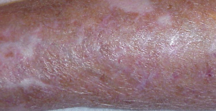
Introduction:
    “A pressure ulcer is any lesion caused by unrelieved pressure resulting in
    damage of underlying tissue. Pressure ulcers are usually located over bony
    prominences and are graded or staged to classify the degree of damage.”1
    Pressure ulcer incidence is sufficiently high, especially among certain high-risk groups, to warrant
    concern among health care providers. Pressure ulcers can be a frequent and costly problem and are
    common in acute care, home care and nursing home environments. An estimated 1.3 to 3 million adults
    have a pressure ulcer and in 2006 a publication stated a cost estimate of $70,000 to manage each
    Stage III or IV pressure ulcer. As prevention of this debilitating condition is strongly considered to be
    less costly than its treatment, guidelines for health care clinicians to prevent and predict pressure ulcer
    occurrence have been written to provide effective prevention measures and thus reduce the incidence
    of pressure ulcers.1

    A series of complex internal causative factors dictate the risk of pressure ulcer formation, including:-
    malnutrition, age, mobility, mental status and neuropathy. Pressure is the external causative factor in
    pressure ulcer formation, however additional factors such as:- shear forces, friction, moisture and any
    other factor that may contribute to the loss of skin integrity should also be considered. Whilst these
    additional factors alone do not account for the underlying tissue damage, they can accelerate ulceration
    i.e. progression from Stage I to Stage II. Additionally, the loss of barrier function of the skin increases
    the complexity of clinical care and increases the risk of infection.

    The NPUAP (US National Pressure Ulcer Advisory Panel) and EPUAP (European Pressure Ulcer Advisory
    Panel) have issued a set of guidelines to assist practitioner and patient decisions about appropriate
    healthcare. Whilst these guidelines may not be appropriate for use in all circumstances, they provide a
    good consensus on best practice and cover a wide range of recommendations, including:-

      • Risk Assessment
      • Skin Assessment
      • Nutrition and Pressure Ulcer Prevention
      • Repositioning for Pressure Ulcer Prevention
      • Support Surfaces
      • Special Population: Patients in the operating room

2                                                                                                            3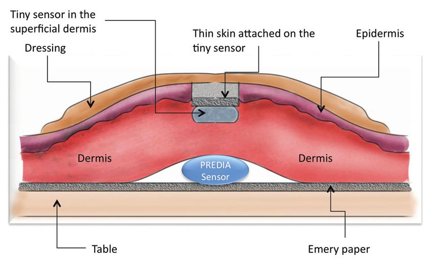
The NPUAP and EPUAP have also agreed on a pressure ulcer classification, Table
12 which classifies pressure ulcers on the level of injury as first proposed by Shea.5
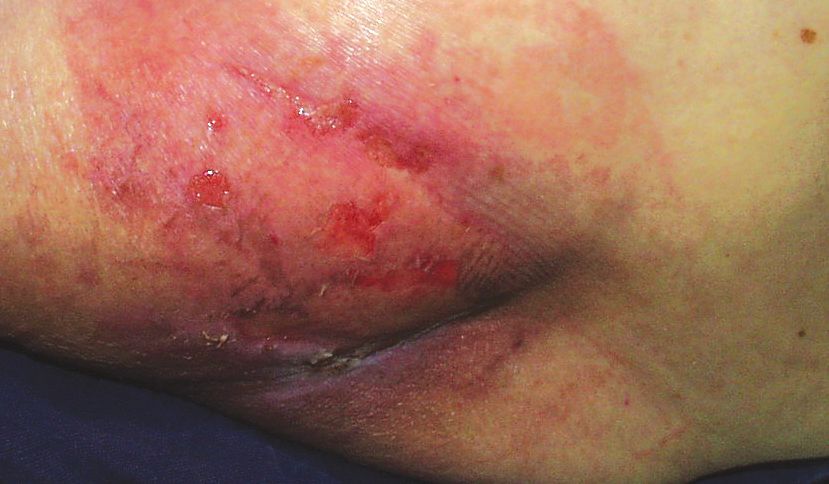
Table 1: Dressings are used in current practice as part of a protocol of care for the management of pressure
ulcers. With a wide range of dressings available, the practitioner will make dressing choices based upon
Category/ Intact skin with non-blanchable redness of a localised area usually over a bony many factors including: ability to absorb exudate, debride sloughy / necrotic tissue, barrier properties,
prominence. Darkly pigmented skin may not have visible blanching; its colour adhesiveness and patient comfort. Hydrocolloid and film dressings are indicated for Stage I pressure
Stage I: may differ from the surrounding area. The area may be painful, firm, soft, warmer,
Non-blanchable ulcers, where the skin is still intact, as stated in table 1, as their function is to protect the vulnerable area
or cooler as compared to adjacent tissue. Category I may be difficult to detect in
Erythema individuals with dark skin tones. May indicate “at risk” persons. from shear force, friction and moisture. The type of adhesive should also be selected carefully so not to
cause any additional skin damage or irritation to the area.
Partial Thickness loss of dermis presenting as a shallow open ulcer with a red / pink
Barrier products, such as protective emollients and film forming barriers, are also commonly used to
Category/ wound bed, without slough. May also present as an intact or open / ruptured serum-
Stage II: filled or sero-sanginous filled blister. Presents as a shiny or dry shallow ulcer without maintain good skin integrity and protect the skin in areas that are at risk of pressure ulcer formation.
Partial slough or bruising.* This category should not be used to describe skin tears, tape The primary purpose of these products is to provide a barrier to excess moisture or urinary / faecal
burns, incontinence associated dermatitis, maceration or excoriation. incontinence and to keep the skin hydrated. Dry skin seems to be a significant independent risk factor
Thickness
* Bruising indicates deep tissue injury for pressure ulcer development.2
Full thickness tissue loss. Subcutaneous fat may be visible but bone, tendon or This document will consider the external factors which influence the formation of pressure ulcers and
muscle are not exposed. Slough may be present but does not obscure the depth the role of a dressing as part of a protocol of care for the prevention of pressure ulcers in areas which
Category/ of tissue loss. May include undermining or tunnelling. The depth of a Category / are considered to be at risk of pressure ulcer formation and in Stage I pressure ulcers.
Stage III: Stage III pressure ulcer varies by anatomical location. The bridge of the nose, ear,
Full Thickness occiput and malleolus do not have (adipose) subcutaneous tissue and Category /
Skin Loss Stage III ulcers can be shallow. In contrast, areas of significant adiposity can Static Pressure
develop extremely deep Category / Stage III pressure ulcers. Bone / tendon is
not visible or directly palpable. The primary cause of pressure ulcers is static pressure applied to both the skin and underlying tissue.
When this pressure is greater than the blood pressure within the capillaries, blood flow is impeded.
Full thickness tissue loss with exposed bone, tendon or muscle. Slough or
eschar may be present, Often includes undermining and tunnelling. The depth
Maintaining interface pressures below capillary closing pressure (for example 32mmHg) is considered
Category/ of a category / stage IV pressure ulcer varies by anatomical location. The bridge to be the gold standard for pressure relief.6 Sustained and sufficient pressure to disrupt blood flow
Stage IV: of the nose, ear, occiput and malleolus do not have (adipose) subcutaneous tissue results in hypoxia, localised ischemia and tissue acidosis, leading to cellular necrosis. Pressure ulcers
Full Thickness and these ulcers can be shallow. Category / Stage IV ulcer can extend into muscle typically occur over bony prominences, however occasionally they can occur in soft tissue areas
Tissue Loss and / or supporting structures (e.g., fascia, tendon or joint capsule) making
osteomyelitis or osteitis likely to occur. Exposed bone / muscle is visible or
due to the effect of foreign objects, such as a medical device. Muscle has been shown to withstand
directly palpable. pressure loads of around 50mmHg for long periods.7
Unstageable/ Full thickness tissue loss in which actual depth of the ulcer is completely obscured
Unclassified: by slough (yellow, tan, grey, green or brown) and / or eschar (tan, brown or black)
in the wound bed. Until enough slough and / or eschar are removed to expose
Full Thickness the base of the wound, the true depth cannot be determined; but it will be either
Skin or Tissue a Category / Stage III or IV. Stable (dry, adherent, intact without erythema or Figure 1: Tissue under Pressure – Adapted
Loss – Depth fluctuance) eschar on the heels serves as “the body’s natural (biological) cover” from Clinical Practice Guidelines # 3, Agency
Unknown and should not be removed. for Health Care Policy and Research, US
Department of Health and Human Services.8
Purple or maroon localised area of discoloured intact skin or blood- filled blister
due to damage of underlying soft tissue from pressure and / or shear. The area
Suspected Deep may be preceded by tissue that is painful, firm, mushy, boggy, warmer or cooler
Tissue Injury – as compared to adjacent tissue. Deep tissue injury may be difficult to detect in
Depth Unknown individuals with dark skin tones. Evolution may include a thin blister over a dark
wound bed. The wound may further evolve and become covered in eschar. Evolution
may be rapid exposing additional layers of tissue even with optimal treatment.
4 5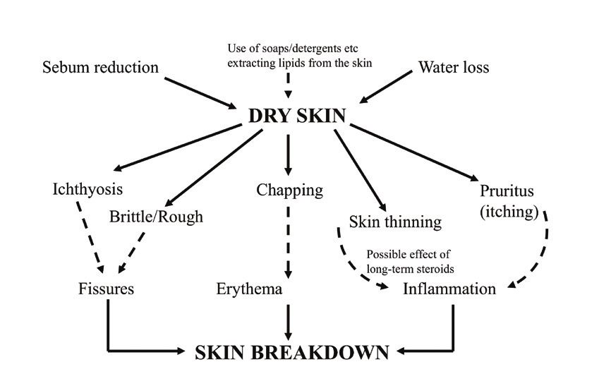
For immobile patients, or patients suffering prolonged periods of immobility, dressings alone have a                     AQUACEL™ Foam dressing is designed to help reduce the risk of
limited (or no) role in alleviating the level of static pressure when compared to the effects of regular                   skin breakdown by absorbing exudate and providing a low
patient re-positioning or specifically designed pressure-relieving devices, such as pressure-relieving                                     co-efficient of friction.
mattresses or pressure off-loading devices. The phenomenon of “bottoming out” of static support
surfaces has been detailed in other papers, and for static foam mattress overlays, thicknesses of
                                                                                                               Nakagami et al11 compared interface pressures and shear forces over the heel in a pressure ulcer
3-4 inches are typical.1
                                                                                                               preventative dressing and a thin film dressing in a clinical setting. In the study 30 hospitalised
Torra I Bou et al9 conducted an experimental study designed to calculate the level of pressure                 elderly patients participated. The results of the study showed that a dressing with a low friction
before and after the application of a Hydrocellular dressing in the area of the sacrum, ischium and            external surface can significantly reduce shear force, however it did not significantly reduce interface
heel of three healthy volunteers. Measurements were taken on two surfaces, a viscoelastic foam                 pressures.
mattress and a conventional hospital mattress. Pressure was determined by a Talley pressure
                                                                                                               Ohura et al12 developed an in vitro model to assess the impact of an external shear force and
monitor and repeated through a range of degrees of inclination. The overall average pressure
                                                                                                               pressure on a superficial layer of skin and subcutaneous tissue with an underlying bony prominence.
reductions after applying the Hydrocellular dressing, on all volunteers, at all inclinations and
                                                                                                               The model incorporated porcine skin, a Predia sensor capable of measuring shear and pressure
on both surfaces were 19.5% in the sacrum, 13.8% in the ischium and 20.15% in the heel.
                                                                                                               simultaneously and a small strain gauge shear sensor buried in the superficial dermis, as illustrated
Dressings may however have a larger role through helping to re-distribute pressure in other                    in Figure 3. An external 1Kg force with a cotton cloth interface was applied to this skin model. This
circumstances, where a specific off-loading device or support mattress may not be appropriate.                 external force was attached to a friction pull tester which pulled at a rate of 10cm per 30 seconds.
For example, this may be worthwhile when the use of another medical device may apply sustained
pressure to the patient (e.g. the use of an oxygen mask).

Shear Force
Shear force is another example of pressure than can be exerted onto the body of a patient.                                                                           Figure 3: Illustration of Ohura et al12
                                                                                                                                                                     pig skin model
Shear forces are produced when surfaces are slid across one another. In the case of a patient,
examples of shear forces are: when the angle of a bed or chair is changed, or when a patient
slides up or down a bed. These movements can result in pulling and stretching of the underlying
tissue and blood vessels.

                                                                                                               Five dressings were evaluated in this model: ALLEVYN™ ADHESIVE (Smith and Nephew), TIELLE™
                                                                                                               (Systagenix), Tegaderm™ (3M Healthcare), Development Opsite™ (product under development, Smith
                                                          Figure 2: Adapted from Clinical Practice             and Nephew) and DuoDERM™ CGF dressing (ConvaTec).
                                                          Guidelines # 3, Agency for Health Care
                                                          Policy and Research, US Department of                The static pressure recorded under the test dressings remained the same as the control (no dressing)
                                                          Health and Human Services.8
                                                                                                               within a range of 6.1 - 7.2mmHg. During weight movement, the pressure of the control raised to
                                                                                                               16.36mmHg, all dressings tested were shown to produce a 26-46% reduction compared to the
                                                                                                               control pressure in the subcutaneous layer.
Friction is created by movement of the patient across surfaces, such as clothes or bed linen.                  The mean control (no dressing) for shear force in the subcutaneous layer was 0.47N. The shear forces
Repeated movements can result in the superficial loss of epidermis and outer layers of the stratum             were reduced by 31 - 45% compared to the control, depending upon which dressing was applied,
corneum. This can result in abrasion-type wounds, which can produce considerable amounts of                    but with no significant differences between dressings. For all dressings tested, the shear force in the
exudate. The contents of this exudate can contribute to the adhesion of skin to a surface and thus             subcutaneous layer was reduced compared to the shear force within the superficial layer. DuoDERM™
can further increase shear forces. Areas subject to friction force are likely to develop wounds (or skin       CGF dressing was shown to have the lowest shear force in the superficial layer, when compared to
breakdown around wounds). Areas identified as at risk of friction forces include the heels, buttocks,          the other dressings tested.
sacrum, elbows and trochanters.10

                                                                                                           6                                                                                                           7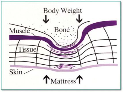
In vitro testing was performed at ConvaTec, Deeside, UK to determine the ability of AQUACEL™ Foam                                                       Just as the skin acts as a barrier, a product that provides a barrier to liquids offers a form of
and Mepilex™ Border dressings to reduce shear force transmitted from a force applied to the dressing                                                    protection to help maintain good skin integrity and to protect skin at risk from the damaging effects
surface through to the underlying tissue.                                                                                                               of incontinence, moisture and friction.

The test method was based upon the work performed by Ohura et al12, using a sled to create the
surface shear force, the speed of the testing and the same Predia pressure and shear force sensor                                                             The European Pressure Ulcer Advisory panel and National Pressure Ulcer Advisory Panel.
were replicated. This model has been limited to comparing the reduction in shear force transmitted                                                            Prevention and Treatment of Pressure Ulcers: Quick Reference Guide recommends
through the dressing alone, thus                                                                                                                              “Use a structured approach to risk assessment that includes a comprehensive
eliminating the variability of using                     8
                                               AQUACEL Foam            ™                                                                                      skin assessment to evaluate any alterations to intact skin. (Strength of
porcine skin. The pressure range               Mepilex Border
                                                         7
                                                                 ™

                                                                                                                                                              Evidence = C) Consider individuals with alterations to intact skin to be at
used within the model to determine                                                                                                                            risk of pressure ulcer development.”
the ability of a dressing to decrease                    6

shear forces with increasing applied                                                                                                                          Alteration in skin condition may include, dry skin, erythema, and other alterations. The
                                       Shear Force (N)

                                                         5
                                                                                                                                                              presence of non-blanching erythema also increases the risk of further pressure ulcer
load was increased.                                      4                                                                                                    development.
Within this in vitro model, the data                     3
                                                                                                                                                              “Protect the skin from exposure to excessive moisture with a barrier product in
shows at a 99.9% confidence level,                                                                                                                            order to reduce the risk of pressure damage. (Strength of Evidence = C)”
                                                         2
significantly less shear force was
transmitted through the AQUACEL™                         1                                                                                                    The mechanical properties of the stratum corneum are changed by the presence of
Foam dressing to the Predia sensor                                                                                                                            moisture and as a function of temperature.
                                                         0

compared to Mepilex™ Border                                  0   500          1000      1500        2000        2500

                                                                                        Weight of Sled plus Sand (g)
                                                                                                                         3000      3500      4000

dressings (p
AQUACEL™ Foam dressing is designed to
Alternatively, to maintain the skin’s normal softness and pliability, a 10-20% water content is needed                                                                                  stay in place for up to 7 days.
within the skin.15 When skin loses moisture, it becomes dry, flaky, chapped and less pliable (Figure 7).
Ulcers are more likely to develop in dry skin.14 If the stratum corneum is removed, the skin’s barrier                               When choosing a dressing, it is important that removal of the dressing does not strip the stratum
capability is lost, increasing water vapour loss (Transepidermal water loss) through the damaged area                                corneum or increase the risk of skin breakdown. In vitro bioadhesion studies18,19 have evaluated
and potentially allowing external fluid or agents (e.g. bacteria) into the tissue. Studies have shown that                           the adherence of fibroblast cells involved in the wound healing process to a wide range of wound
if the stratum corneum is removed, that the water vapour loss of forearm skin increases 100-fold, to                                 care products. To mimic peri-wound adherence, a modification of these studies has used epidermal
approximately that of a water layer alone.15                                                                                         keratinocyte cells to assess the potential for a dressing to cause skin trauma upon removal.

                                                                                                                                     Adult human keratinocyte cells were obtained and transferred into a sterile dish containing Hank’s
                                                                                                                                     balanced salt solution (HBSS). The tissue was washed and cut into small (3–5mm2) pieces which were
                                                             Figure 7: Dry skin                                                      then placed into 25cm2 tissue culture flasks containing media (Dulbecco’s Modified Eagle Medium
                                                                                                                                     (DMEM), supplemented with 10% foetal calf serum (FCS) (Sigma, UK), 20 mM Hepes buffer,
                                                                                                                                     100μg/ml gentamicin and 0.5μg/ml amphotericin B). Cell cultures were incubated at 37°C in a 5%
                                                                                                                                     CO2 / 95% air environment. Readiness for sub-culturing was determined by the extent of keratinocyte
                                                                                                                                     cell outgrowth (5–10 days). Cells were farmed successively in a 1:4 split ratio to passage 3–8 before
                             AQUACEL™ Foam dressing provides a controlled                                                            experimental use. Keratinocytes were harvested from stock dishes and plated out at 2x10⁵ cells/ml
                             Moisture Vapour Transmission Rate,17 which may                                                          in 6 well plates. A 1cm2 piece of each test dressing was cut from adhesive area and applied dry. All
                            help to reduce Trans Epidermal Water Loss (TEWL)*                                                        cut dressings were placed onto the monolayer of keratinocytes and pressed gently in place. After
                                                                                                                                     24 hours, the dressings were carefully removed from the surface of the culture, using minimal force
                                                                                                                                     to avoid damaging the cells or causing any additional cells to detach from the dressing. The cell
                                                                                                                                     numbers on each dressing were determined through trypsinisation and counting using a Neubauer
                                                                                                                                     cell counting chamber.

                                                                                                                                                                   90

                                                                                                                                                                   80

                                                                                                                                                                   70

                                                                                                                                            Cell Number x 1000
            Figure 8: Schematic of a WET skin environment and how       Figure 9: Schematic of a DRY skin environment and how
                                                                                                                                                                   60
            internal / external factors can cause skin breakdown.       internal / external factors can cause skin breakdown.
                                                                                                                                                                   50
Practitioners have a plethora of product choices to protect the skin from faecal and urinary
                                                                                                                                                                   40
incontinence, including petrolatum-based barrier creams and film-forming barrier creams which
can increase the hydration of at-risk skin.                                                                                                                        30

                                                                                                                                                                   20
Wound dressings with low friction, waterproof barriers offer an alternative option to the
practitioner. Dressings can be indicated for up to 7 days wear time, meaning that the practitioner                                                                 10
does not need to regularly re-apply barrier products such as emollients, which may rub off on bed                                                                  0
linen or clothing. Furthermore, when faecal or urinary incontinence is present, the dressing backing                                                                       AQUACEL™ Foam       Allevyn™ GENTLE Border     Mepilex™ Border   Biatain™ Silicone Pad

may be wiped clean, negating the need to use cleaning products which may themselves cause                                                                        Figure 10: Number of Keratinocyte cells adhering to dressing.
skin damage, together with the cleansing process itself potentially causing further breakdown of
the stratum corneum.                                                                                                                 In this in vitro model, significantly less keratinocyte cells were observed to adhere to the
                                                                                                                                     AQUACEL™ Foam dressing than to the other dressings tested.20
*As demonstrated in vitro
                                                                                                                                10                                                                                                                                  11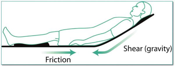
The information contained within Figure 11 is to help understand the risk factors associated with skin
AQUACEL™ Foam dressing is designed to help reduce breakdown and to develop care plans to reduce the likelihood that a clinically and economically costly
pain and trauma upon removal.20, 25 wound will develop.
In addition to the risk factors identified by ConvaTec Solutions™ Algorithms, other factors may pose
further risk for the development of chronic ulcers.1 Several scales may be used to assess an individual’s
The European Pressure Ulcer Advisory panel and National Pressure Ulcer Advisory Panel. risk, including the well-known Braden and Norton scales, or the lesser known Gosnell scale.24
Prevention and Treatment of Pressure Ulcers: Quick Reference Guide recommends
“Inspect skin regularly for signs of redness in individuals identified as being at risk
of pressure ulceration. The frequency of inspection may need to be increased in Medical Diagnosis High risk for impairment of skin integrity
response to any deterioration in overall condition. (Strength of evidence = B)” Assessments Reddened, intact skin
( ) Indicates the next step to take based
Ongoing assessment of the skin is necessary to detect early signs of pressure damage. on the assessment/intervention
Blanching Nonblanching
( ) Indicates that a decision has to be
made about which step to take
Risk factors*
One potential drawback of the use of dressings for skin protection in the case of pressure ulcer * Risk factors include, but are not limited to:
alteration in mobility and/or nutrition, pressure,
shear, friction, excess moisture (incontinence),
prevention is the balance between skin inspection and the cost of dressing removal/replacement. Yes and limited moisture (dry skin).
†
onblanching hyperemia may be indicative of
N
With the advancement of adhesive technologies, certain dressings can now be re-positioned,19* deep tissue damage that may be irreversible.
Goals of Nursing
allowing regular skin inspection without the increased cost of dressing changes. Care Plan
Maintain intact skin Maintain intact skin† This algorithm is based on currently accepted
standards of care and is intended for use
only as a guideline. Final treatment decisions
Nursing Action should always be consistent with the overall
Assess knowledge of patient/caregiver goal of patient care and be based upon
AQUACEL Foam dressing is designed to be
™ on risk of skin breakdown and
methods of prevention
consultation with a healthcare professional.
Adapted with permission from van Rijswijk L.
re-positionable,21 allowing inspection of intact skin.
Wound Care Policies and Procedures Manual.
Skillman, NJ: ConvaTec House Calls Total
Wound Management Program, 1995.
Reduce effect of risk factors
Clinical Practice Pressure Regular repositioning and pressure redistribution (pillows, devices)
Apply skin care products to lubricate (ConvaTec options: Aloe Vesta™ Skin
Conditioner, Sensi-Care™ Moisturizing Body Cream) or moisture-retentive
A clinical study implementing a comprehensive prevention program has been shown to reduce Shear/Friction dressings to reduce friction (ConvaTec options: DuoDERM™ Extra Thin dressings
pressure ulcer incidence by 87%. Since the current cost of treatment is estimated at $500 to $50,000 or DuoDERM Signal™ dressings)
Protect with skin barrier products (ConvaTec options: Aloe Vesta™ Protective
per ulcer, such prevention programs could significantly reduce healthcare costs.22 Excess moisture
Ointment, Aloe Vesta™ Protective Barrier Spray) or moisture-retentive dressings
to protect (ConvaTec options: DuoDERM™ Extra Thin dressings or DuoDERM Signal™
dressings) and incontinence products (ConvaTec option: Flexi-Seal™ FMS)
According to a publication from the US Agency for Healthcare Research and Quality (AHRQ), the Use moisturizing bathing (ConvaTec options: Aloe Vesta™ Body Wash &
ConvaTec Solutions™ Algorithm for Pressure Ulcer Prevention was described as a proven technique Dryness
Shampoo, Aloe Vesta™ Cleansing Foam, Aloe Vesta™ Bathing Cloths) and
skin conditioning products (ConvaTec options: Aloe Vesta™ Skin Conditioner,
that can be used to enhance patient outcomes.23 Sensi-Care™ Moisturizing Body Cream)
N u t r i t i o n a l t e r e d Nutritional support
Organisations are encouraged to develop a comprehensive program to prevent the development of
chronic wounds. These wounds can be quite costly, both financially and in terms of their impact on a
patient’s quality of life.
Expected Outcome
Skin remains intact without
signs of hyperemia
*Skin inspection without dressing change would only be recommended for intact skin. Figure 11: ConvaTec Solutions™ Algorithm (An updated pressure ulcer prevention algorithm is being validated and will be available in 2013.)
12 13Conclusions References
1. Clinical Practice Guidelines: Number 15 Treatment of Pressure Ulcers. Rockville , Md:US Department of Health and Human Services,
The incident rates, economics, and impact on the quality of life to the patient for pressure ulcers Agency for Health Care Policy and Research; 1994.AHCPR publication 95-06542.
are well documented and understood. As the primary cause of pressure ulcer formation is static 2. European Pressure Ulcer Advisory panel and National Pressure Ulcer Advisory Panel. Prevention and Treatment of Pressure Ulcers: Quick
Reference Guide. Washington DC: National Pressure Ulcer Advisory Panel; 2009.
pressure, the caregiver’s primary concern is to remove pressure from at-risk areas in order to help
prevent pressure ulcer formation, however several other factors also have a role to play in pressure 3. Lyder CH. Pressure Ulcer prevention and Management. JAMA. 2003 Jan ;289(2):223-225
ulcer formation. 4. Reddy M, Gill SS, Rochon PA. Preventing pressure Ulcers a systematic review. JAMA. 2006;296(8):974-984.
5. Shea, JD. Pressure sores classification and management. Clin Orthop Relat Res 1975, (112):89-100
The roles of shear forces and excess moisture as secondary contributing factors are well
6. Chronic Wound Care, Third Edition 2001 (P. 620). Krasner, Rodeheaver, Sibbald
documented. Whilst these factors alone do not directly cause pressure ulcers, they do soften
and damage the skin, making it more susceptible to further damage. 7. Gefen A. Reswick and Rogers pressure time curve for pressure ulcer risk. Part 2. Nursing Standard 2009; 23: 40-4
8. Clinical Practice Guidelines Number 3, Consumer Version. Preventing Pressure Ulcers. Rockville, Md:US Dept of Health and Human
Hydrocolloid Dressings and skin care products have played a role in the prevention of pressure Services, Agency for Healthcare Policy and Research:1992. AHCPR publication 92-0048
ulcers for many years, with their primary functions being either to protect the skin from friction 9. Torra I Bou JE, Reuda Lopez J, Ramon Canton, C. Experimental study Reduction of pressure in areas of risk of developing pressure ulcers
and excess moisture by forming a barrier, or to maintain skin moisturisation so that the skin with a hydrocellular dressing. Rev Enferm 200023(3):211-8
maintains its natural barrier function. 10. Bryant RA, Clark RAF. Skin pathology and types of damage. Bryant RA, Nix DP. Acute and Chronic Wounds. Current Management
Concepts. 3rd ed. St Lois, Mo;Mosby; inc; 2007:100-129
Dressing technologies have further now developed, with newer adhesives that are designed to ensure 11. Nakagami G, Sanada H, Konyo C et al Comparison of Two Pressure Ulcer Preventative Dressings for Reducing Shear Force on the Heel.
low trauma upon removal and which have the ability to be re-positioned.22 These new technologies 12. Ohura. N, Takahashi. M,Ohura Jnr N, Influenece of External Forces (Pressure and shear force) on superficial layer and subcutis of porcine
allow the caregiver further product choices to reduce the skin damage caused by friction and excess skin and effects of dressing materials: Are dressing materials beneficial for reducing pressure and shear force in tissues? Wound Repair
and Regeneration 2008 16(1):102-7
moisture with the confidence that an at-risk area of intact skin can be inspected without causing
further skin damage or incurring the cost of a dressing change upon each inspection. 13. Comparative Assessment of in vitro Shear Force Reduction through AQUACEL® Foam and Mepilex® Border Dressings. WHRI3783 TA290.
Data on File ConvaTec inc.
14. Moreau D, ed dir; Philadelphia Publishing Company Staff, eds. Wound Care Made Incredibly Easy. Philadelphia Pa: Lippincott Williams &
AQUACEL™ Foam dressing is designed to protect against skin Witkins; 2003
breakdown caused by excess moisture, friction or shear force and may 15. Idson B; Hydration and Percutaneous Absorption, Curr. Probl, vol, pp 132-141
be used as a part of a comprehensive protocol of care to 16. AQUACEL™ foam Dressing - Waterproofness, Viral and Bacterial Barrier, WHRI3538 MS069. Data on file, Convatec Inc.
protect at-risk areas and help prevent skin damage. 17. In vitro testing of AQUACEL™ foam and Competitor Dressings - Fluid Handling Capacity, WHRI3533 MS067. Data on file, ConvaTec Inc.
18. Cochrane C, Rippon MG, Rogers A, Walmsley R, Knottenbelt D, Bowler P, 1999. Application of on in vitro model to evaluate bioadhesion of
fibroblasts and epithelial cells to two different dressings. Biomaterials 20: 1237-1244.
19. Walker M, Lam S, Pritchard D, Cochrane CA, 2010. Biophysical properties of a Hydrofiber® cover dressing. Wounds UK; 6: 16-29.
20. Evaluation of Keratinocyte adhesion to Wound Dressings. CCA085. Data on File. ConvaTec Inc
21. In vitro testing of AQUACEL™ foam Dressings adhesion characteristics. WHRI 3539 MS070. Data on File. ConvaTec Inc
22. Lyder CH, A comprehensive program to prevent pressure ulcers in long-term care: exploring costs and outcomes. Ostomy and Wound
Management. 2002 Apr; 48(4):52-62
23. Agency for Healthcare research and Quality. Patient Safety and Quality. An Evidence Based Handbook for Nurses.Available at http://www.
ahrq.gov/qual/nurseshdbk/. Accessed: October 1,2010
24. Maklebust J, Sieggreen MY, Assessment.In:Pressure Ulcers: Guidelines for Prevention and Nursing Management. 2nd Ed. Springhouse,
Pa: Springhouse corp; 1996
25. Global Clinical Case Study Compendium. A Next Generation Foam: AQUACEL™ Foam Dressing. AP-013181-MM. February, 2013.
14 15AQUACEL, DuoDERM, DuoDERM Signal, Sensi-Care, and Aloe Vesta are trademarks of ConvaTec Inc.
All other trademarks are the property of their respective owners.
© 2013 ConvaTec Inc. AP-013714-MM
16You can also read