Yield of EEG After a First Unprovoked Seizure
←
→
Page content transcription
If your browser does not render page correctly, please read the page content below
Yield of EEG After a First Unprovoked Seizure

                                                                    Loraine Fisch, Margitta Seeck and Francesca Pittau
                                                                    Unité d’EEG et d’exploration de l’épilepsie, Service de
                                                                    Neurologie, Hôpitaux Universitaires de Genève

      Summary                                                       (EA) im Elektroenzephalogramm (EEG). In diesem Ar-
                                                                    tikel diskutieren wir das Risiko eines Rückfalls nach ei-
          New diagnostic criteria define epilepsy as a disorder     nem ersten nicht-provozierten Anfall und den Ertrag
      of the brain characterized by an enduring predisposi-         von Standard- und Schlaf-EEG zur Identifizierung von
      tion to generate epileptic seizures. Two factors are as-      EA und/oder unspezifischen EEG-Verlangsamungen.
      sociated with an increased risk of relapse: the presence      Sensitivität ist definiert als die Fähigkeit des EEGs, EA
      of a cerebral lesion and epileptiform abnormalities           zu entdecken, wenn eine Epilepsie vorhanden ist; Spezi-
      (EA) in the electroencephalogram (EEG). In this paper         fizität bezieht sich auf die Wahrscheinlichkeit, Epilepsie
      we focus on the risk of relapse after a first unprovoked      zu diagnostizieren, wenn das EEG EA zeigt. Haupter-
      seizure; we review the yield of standard and sleep EEG        gebnisse: 1) Sensitivität und Spezifizität von EA sind 17
      to identify EA and/or abnormal but unspecific slowing.        % und 95 % für Erwachsene sowie 58 % und 70 % für
      Sensitivity is defined as the percentage of EEG with EA,      Kinder. Ein Erwachsener hat eine Rückfallwahrschein-
      when epilepsy is present; specificity as the percentage       lichkeit von 77 %, wenn das EEG EA zeigt, und 47 %,
      of presence of epilepsy, when EEG shows EA. Main find-        wenn das nicht der Fall ist. Die Zahlen für Kinder sind
      ings are: 1) Sensitivity and specificity of interictal EA     etwas niedriger (66 % und 38 %). 2) Der Gewinn eines
      are: 17% and 95% for adults, and 58% and 70% for chil-        Standard-EEGs ist höher, wenn es innerhalb von 24 h
      dren. An adult presenting with a first unprovoked sei-        nach dem Anfall durchgeführt wird (51 % in einer ge-
      zure has a 77% post-test probability of relapse if routine    mischten Patientenpopulation von Kindern und Er-
      EEG includes EA and 47% if it does not (focal and gener-      wachsenen). 3) Die Wahrscheinlichkeit, doch noch EA
      alized discharges confounded). Percentages for children       zu finden, wenn das 3. Standard-EEG normal ist, ist ext-
      are slightly lower than adults (66% and 38%). 2) There is     rem niedrig. Die Ausbeute kann deutlich erhöht werden
      an increased yield if routine EEG is performed within 24      durch ein Schlaf-EEG, d.h. bei 23 - 50 % mehr Patienten
      hours after seizure (51% in a mixed population of chil-       kann eine Epilepsie diagnostiziert werden. Das Stan-
      dren and adults). 3) Identification of EA after the third     dard-EEG enthält wertvolle Informationen bezüglich
      normal standard wake EEG is extremely low. Sleep EEG          des zugrundeliegenden Syndroms und Rückfallrisikos.
      increases significantly the likelihood to detect EA, i.e.     Falls negativ, empfehlen wir, ein Schlaf-EEG durchzu-
      with up to 50%. Standard EEG carries valuable informa-        führen, welches alle Schlafstadien sowie die ersten 2
      tion with respect to the underlying syndrome and risk         Stunden nach dem Erwachen umfasst.
      of relapse. If negative, we propose to obtain a sleep re-
      cording, including the first 2 hours after awakening.         Schlüsselwörter: Erstanfall, Rückfallrisiko, medikamen-
                                                                    töse Behandlung, MRT
                                Epileptologie 2016; 33: 216 – 222

      Key words: First seizure, relapse risk, drug treatment,       Contribution de l’EEG au diagnostic épileptique
      MRI                                                           après une première crise non provoquée

                                                                        L’épilepsie est une affection cérébrale caractérisée
      EEG nach erstem unprovoziertem Anfall – wel-                  par une prédisposition durable à générer des crises
      che Zusatzinformation können wir erwarten?                    d’épilepsie. Deux facteurs sont associés à une aug-
                                                                    mentation des récidives : la présence d’une lésion
         Die neuen diagnostischen Kriterien für Epilepsie           cérébrale et une anomalie épileptiforme (AE) à l’élec-
      definieren diese Erkrankung als eine andauernde Prä-          troencéphalogramme (EEG). Dans ce papier, nous met-
      disposition, Anfälle zu generieren. Zwei Faktoren sind        tons l’accent sur le risque de récidive après une crise
      mit einem Rückfallrisiko assoziiert: das Vorhandensein        non provoquée et revoyons la place de l’EEG standard
      einer zerebralen Läsion und epileptogene Anomalien            et de l’EEG de sommeil dans l’identification des AE et/

216    Epileptologie 2016; 33                                        Yield of EEG After a First Unprovoked Seizure | L. Fisch, M. Seeck, F. Pittau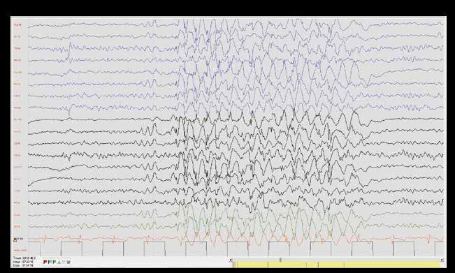
ou autres anomalies non spécifiques. La sensibilité est                         The same is true for distinct epileptic syndromes (like
définie comme la capacité de l’EEG à détecter les AE                            juvenile myoclonic epilepsy), reflex epilepsy or a single
lorsque la maladie est présente; la spécificité est défi-                       symptomatic seizure of a focal cortical dysplasia [5].
nie comme le risque d’avoir la maladie lorsque l’EEG ré-                            Regarding these considerations, in 2014, the task
vèle une AE. Voici nos conclusions principales: 1) la sen-                      force of the ILAE re-considered the diagnosis of epilepsy
sibilité et la spécificité d’une AE interictale sont : 17% et                   by any of the following conditions [6]:
95% pour les adultes, 58% et 70% pour les enfants. Un
adulte se présentant avec une première crise non-pro-                           •   At least two unprovoked seizures occurring more
voquée a une probabilité post-test de récidive de 77%                               than 24 hours apart
lorsque l’EEG montre une AE et de 47% en l’absence                              •   One unprovoked seizure and a probability for fur-
d’AE (décharges focales et généralisées confondues). Le                             ther seizures similar to the general recurrence risk
pourcentage chez les enfants est discrètement plus bas                              after two unprovoked seizures (at least 60% )
(66% et 38%). 2) L’EEG de routine est de meilleur ren-                          •   At least two seizures in a setting of reflex epilepsy
dement lorsqu’il est réalisé dans les 24 heures après
la crise (51% d’anomalies dans une population mixte                                 The threshold of 60% is considered as estimation
d’adultes et d’enfants). 3) L’identification d’une AE après                     and not as strict cut-off. This number is based on the
le troisième EEG est extrêmement faible. L’EEG de som-                          risk of relapse after two unprovoked seizures, which is
meil augmente significativement la probabilité de dé-                           about 60% at 2 years and 70 - 75% at 5 years of follow-
tecter une AE et ceci jusqu’à 50%. L’EEG standard nous                          up. It requests a workup to calculate the individual risk
informe surtout sur la présence d’un syndrome épilep-                           of predisposition for further seizures.
tique et du risque de récidive. Si ce dernier est négatif,                          As we will discuss below (“Risk of relapse”), two fac-
un EEG de sommeil, incluant les premières heures après                          tors are consistently associated with an increased risk
l’éveil, est de mise.                                                           of relapse: the presence of cerebral lesion and epilep-
                                                                                tiform abnormalities in the EEG. Seizures clustering
Mots clés : Première crise, risque de récidive, traitement                      within 24 hours confer approximately the same risk for
médicamenteux, IRM                                                              later seizure as a single seizure [7]. Thus, two or more
                                                                                seizures occurring in a 24-hour period are considered to
                                                                                be a single unprovoked seizure.
1. First seizure and epilepsy: current definition                                   High risk for recurrence after a single seizure should
and epidemiology                                                                lead to the consideration of starting an antiepileptic
                                                                                treatment already after the first seizure. In that case,
    Epilepsy is one of the most frequent neurological                           the risk of recurrent seizures, at least during the first 2
diseases, affecting between 0.5 - 1% of the popula-                             years, is significantly reduced by an average of 34% [8].
tion, i.e, approximately 50 Mio people worldwide [1]. In                        However, the long-term prognosis is not changed; for
2005, a task force directed by the International League                         this reason, when a lesion is present, the possibility of
Against Epilepsy (ILAE) and the International Bureau for                        surgery should be brought up already during the first
Epilepsy defined epilepsy as “A disorder characterized                          consultation.
by an enduring predisposition of the brain to generate
epileptic seizures and by the neurobiologic, cognitive,
psychological and social consequences of this condi-                            2. Routine EEG in first seizure
tion” [2]. A commonly used operational definition em-
ployed for epidemiological purposes considers a diag-                               Routine EEG should be performed within 24 hours
nosis of epilepsy after 2 unprovoked seizures occurring                         of the first seizure. Indeed a prospective study on 300
at least 24 hours apart [3]. Studies showed that after                          consecutive patients showed that interictal epilepti-
2 unprovoked non-febrile seizures, the probability of                           form abnormalities (EA) were present in 51% of pa-
having another seizure was 73% [3] at 5 years (95% CI                           tients who underwent an EEG within the first 24 hours,
is 59 - 87%) versus 40 - 52% after a single unprovoked                          compared to 34% of the patients who had a later EEG
seizure [1].                                                                    [9]. However, it is of note that this study included many
    Nowadays, the “two unprovoked seizures” defini-                             children, which differ from adults in terms of occur-
tion appears to be inadequate in several clinical cir-                          rence likelihood of discharges. Several studies have
cumstances. In 2009, Hesdorffer showed that a patient                           shown that interictal EA are more frequent after sei-
who presented with a single unprovoked seizure after a                          zures (postictal activation) [10, 11]. Although these
remote brain insult, such as stroke, tumor, central nerv-                       studies were performed on chronic epilepsy, it seems
ous system infection or trauma is at high risk of a sec-                        that this increased frequency also applies to new-onset
ond unprovoked seizure. This risk is comparable to the                          epilepsies [9]. Unfortunately in many cases, scheduling
risk for further seizures after 2 unprovoked seizures [4].                      of early EEG is not feasible. On the other hand, very ear-
                                                                                ly EEG may show transient, less specific abnormalities,
                                                                                like postictal slowing, which must be interpreted cau-

Yield of EEG After a First Unprovoked Seizure | L. Fisch, M. Seeck, F. Pittau                                          Epileptologie 2016; 33   217
tiously, as they can also result from the presence of a 3. Sleep EEG
lesion and are not necessarily a sign of epileptogenicity
[12]. An exception are rhythmic delta, extratemporal or The yield of EEG can be significantly increased in all
temporal, which usually indicates the presence of sei- age groups by the use of sleep recording. Indeed sleep
zures [13]. states influence the presence of interictal and ictal
Routine EEG should be performed with at least 21 epileptic activity. Particularly, non-rapid eye movement
electrodes, placed according to the standard 10 - 20 (NREM) sleep has been characterized as a state of rela-
system and last at least 20 minutes. It is recommended tive “neuronal synchronization”. Such coordinated syn-
that hyperventilation of 3 minutes and intermittent aptic activity allows the recruitment of a critical mass
photic-stimulation at 1 - 50 Hz with the eyes open and of neurons, necessary to initiate and sustain epileptic
closed at each frequency are carried out. The placement activity [18]. This is why interictal (mainly focal) EA are
of additional inferior temporal electrodes (F9, T9, P9 more common in NREM sleep than in awake record-
and F10, T10, P10) is of extreme importance in particu- ings. Carpay et al. [14] reported that 60 of 177 (34%)
lar if temporal seizures are searched, a frequent con- children with normal findings during a standard re-
stellation in adults. cording showed EA after sleep deprivation (mostly dur-
Accurate classification of seizure type will help cli- ing sleep). Similarly, King et al. [9] reported that 35%
nicians in diagnostic and therapeutic decisions. Clinical of adults and children whose initial EEG findings were
history is fundamental, but unfortunately, after a first normal, showed EA in a subsequent study performed
episode, this is fraught with limitations due to the lack during sleep. Overall, the literature suggests that sleep
of witnesses, or peri-ictal amnesia. King et colleagues EEG increases the yield of significant EEG abnormalities
[9], on a population of 300 patients (20% below 16 by 30 - 35%.
years, range 5 - 83 years), were able to classify seizures Whereas NREM sleep may “unmask” the EA that are
into focal versus generalized in just 47% of cases after not present on awake state, REM sleep is reported to
considering medical history and physical examination show fewer EA. However, REM recordings show a more
findings alone. When EEG findings were also taken limited electric field of EA, i.e. corresponding to the true
into account, correct classification was possible in an irritative region and thus contributing to localization of
additional 30%; thus, in their study group, only 23% the epileptogenic focus [19, 20]. Shinnar et al. [16] de-
of seizures remained unclassified. Specific syndromes scribed 148 children with unprovoked first seizure who
also influence the likelihood of seeing EA on EEG, with had both sleep and wakefulness recorded on a single
higher rates in patients with absence seizures (92%) EEG. EA were identified either only while awake or only
and atonic or myoclonic seizures (85%) compared with while asleep in 30% of subjects, and in both states in
focal seizures (59%) [14]. 70% of subjects. While generalized discharges are more
What is the relevance of non-epileptiform abnor- common during the awake state, focal discharges are
malities, such as focal slow activity, regional attenua- more easily detected during the sleep state.
tion, or abnormalities of background cerebral rhythms? Sleep recording can be also useful to detect epilep-
They are much less specific risk predictors than EA, al- tic seizures, of which patients can be unaware. NREM
though they can imply localized structural pathology sleep activates frontal lobe seizures more than tempo-
underlying the seizure disorder, or diffuse cortical dys- ral lobe seizures, and temporal lobe seizures are more
function as in symptomatic generalized epilepsies [15]. likely to secondarily generalize during sleep than during
Non-epileptiform abnormalities are more common in wakefulness [21, 22]. A variety of epilepsy syndromes
symptomatic cases (25%) than in idiopathic epilepsy occur predominantly or exclusively during NREM sleep,
syndromes (7%) [14, 16]. As stated above, rhythmic fo- or during awaking phases. For example, EEG in patients
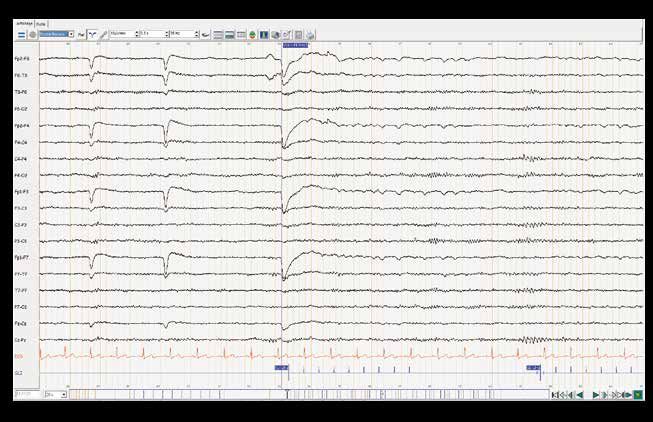
cal delta usually indicate active epileptogenicity. suffering from “grand mal on awakening” may have a
What happens if the first routine EEG is normal? A completely normal routine EEG, but very active and fre-
retrospective study on 619 patients reveals that the cu- quent EA just before awakening (Figures 1 and 2). Oth-
mulative yield of EA is 39% after the first EEG study and er striking examples are the syndrome of continuous
68% after the third. Beyond the 3rd EEG, the probability spike-wave activity during slow-wave sleep, defined by
to find epileptiform abnormalities is very low. Thus, at an EEG pattern consisting of generalized slow-spike-
this point a sleep EEG should be requested if this was wave discharges present for 85 - 90% of slow-wave
not yet done before [17]. sleep and relatively suppressed during REM sleep and
wakefulness, and the Landau-Kleffner syndrome. These
syndromes, characterised by a continuous spike-wave
in slow sleep, start in early to mid-childhood and lead
to cognitive regression and seizures. Early, appropriate
treatment is indicated to attempt to ameliorate the
electrical status and improve the child’s cognitive func-
tion.
218 Epileptologie 2016; 33 Yield of EEG After a First Unprovoked Seizure | L. Fisch, M. Seeck, F. PittauFigure 1. 19 y.o. patient with a first unprovoked generalized seizure during wakefulness. Routine EEG was performed
4. Relevance of ictal recordings in the 1st EEG              presence in healthy subjects is extremely rare, with an
                                                                   incidence of 0.5% [29].
          It is possible that the first seizure which comes to          Although interictal EA have been associated with a
      medical attention, is not the patient’s true first seizure   higher risk of relapse [1, 30 - 32] their diagnostic value
      [9, 23]. Patients presenting at emergency often have a       has been unclear for a long time. Indeed a meta-analy-
      history of more subtle seizures (e.g., absence seizures      sis of 2003 [32] showed that sensitivity and specificity
      or myoclonic or simple partial seizures) that were not       of interictal EA for seizure relapse after a first seizure
      identified by the patient or its entourage. These types      varies widely among published studies, with a range
      of seizures could be observed already during the first       from 20% to 80% for sensitivity, and 41% to 99% for
      routine EEG. For this reason the facilitating techniques     specificity. Just recently, a Cochrane [33] systematic re-
      are fundamental during EEG: for instance, hyperventi-        view and meta-analysis about diagnostic accuracy of
      lation can trigger absences in children with untreated       routine EEG on 1799 patients with first seizure and 1
      childhood or juvenile absence epilepsy; photic stimula-      year of follow-up was published [34]. In adults, sensi-
      tion can induce myoclonic jerks in patients with juve-       tivity (defined as the percentage of EEG with EA, when
      nile myoclonic epilepsy. Focal seizures occur more rarely    epilepsy is present) is 17.3% (range 7.9 - 33.8) and spec-
      during standard EEG, and if they do, they are rather an      ificity (percentage of presence of epilepsy, when EEG
      alarming sign for a very active epileptic condition and      shows EA) is on average 94.7% (range 73.7 - 99.1). In
      hospitalisation should be considered. In any case, indi-     children, a sensitivity value of 58 % (range 49.7 - 65.6)
      vidualized and specialized care and appropriate anti-        and a specificity of 70% (range 57.5 - 79.5) were iden-
      epileptic medication should be initiated. If the routine     tified. The same study revealed that an adult present-
      EEG shows an electrical, or non-convulsive, status epi-      ing with a first unprovoked seizure has a 77% post-test
      lepticus, injection of antiepileptic drugs under EEG con-    probability of relapse if routine EEG includes EA (posi-
      trol and hospitalisation is strongly recommended.            tive likelihood ratio) and 47% if it does not (negative
                                                                   likelihood ratio). Similary, a child has a 66% post-test
                                                                   probability of relapse if routine EEG includes EA and
      5. Risk of relapse                                           38% if it does not. These observations are extremely
                                                                   important, considering that a patient with a first un-
          In 2014, a meta-analysis estimated the risk of re-       provoked seizure should be treated if the probability of
      lapse after the first event in patients who were treat-      relapse is >60% at 10 years [6].
      ed immediately or with delay and showed a risk of                 Other factors carry important information regard-
      15%, 8%, 6% and 7% of relapses after 6, 12, 18 and 24        ing the overall prognosis, as the underlying syndrome.
      months in patients who were treated immediately. If          Idiopathic generalized (or genetic generalized, as it is
      an observational attitude was chosen and treatment           named in the new classification) epilepsy achieves re-
      postponed, these numbers increased to 18%, 10%, 9%           mission in 80 to 85% compared to focal epilepsy in 40
      and 7%. The risk of relapse was higher in patients with      - 65% [3]. Multiple seizure types in the same patient are
      an abnormal EEG than with an abnormal imaging, giv-          associated with higher seizure recurrence [26]. Younger
      en that not all epilepsy syndromes are related to cer-       age at onset has also been described as predictor of
      ebral lesions [24]. Several studies with long follow-up      worse outcome. Onset of epilepsy before the age of 12
      showed that 80 to 90% of individuals recur within two        months is a poor prognostic factor. Best prognosis is
      years of the initial seizure [25].                           noted if onset occurs after the age of three years [25].
          However, while early antiepileptic treatment de-         A prospective observational study of over 1000 adults
      creased the number of further seizures, it did not           presenting with a first unprovoked seizure showed a
      change relapse rate beyond 2 years disease duration.         similar likelihood of seizure recurrence in older (> 65
      Indeed the two multi-centre randomized trials (FirST,        years) compared with younger adults (53 versus 48
      MESS) failed to show any change in long-term prog-           percent). However, by five years, the cumulative risk
      nosis in patients with early treatment versus delayed        of recurrence was higher in older adults (75 versus 61
      treatment after further seizures [26].                       percent). This relates to a greater likelihood of a remote
          EEG and brain imaging are considered essential as        symptomatic etiology rather than age itself [35]. An-
      part of the neuro-diagnostic evaluation of adults pre-       other powerful predictor of the long-term prognosis is
      senting with an apparently unprovoked first seizure, as      the early response to treatment. Several studies found
      suggested by the practice parameter from the Ameri-          that the response to the first antiepileptic drug showed
      can Academy of Neurology [27]. A prospective study on        to be the strongest predictor of good long-term out-
      208 consecutive patients with first seizure followed for     come in adults and children. Along the same line, pa-
      5 years [28], showed that an EEG with epileptiform ab-       tients who are not seizure-free after ≥ 2 antiepileptic
      normalities was associated with a relative increase for      drugs should be referred to specialized centre to deter-
      seizure recurrence at 1 to 5 years of 2.16 (95% CI 1.07      mine the reasons for lack of response and/or search for
      - 4.38) as compared to patients without such EEG ab-         the possibility of epilepsy surgery [36].
      normalities. It is important to remember that the EA

220    Epileptologie 2016; 33                                       Yield of EEG After a First Unprovoked Seizure | L. Fisch, M. Seeck, F. Pittau
Recently, Fisch et al. showed that patients with in- 8. Wiebe S, Tellez-Zenteno JF, Shapiro M. An evidence-based approach to
stalled medical follow-up are significantly more likely the first seizure. Epilepsia 2008; 49(Suppl 1): 50-57
to receive a precise diagnosis and increased delay to 9. King MA, Newton MR, Jackson GD et al. Epileptology of the first-seizure
the next unprovoked seizure in comparison with pa- presentation: a clinical, electroencephalographic, and magnetic reso-
tients without organized medical care (p=0.008). The nance imaging study of 300 consecutive patients. Lancet 1998; 352:
study emphasized the need of specialized care starting 1007-1011
already in the emergency room, provided by epileptolo- 10. Marsan CA, Zivin LS. Factors related to the occurrence of typical paro-
gists. After a first evaluation, important exams such as xysmal abnormalities in the EEG records of epileptic patients. Epilepsia
EEG and MRI are rapidly and reliably scheduled and re- 1970; 11: 361-381
sults can be discussed at the next appointment. Early- 11. Gotman J, Marciani MG. Electroencephalographic spiking activity, drug
specialized improved not only the diagnostic accuracy, levels, and seizure occurrence in epileptic patients. Ann Neurol 1985; 17:
but also adherence to follow-up consultations, proba- 597-603
bly because patients better understood their condition 12. Wirrell EC. Prognostic significance of interictal epileptiform discharges
and the importance of compliance and lifestyle adjust- in newly diagnosed seizure disorders. J Clin Neurophysiol 2010; 27: 239-
ments [37]. 248
Psychiatric and neuropsychological comorbidities 13. Trinka E, Leitinger M. Which EEG patterns in coma are nonconvulsive sta-
are associated with a lower response to drug treatment tus epilepticus? Epilepsy Behav 2015; 49: 203-222
and higher risk of failure of remission. In these cases, 14. Carpay JA, de Weerd AW, Schimsheimer RJ et al. The diagnostic yield of a
specialized consultations, relevant non-epileptic treat- second EEG after partial sleep deprivation: a prospective study in child-
ment and/or increased frequency of follow-up appoint- ren with newly diagnosed seizures. Epilepsia 1997; 38: 595-599
ments should be scheduled, at least initially [38, 39]. 15. Smith SJ. EEG in the diagnosis, classification, and management of pa-
To conclude, EEG is a fundamental test to diagnose tients with epilepsy. J Neurol Neurosurg Psychiatry 2005; 76(Suppl 2):
the presence or absence of epilepsy after a first seizure. ii2-7
Ideally, it should be performed as fast as possible after 16. Shinnar S, Kang H, Berg AT et al. EEG abnormalities in children with a first
the event, if possible within 24 hours. Proper and cor- unprovoked seizure. Epilepsia 1994; 35: 471-476
rect diagnosis of the type of epilepsy is fundamental in 17. Baldin E, Hauser WA, Buchhalter JR et al. Yield of epileptiform electroen-
order to offer optimal treatment and prognostic infor- cephalogram abnormalities in incident unprovoked seizures: a popula-
mation regarding seizure relapse. It is self-evident that tion-based study. Epilepsia 2014; 55: 1389-1398
such information is of utmost importance for the medi- 18. Steriade M, Contreras D, Amzica F. Synchronized sleep oscillations and
cal and socio-professional wellbeing of each patient. their paroxysmal developments. Trends Neurosci 1994; 17: 199-208
It should not be forgotten that with each initiation of 19. Sammaritano M, Gigli GL, Gotman J. Interictal spiking during wakeful-
treatment the possibility and timing of withdrawal of ness and sleep and the localization of foci in temporal lobe epilepsy. Neu-
antiepileptic medication should be discussed with the rology 1991; 41: 290-297
patient, if possible early in the course of the disease 20. Adachi N, Alarcon G, Binnie CD et al. Predictive value of interictal epilep-
to avoid “autonomous” withdrawals which end in the tiform discharges during non-REM sleep on scalp EEG recordings for the
emergency room. lateralization of epileptogenesis. Epilepsia 1998; 39: 628-632
21. Bazil CW, Walczak TS. Effects of sleep and sleep stage on epileptic and
nonepileptic seizures. Epilepsia 1997; 38: 56-62
References 22. Crespel A, Coubes P, Baldy-Moulinier M. Sleep influence on seizures and
epilepsy effects on sleep in partial frontal and temporal lobe epilepsies.
1. Berg AT, Shinnar S. The risk of seizure recurrence following a first unpro- Clin Neurophysiol 2000; 111(Suppl 2): S54-59
voked seizure: a quantitative review. Neurology 1991; 41: 965-972 23. Hamiwka LD, Singh N, Niosi J, Wirrell EC. Diagnostic inaccuracy in child-
2. Fischer MJ, Scheler G, Stefan H. Utilization of magnetoencephalography ren referred with “first seizure”: role for a first seizure clinic. Epilepsia
results to obtain favourable outcomes in epilepsy surgery. Brain 2005; 2007; 48: 1062-1066
128: 153-157 24. Bonnett LJ, Marson AG, Johnson A et al. External validation of a pro-
3. Hauser WA, Rich SS, Lee JR et al. Risk of recurrent seizures after two un- gnostic model for seizure recurrence following a first unprovoked seizure
provoked seizures. N Engl J Med 1998; 338: 429-434 and implications for driving. PLoS One 2014; 9: e99063
4. Hesdorffer DC, Benn EK, Cascino GD, Hauser WA. Is a first acute sympto- 25. Shinnar S, Berg AT, Moshe SL et al. The risk of seizure recurrence after a
matic seizure epilepsy? Mortality and risk for recurrent seizure. Epilepsia first unprovoked afebrile seizure in childhood: an extended follow-up.
2009; 50: 1102-1108 Pediatrics 1996; 98: 216-225
5. Fauser S, Huppertz HJ, Bast T et al. Clinical characteristics in focal cortical 26. Su L, Di Q, Kwan P et al. Prediction for relapse and prognosis of newly
dysplasia: a retrospective evaluation in a series of 120 patients. Brain diagnosed epilepsy. Acta Neurol Scand 2013; 127: 141-147
2006; 129: 1907-1916 27. Krumholz A, Wiebe S, Gronseth G et al. Practice Parameter: evaluating an
6. Fisher RS, Acevedo C, Arzimanoglou A et al. ILAE official report: a practi- apparent unprovoked first seizure in adults (an evidence-based review):
cal clinical definition of epilepsy. Epilepsia 2014; 55: 475-482 report of the Quality Standards Subcommittee of the American Acade-
7. Neligan A, Bell GS, Giavasi C et al. Long-term risk of developing epilepsy my of Neurology and the American Epilepsy Society. Neurology 2007;
after febrile seizures: a prospective cohort study. Neurology 2012; 78: 69: 1996-2007
1166-1170
Yield of EEG After a First Unprovoked Seizure | L. Fisch, M. Seeck, F. Pittau Epileptologie 2016; 33 22128. Hauser WA, Rich SS, Annegers JF, Anderson VE. Seizure recurrence after
a 1st unprovoked seizure: an extended follow-up. Neurology 1990; 40:
1163-1170
29. Gregory RP, Oates T, Merry RT. Electroencephalogram epileptiform ab-
normalities in candidates for aircrew training. Electroencephalogr Clin
Neurophysiol 1993; 86: 75-77
30. Camfield PR, Cramfield CS, Dooley JM et al. Epilepsy after a first unpro-
voked seizure in childhood. Neurology 1985; 35: 1657-1660
31. Berg AT. Risk of recurrence after a first unprovoked seizure. Epilepsia
2008; 49(Suppl 1): 13-18
32. Gilbert DL, Sethuraman G, Kotagal U, Buncher CR. Meta-analysis of EEG
test performance shows wide variation among studies. Neurology 2003;
60: 564-570
33. Leeflang MM, Deeks JJ, Gatsonis C et al. Systematic reviews of diagnostic
test accuracy. Ann Intern Med 2008; 149: 889-897
34. Bouma HK, Labos C, Gore GC et al. The diagnostic accuracy of routine
electroencephalography after a first unprovoked seizure. Eur J Neurol
2016; 23: 455-463
35. Lawn N, Kelly A, Dunne J et al. First seizure in the older patient: clinical
features and prognosis. Epilepsy Res 2013; 107: 109-114
36. Mohanraj R, Brodie MJ. Early predictors of outcome in newly diagnosed
epilepsy. Seizure 2013; 22: 333-344
37. Fisch L, Lascano AM, Vernaz Hegi N et al. Early specialized care after a
first unprovoked epileptic seizure. J Neurol 2016; Sept 7 Epub ahead of
print
38. Hitiris N, Mohanrai R, Norrie J et al. Predictors of pharmacoresistant epi-
lepsy. Epilepsy Res 2007; 75: 192-196
39. Petrovski S, Sczoeke CE, Jones NC et al. Neuropsychiatric symptomato-
logy predicts seizure recurrence in newly treated patients. Neurology
2010; 75: 1015-1021
Address for correspondence:
Dr Francesca Pittau, MD PhD
Neurology Department
Geneva University Hospitals
4 Rue Gabrielle-Perret-Gentil
CH 1211Geneva 14
Tél. 0041 79 553 22 57
Fax 0041 22 372 83 40
francesca.pittau@hcuge.ch
222 Epileptologie 2016; 33 Yield of EEG After a First Unprovoked Seizure | L. Fisch, M. Seeck, F. PittauYou can also read