A Smartphone based Application for Skin Cancer Classification Using Deep Learning with Clinical Images and Lesion Information - arXiv
←
→
Page content transcription
If your browser does not render page correctly, please read the page content below
A Smartphone based Application for Skin Cancer
                                              Classification Using Deep Learning with Clinical
                                                        Images and Lesion Information
                                                      Breno Krohling                       Pedro B. C. Castro           Andre G. C. Pacheco               Renato A. Krohling
                                            Bio-inspired Computing Lab Bio-inspired Computing Lab Bio-inspired Computing Lab Bio-inspired Computing Lab
                                                 LABCIN - UFES               LABCIN - UFES             LABCIN - UFES               LABCIN - UFES
                                                   Vitória, Brazil           Vitória, Brazil           PPGI - UFES                PPGI - UFES
                                            breno.krohling@aluno.ufes.br pedrobccastro@gmail.com         Vitória, Brazil           Vitória, Brazil
arXiv:2104.14353v1 [eess.IV] 28 Apr 2021

                                                                                                     agcpacheco@inf.ufes.br      rkrohling@inf.ufes.br

                                              Abstract—Over the last decades, the incidence of skin cancer,           around 78% of the population have their own smartphone [18].
                                           melanoma and non-melanoma, has increased at a continuous                   Therefore, a smartphone-based application to assist clinicians
                                           rate. In particular for melanoma, the deadliest type of skin               to diagnose skin cancer during the screening process seems to
                                           cancer, early detection is important to increase patient prognosis.
                                           Recently, deep neural networks (DNNs) have become viable to                be feasible.
                                           deal with skin cancer detection. In this work, we present a                   Phillips et al. [19] developed an Android application to
                                           smartphone-based application to assist on skin cancer detection.           distinguish melanoma and non-melanoma using a Support
                                           This application is based on a Convolutional Neural Network                Vector Machine (SVM) trained on a dataset composed of 20
                                           (CNN) trained on clinical images and patients demographics,                images of 3 types of skin lesions. As the model was trained
                                           both collected from smartphones. Also, as skin cancer datasets
                                           are imbalanced, we present an approach, based on the mutation              using few samples, its performance is quite limited. Ly et al.
                                           operator of Differential Evolution (DE) algorithm, to balance              [20] proposed a deep learning model based on convolutional
                                           data. In this sense, beyond provides a flexible tool to assist doctors     neural network (CNN) for Android and iOS platforms. Their
                                           on skin cancer screening phase, the method obtains promising               model was tested on the grand challenge PHDB melanoma
                                           results with a balanced accuracy of 85% and a recall of 96%.               dataset [21] and outperformed the known baseline model in
                                              Index Terms—skin cancer detection, smartphone application,
                                           deep learning, convolutional neural network                                terms of accuracy and computational efficiency. Dai et al. [22]
                                                                                                                      presented an iOS mobile application for skin cancer also using
                                                                       I. I NTRODUCTION                               a CNN. The model was trained on the HAM10000 dataset
                                              The skin cancer occurrence, melanoma and non-melanoma,                  [23], which contains 10,000 dermoscopy images clustered into
                                           has increased at a constant rate over the last decades [1].                7 different types of skin lesions. Both [20] and [22] are based
                                           The World Health Organization (WHO) estimates that 2-3                     on dermoscopy images, which means to use their smartphone
                                           million non-melanoma cancers and 132,000 melanomas occur                   application one needs a special dermatoscope attached to it.
                                           every year in the world [2]. The presence of skin cancer is                This is a limitation since this device is expensive and not often
                                           strongly related to the incidence of ultraviolet radiation caused          available in remote areas. In addition, both applications do not
                                           by sunlight exposure [3]. Due to the lack of pigmentation,                 take into account patient demographics.
                                           caucasian people are under the highest risk [4]. Early detection              Pacheco and Krohling [24] proposed a skin lesion classifier
                                           is important to increase patient prognosis [5].                            for six common skin diseases. The classifier is based on a
                                              Several computer-aided diagnoses (CAD) have been pro-                   deep learning model that take into account clinical images and
                                           posed to tackle automated skin cancer detection [6]–[15].                  patient demographics, both collected from smartphones. Next,
                                           Nowadays, most approaches are based on Convolutional Neu-                  Castro et al. [25] developed an approach based on the mutation
                                           ral Networks (CNN) trained on dermoscopy images [6]–[12].                  operator of Differential Evolution (DE) algorithm to handle
                                           However, in emerging countries such as Brazil, in particular               the data imbalance problem. In addition, they implement an
                                           in the countryside [16], there is a lack of dermatologists and             App for classification of melanoma and non-melanoma skin
                                           dermatoscopes1 , which constraints the use of a CAD system                 lesions. In this paper, we extend their work in the following
                                           based on dermoscopy images. In this context, smartphones                   points:
                                                                                                                         • We include more skin lesions by using the PAD-UFES-20
                                           may be useful devices to handle this problem. According to
                                           the Ericsson report [17], in 2019 the total number of mobile                     dataset [26].
                                                                                                                         • We train and validate the model to discriminate between
                                           subscriptions around the world was around 8 billion. In Brazil,
                                                                                                                            skin cancer and non-skin cancer. The tested CNN model
                                             1a   medical device that magnifies the lesion for better visualization         is used in a smartphone-application to assist clinicians to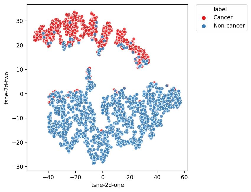
diagnose skin cancer during the screening process.                                      III. M ETHODS
The remainder of this paper is organized as follows: in section         In this section, we describe an approach to combine clinical
2, we present a literature review of deep learning methods           images and lesion clinical information using a CNN [24].
applied to skin cancer classification. In section 3, we describe     Next, we describe the data balancing methods employed in
our previous work and extensions to the data balance approach.       this work.
In section 4, we present the technologies used to develop            A. Deep model to classify skin cancer
the smartphone application. In section 5, we present the
                                                                        Pacheco and Krohling [24] introduced a new dataset com-
experimental results and discussions. Lastly, in section 6, we
                                                                     posed of clinical images and lesion clinical information as well
draw some conclusions.
                                                                     as an approach to combine both types of data. Each sample
                                                                     in the dataset has a clinical diagnosis, an image, and eight
                     II. R ELATED WORKS                              clinical features: the patient’s age, the part of the body where
                                                                     the lesion is located, if the lesion itches, bleeds or has bled,
                                                                     hurts, has recently increased, has changed its pattern, and if
   Recently, open datasets containing dermoscopy skin lesion
                                                                     it has an elevation. We encode the clinical information in 22
images have significantly increased the number of samples
                                                                     variables: 15 bits for region of the body, 1 integer for age, and
[21], [23], [27]. As a consequence, deep learning models have
                                                                     6 bits for the remaining features. These features are based on
become viable to tackle skin cancer detection. Remarkable
                                                                     common questions that dermatologists ask patients during an
works such as the ones proposed by Esteva et al. [12] and
                                                                     appointment [24].
Brinker et al. [9] showed that deep learning techniques achieve
                                                                        In order to combine clinical images and lesion clinical infor-
similar performances to dermatologists. Consequently, many
                                                                     mation, Pacheco and Krohling [24] proposed a straightforward
other deep learning approaches have been proposed to classify
                                                                     mechanism to control the contribution of features extracted
skin cancer.
                                                                     from images (FI) and clinical information (CI). We applied the
   Different works such as Arik et al. [28], Shihadeh et             same approach, but now for skin cancer classification. Figure 1
al. [29], and Demir et al. [30] trained and applied com-             shows a schematic diagram of the modified approach proposed
mon convolutional neural networks (CNNs) architectures, e.g.,        in this work:
ResNet [31] and Inception-v3 [32], on dermoscopy images                 It is possible to assign more importance for FI or CI by
to detect melanoma. Moldovan [33] and Pai and Giridharan             changing the number of features of each one. As the number
[34] presented similar approachs, however, they classify seven       of clinical data NCI is fixed, one can manipulate the number
different skin diseases. Other works, such as Alquran et al.         of features extracted from the image NF I . In Equation (1) is
[14] and Mahbod et al. [35] changed the CNN workflow by              described how to calculate the NF I given the NCI and the
including a Support Vector Machine (SVM) to work as the              contribution factor (λ) of NCI from all the features.
model’s classifier. In addition, ensemble of CNNs are also
common applied for this task [36]–[38].                                                     NCI
                                                                                   NF I =       − NCI , λ ∈ [0, 1].               (1)
   Deep learning approaches to extract the region of interest                               1−λ
(ROI) of the lesions were also proposed by De Angelo et              B. Data Balancing
al. [39] and Ali et al. [40]. Both works are based on the               A dataset is imbalanced when the number of samples
U-Net [41] architecture to segment the skin lesions borders.         for each class is not uniform distributed among the classes.
Nonetheless, they apply the conditional random fields (CRF)          Classifiers tend to perform worse on imbalanced dataset since
[42] algorithm and a fuzzy filter approach, respectively, as         they are designed to generalize from data [48]. To deal with
pos-processing methods to improve the segmentation.                  imbalanced data, we have applied the standard data balancing
   Other efforts to improve the performance of the deep              technique weighted loss function. In addition, we present
learning methods were also proposed. Namozov et al. [43]             a competitive approach based on the mutation operator of
investigated the impact of the activation function on skin           Differential Evolution (DE). In the following, we described
lesion classification. Barata et al. [44] proposed a hierarchical    the methods used for data balancing.
structure, which mimic dermatologists when diagnosing skin              1) Weighted loss function: This technique does not change
lesions, to train and perform a deep learning models. Adegun         the frequency of samples on datasets. It consists of using a
and Viriri [45] proposed a deep convolutional autoencoder            weighted loss function based on a strategy that penalizes miss
based model to melanoma detection. Dos Santo and Ponti               classification of minority classes. In this paper, we applied the
[46] presented an analysis of the impact of dimensionality           weighted cross-entropy as a loss function. The weight assigned
reduction, colors space contraction, and noisy effects in the        to each label is described by:
feature space in DNNs applied to skin cancer classification.
Lastly, Aggarwal et al. [47], proposed an attention mechanism                                     N
                                                                                                Wi =                     (2)
that helps CNNs to learn filters that activate pixels of important                                ni
regions on the skin lesion image to classify three different         where N is the total of samples and ni is the number of
types of skin cancer.                                                samples of class i.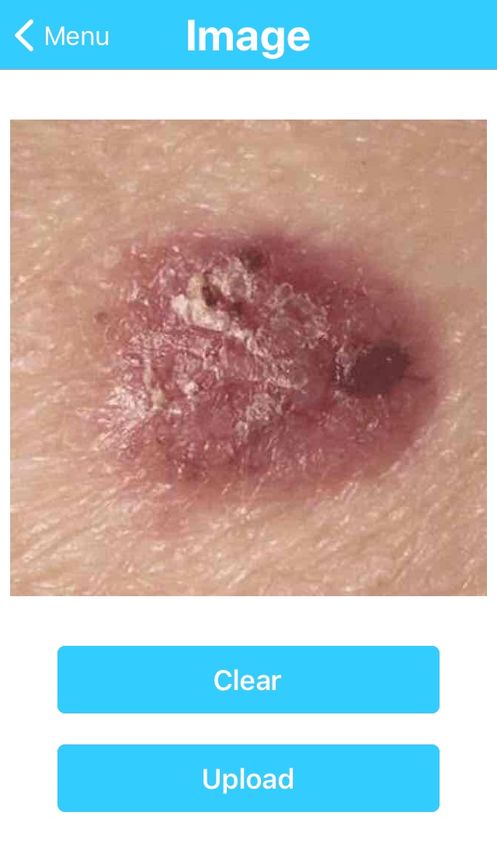
Fig. 1: The illustration of the model proposed by Pacheco and Krohling [24]. In this work, we modified the last layer for skin
cancer classification

  2) Differential Evolution (DE): Inspired by the mutation
operator from the differential evolution algorithm [49], the
proposed method combines 3 images resulting in a new image.
The operator is defined as follows:

                        X4 = X1 + α(X2 − X3 )                      (3)

where X is a set of images and α is a factor ranging from
-0.5 to 0.5, a new value for α is chosen in each combination
according to a uniform probability distribution. Regarding
clinical information used, for each combination generated, the
clinical information is randomly chosen between one of the
three base images. This technique is applied only for the same
kind of skin lesion, which belongs to the same class.

                        IV. A PP DEVELOPMENT
   In order to assist on skin cancer classification, we developed
a multi-platform smartphone application. The app’s purpose
is to assist clinicians who have no or low dermatological
experience or do not have access to a dermatoscope. Using
the app, clinicians may prioritize patients with possible skin
cancer on screening process, leading them to a specialist.               Fig. 2: Schematic diagram of the smartphone app to skin
   Embedding a CNN in a smartphone presents two main                     cancer detection
requirements: 1) the weight’s size that can be too large and
does not fit on the device’s memory; and 2) the need of
computational resource to perform the model. Since not all                  The first back-end layer is based on the java web server
smartphones can fulfill these requirements, we decided to                Tomcat that implements a Rest API to be consumed by the
deploy the CNN model on a server. Figure 2 shows a schematic             user as a service. All user information, for log purpose, is
diagram of the developed system.                                         stored in a MySQL database. The second layer is based on
   On the client side, we have a mobile application devel-               Flask4 , a framework based on Python that is designed for
oped using React Native2 framework, and Expo SDK 3 . The                 micro applications. It makes a direct execution of the machine
application sends skin lesion images along with their clinical           learning models, which were developed also in Python. Every
information to the server. The server performs the CNN model             request for processing a new clinical image with its clinical
and replies the diagnosis prediction. Finally the app displays           information that arrives at Flask is queued in Redis 5 , a NoSQL
it on the screen.                                                        key-based database. If no data is being processed then the

  2 https://facebook.github.io/react-native/docs/getting-started           4 https://flask-doc.readthedocs.io/en/latest/
  3 https://docs.expo.io/versions/latest/                                  5 https://redis.io/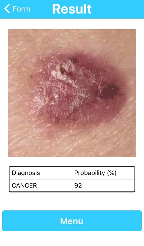
first available data of the Redis queue is read and sent to
a previous trained model. The result of the model is then
stored on Redis that will be further consulted by the user.
Next is presented screenshots of the application to illustrate
its workflow. Figure 3 shows the main screen and the log in
screen, respectively. Next, Figure 4 shows the menu and the
image upload process, respectively. Last, Figure 5 shows the
form to collect clinical information and the image of the lesion
itself with the diagnosis prediction, respectively.

                                                                        (a) App clinical information        (b) App result
                                                                   Fig. 5: App’s clinical information screen and result’s screen

                                                                                  V. R ESULTS AND DISCUSSIONS
                                                                      In this section, we present the dataset used to train the CNN,
                                                                   the criteria used in the networks evaluation, a visualization of
                                                                   the features extracted from the images, the the results obtained
                                                                   from simulations and some discussions.

                                                                   A. Dataset
                                                                      The PAD-UFES-20 dataset [26] used in this work is com-
                                                                   posed by a set of common skin diseases. Each sample within
                                                                   the dataset is composed by a clinical image and a set of
                                                                   metadata related to the lesion. In our experiments, skin cancer
         (a) App home screen          (b) App log in
                                                                   consists of Melanoma (MEL), Basal Cell Carcinoma (BCC),
        Fig. 3: App’s home screen and log in screen                and Squamous Cell Carcinoma (SCC). The non-cancer skin
                                                                   lesions are Actinic Keratosis (ACK), Seborrheic Keratosis
                                                                   (SEK), and Nevus (NEV). We added one more disease class
                                                                   labeled as Others, which includes lesions that were not rep-
                                                                   resented in the previous classes. Next, we split the data into
                                                                   skin cancer and non-cancer as presented in Table I.
                                                                               Disease*      Images*     Diseases    Images
                                                                                MEL              67
                                                                                 BCC            442       Cancer      658
                                                                                 SCC            149
                                                                                 ACK            543
                                                                                 SEK            215
                                                                                                        Non-cancer    1399
                                                                                 NEV            196
                                                                              OTHERS**          445
                                                                                Total                     2057
                                                                              *Original Dataset. **New Class

                                                                   TABLE I: The frequency of each skin lesion on the extended
                                                                   dataset

                                                                   B. Evaluation criteria
                                                                      As evaluation criteria, we aimed first at a high recall,
                                                                   followed by a high accuracy, and last for a high precision.
                                                                   This choice is justified since the recall is directly related to
                                                                   the number of false negative, i.e., the number of skin cancers
            (a) App menu        (b) App image acquisition          classified by the network as non-cancer. A false negative is the
                                                                   worst scenario for skin cancer classification since the clinician
 Fig. 4: App’s menu screen and image acquisition’s screen          assumes that the lesion is a non-cancer. The precision is related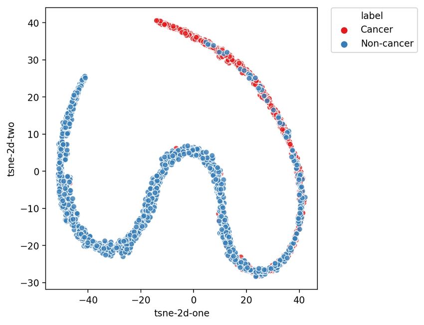
to the number of false positive, meaning the number of non- 2) The impact of clinical information: For the study of
cancer lesions classified as skin cancer by the network. In this the impact of clinical information, the number of features
case, although the patient would be worried, the clinician will extracted from image is equal to the best results obtained in
send the patient to a specialist. Sec. V-C1. Also, we simulated with the five values of beta
used previously in order to find the best results as listed in
C. Results Table VI. From Table VI and Sec. V-C1, we can compare
the performance of the network with and without clinical
The results are divided according to the type of simulation information. Table VII presents these results. From Table VII,
performed. First, we present a sensitivity study used to find we can notice that the use of clinical information provided
the best setup for the model. Second, we review the impact of an average increase in BACC, precision and recall of 1.41%,
clinical information combined with image on the CAD perfor- 1.14% and 2.39%, respectively.
mance. Finally, we investigated the impact of data balancing
Metrics
on the model’s performance. Beta
BACC PR REC
For all tests, a ResNet50 was trained using the architec- 1 88.12 ± 1.39 81.64 ± 4.06 85.67 ± 4.07
ture described in Pacheco and Krohling [24], i.e., combin- 3 88.23 ± 3.01 75.80 ± 6.75 90.74 ± 2.57
5 84.09 ± 2.73 63.95 ± 5.23 94.03 ± 3.13
ing features extracted from the images with lesion clinical 7 85.83 ± 1.62 68.53 ± 2.60 91.94 ± 2.42
information using a 5-fold cross-validation. ResNet50 was 10 85.83 ± 3.26 68.5 ± 3.56 91.94 ± 5.05
used due to its effective performance [24]. We performed
TABLE III: Model’s performance for F-measure regarding
the training phase for 100 epochs using Stochastic Gradient
varying beta without clinical data in the classification process
Descent (SGD) optimizer with a learning rate equal to 0.01
that decreases by a factor of 1/2 or 1/5 every 20 epochs,
alternately. We applied a standard data augmentation [50]
Metrics
and used the presented techniques to deal with imbalanced Beta
BACC PR REC
dataset. All images were resized to 224×224×3. The evaluation 3* 85.50 ± 2.47 65.09 ± 5.80 96.42 ± 2.77
metrics were: balanced accuracy (BACC), precision (PR), 5 84.09 ± 2.73 63.95 ± 5.23 94.03 ± 3.13
*With clinical information.
recall (REC), and F-measure. The F-measure was used because
of the imbalanced dataset. TABLE IV: Model’s performance for the extended dataset to
1) Best Setup: In order to find the best setup, we used compare the impact of clinical data in the classification process
standard data augmentation and weighted loss to deal with
the imbalanced dataset, as proposed in [24]. The goal is to 3) Visualization: We applied the t-Distributed Stochastic
find the network’s best configuration of hyperparamters, so Neighbor Embedding (t-SNE) [51], which is a tool to visualize
we can apply it to further experiments. In order to increase high-dimensional data. In total, 2048 features were extracted
the system’s recall, we also introduced the use of F-measure from all dataset samples after the last ResNet50 convolutional
defined by: layer. These features were reduced to two dimensions using
t-SNE and shown in Figure 6. From the figure, we can observe
P R.REC that some samples of cancer are overlapped with non-cancer
Fβ = (1 + β 2 ) (4) ones.
(β 2 .P R) + REC
We carried out a sensitivity analysis, changing the value of
β in (4) along with 5 combinations of features (C) extracted
from the image (FI) and clinical information (CI). Table II
presents all 5 combinations of FI and CI.
C FI x CI NF I NCI
1 90% x 10% 198
2 80% x 20% 88
3 70% x 30% 22 51
4 60% x 40% 33
5 50% x 50% 22
TABLE II: Sensitivity analysis taking into account the impor-
tance of the FI and CI
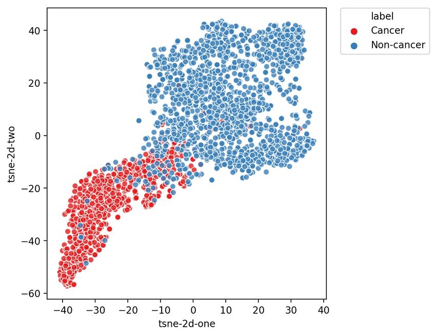
Fig. 6: Visualization of the features extracted by ResNet50
from all samples using t-SNE.
From these experiments, we obtained the best setup with β
equal to 7 and 70% of FI and 30% of CI.. The metrics using For each network, all the samples were used in order
this setup is a BACC of 85.50 ± 2.47, a precision of 65.09 ± to generate a matrix containing all the reduced features by
5.80 and a recall of 96.42 ± 2.77. ResNet50 before the classification layer. From the reduced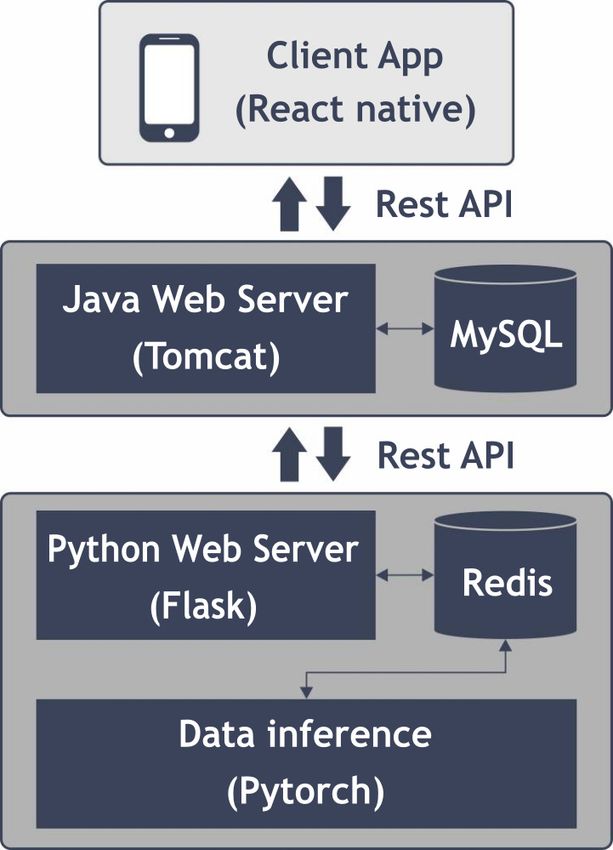
features matrix, a visualization of these features was obtained        model achieved a slightly lower performance when compared
using t-SNE and shown in Figure 7. From Figure 7, it is                with Pacheco and Krohling [24] results. It occurs because we
possible to notice the difference in the representation of the         grouped together lesions that were confused with each other,
features.                                                              without the use of clinical information. For example, the model
                                                                       tends to mistake SEK and NEV, which have similar image
                                                                       features. But when we analysis their clinical information, we
                                                                       notice that NEV median age is slower that SEK [24], which
                                                                       can help in the classification task. Since they were grouped
                                                                       under the same label, non-cancer, the addition of the clinical
                                                                       information does not represent a real gain on the classification
                                                                       as reported in Pacheco and Krohling [24].
                                                                          Regarding the impact of data balancing, the approach based
                                                                       on the mutation operator of DE provided similar results to
  (a) With clinical information     (b) Without clinical information   those obtained using weighed loss function.
Fig. 7: Visualization of the features extracted by ResNet50                                  VI. C ONCLUSION
from all samples using t-SNE for both networks                            In this paper, we presented a smartphone based application
                                                                       to support the diagnostic of skin cancer using convolutional
   4) The impact of data balancing: The impact of balancing            neural networks. The results obtained with clinical information
techniques is assessed by comparing the 2 balancing ap-                presents an average balanced accuracy of 85% and a recall
proaches, i.e., weighted loss function (WGT) and DE. The               of 96%. The study of the impact of clinical information has
setup used was the best found in Sec. V-C1. Table VIII                 shown that clinical information is relevant to cancer detection
presents the obtained results. The results with the weighted           since it improved on average the balanced accuracy, preci-
loss function presented the best result in terms of balanced           sion and recall in about 1.4%, 1.1% and 2.4%, respectively.
accuracy and recall. The approach based on the mutation                Regarding the data balancing approach, the weighted loss
operator of DE provided the best result in terms of precision.         function presented the best results but the approach based on
                                     Metrics                           the mutation operator of differential evolution is competitive.
        BAL
                   BACC                PR            REC               It is worth mentioning that these results are promising but yet
        WGT     85.50 ± 2.47      65.09 ± 5.80   96.42 ± 2.77          preliminary since our collected dataset is small. The next phase
         DE     84.84 ± 5.63      65.27 ± 9.65   96.12 ± 2.60
                                                                       consists of applying this approach to a real world scenario to
TABLE V: Model’s performance for the extended dataset for              assist doctors in the screening process. We also continue to
each data balance method                                               collect more data to improve our results.
                                                                                        VII. ACKNOWLEDGMENT
D. Discussion                                                             B. Krohling, P. B. C. Castro, and R. A. Krohling would like
   Observing the results in the previous section, we presented         to thank the financial support of the Fundação de Amparo a
an app that may help clinicians with no dermatological ex-             Pesquisa e Inovação do Espı́rito Santo (FAPES) - grant n.
perience. The experiment regarding cancer and non-cancer               575/2018. R. A. Krohling also thanks the Conselho Nacional
classification, indicates that it can be a promising tool in the       de Desenvolvimento Cientı́fico e Tecnológico (CNPq) - grant
screening process, since our preliminary results achieved a            n.309729/2018-1. We also thank all the members of the
balanced accuracy and recall of 85% and 96%, on average.               Dermatological Assistance Program (PAD-UFES), specially
However, those experiments were performed on development               prof. P. L. Frasson, and the support of J. G. M. Esgário, a
process, so further experiments in deployment phase are also           former Labcin member.
necessary.                                                                                              Metrics
                                                                               Beta
   Regarding the classification problem, the presented results                           BACC             PR            REC
                                                                                1     88.12 ± 1.39   81.64 ± 4.06   85.67 ± 4.07
confirms the hypothesis raised by Brinker et. al [10] that                      3     88.23 ± 3.01   75.80 ± 6.75   90.74 ± 2.57
patient clinical information tends to improve deep learning                     5     84.09 ± 2.73   63.95 ± 5.23   94.03 ± 3.13
performance for skin cancer classification. For our particular                  7     85.83 ± 1.62   68.53 ± 2.60   91.94 ± 2.42
                                                                                10    85.83 ± 3.26   68.5 ± 3.56    91.94 ± 5.05
case, the use of clinical information improved for all the
used metrics, achieving a balanced accuracy of 85.5% and               TABLE VI: Model’s performance for F-measure regarding
a recall of 96.42% in the best scenario. These results can             varying beta without clinical data in the classification process
be corroborated from the difference in the representation of
the reduced features using t-SNE with and without clinical
information, where the network using clinical information was
able to provide more distinguished features than the one with-
out, as shown in Figure 7. Nevertheless, we observe that the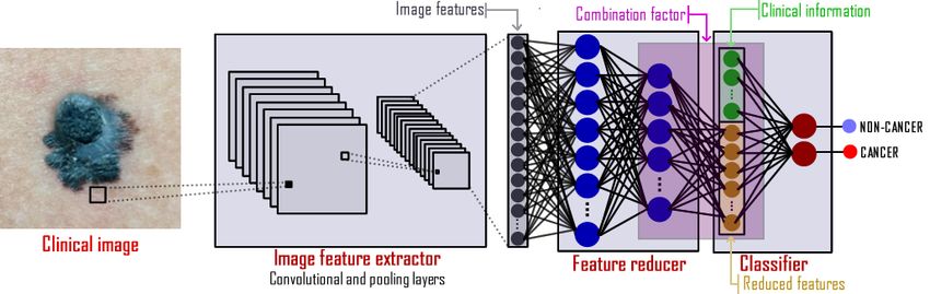
Metrics
          Beta                                                                            Conference on Applied Electrical Engineering and Computing Technolo-
                      BACC              PR                 REC
                                                                                          gies (AEECT). IEEE, 2017, pp. 1–5.
           3*     85.50 ± 2.47     65.09 ± 5.80        96.42 ± 2.77
            5     84.09 ± 2.73     63.95 ± 5.23        94.03 ± 3.13                [15]   M. Q. Khan and et al., “Classification of melanoma and nevus in digital
          *With clinical information.                                                     images for diagnosis of skin cancer,” IEEE Access, vol. 7, pp. 90 132–
                                                                                          90 144, 2019.
TABLE VII: Model’s performance for the extended dataset to                         [16]   H. Feng, J. Berk-Krauss, P. W. Feng, and J. A. Stein, “Comparison of
                                                                                          dermatologist density between urban and rural counties in the united
compare the impact of clinical data in the classification process                         states,” JAMA Dermatology, vol. 154, no. 11, pp. 1265–1271, 2018.
                                                                                   [17]   Ericsson. (2019) Ericsson mobility report. [Online]. Avail-
                                        Metrics                                           able:     https://www.ericsson.com/4aacd7e/assets/local/mobility-report/
          BAL                                                                             documents/2019/emr-november-2019.pdf
                      BACC                PR               REC
          WGT      85.50 ± 2.47      65.09 ± 5.80      96.42 ± 2.77                [18]   IBGE - Instituto Brasileiro de Geografia e Estatı́stica. (2017) Acesso
           DE      84.84 ± 5.63      65.27 ± 9.65      96.12 ± 2.60                       à internet e à televisão e posse de telefone móvel celular para
                                                                                          uso pessoal 2017. [Online]. Available: https://biblioteca.ibge.gov.br/
TABLE VIII: Model’s performance for the extended dataset                                  visualizacao/livros/liv101631 informativo.pdf
for each data balance method                                                       [19]   K. Phillips, O. Fosu, and I. Jouny, “Mobile melanoma detection ap-
                                                                                          plication for android smart phones,” in 2015 41st Annual Northeast
                                                                                          Biomedical Engineering Conference (NEBEC), 2015, pp. 1–2.
                                                                                   [20]   P. Ly, D. Bein, and A. Verma, “New compact deep learning model for
                                                                                          skin cancer recognition,” in 9th IEEE Annual Ubiquitous Computing,
                               R EFERENCES                                                Electronics Mobile Communication Conference (UEMCON), Nov 2018,
                                                                                          pp. 255–261.
 [1] R. L. Siegel, K. D. Miller, and A. Jemal, “Cancer statistics, 2019,” CA:      [21]   M. Berseth, “ISIC 2017 - skin lesion analysis towards melanoma
     a Cancer Journal for Clinicians, vol. 69, no. 1, pp. 7–34, 2019.                     detection,” 2017.
 [2] WHO-World Health Organization. (2019) How common is the skin                  [22]   X. Dai, I. Spasić, B. Meyer, S. Chapman, and F. Andres, “Machine learn-
     cancer? [Online]. Available: https://www.who.int/uv/faq/skincancer/en/               ing on mobile: An on-device inference app for skin cancer detection,”
     index1.html                                                                          in Fourth International Conference on Fog and Mobile Edge Computing
 [3] WHO - World Health Organization. (2019) Health effects of                            (FMEC), 2019, pp. 301–305.
     UV radiation. [Online]. Available: https://www.who.int/uv/health/uv           [23]   P. Tschandl, “The HAM10000 dataset, a large collection of multi-
     health2/en/index1.html                                                               source dermatoscopic images of common pigmented skin lesions,”
 [4] WHO - World Health Organization . (2019) Who is most at risk of                      2018. [Online]. Available: https://doi.org/10.7910/DVN/DBW86T
     getting skin cancer? [Online]. Available: https://www.who.int/uv/faq/         [24]   A. G. C. Pacheco and R. A. Krohling, “The impact of patient clinical
     skincancer/en/index2.html                                                            information on automated skin cancer detection,” Computers in Biology
 [5] D. Schadendorf, A. C. van Akkooi, C. Berking, K. G. Griewank,                        and Medicine, vol. 116, p. 103545, 2020.
     R. Gutzmer, A. Hauschild, A. Stang, A. Roesch, and S. Ugurel,                 [25]   P. B. C. Castro, B. A. Krohling, A. G. C. Pacheco, and R. A. Krohling,
     “Melanoma,” The Lancet, vol. 392, no. 10151, pp. 971–984, 2018.                      “An app to detect melanoma using deep learning: An approach to handle
 [6] N. Zhang, Y.-X. Cai, Y.-Y. Wang, Y.-T. Tian, X.-L. Wang, and                         imbalanced data based on evolutionary algorithms,” in International
     B. Badami, “Skin cancer diagnosis based on optimized convolutional                   Joint Conference on Neural Networks. IEEE, 2020, pp. 1–8.
     neural network,” Artificial Intelligence in Medicine, vol. 102, p. 101756,    [26]   A. G. Pacheco, G. R. Lima, A. S. Salomão, B. Krohling, I. P. Biral,
     2020.                                                                                G. G. de Angelo, F. C. Alves Jr, J. G. Esgario, A. C. Simora, P. B.
 [7] A. Hekler, J. S. Utikal, A. H. Enk, A. Hauschild, M. Weichenthal, R. C.              Castro et al., “Pad-ufes-20: A skin lesion dataset composed of patient
     Maron, C. Berking, S. Haferkamp, J. Klode, D. Schadendorf et al.,                    data and clinical images collected from smartphones,” Data in Brief,
     “Superior skin cancer classification by the combination of human and                 vol. 32, p. 106221, 2020.
     artificial intelligence,” European Journal of Cancer, vol. 120, pp. 114–      [27]   I. I. S. I. Collaboration, “Isic 2018: Skin lesion analysis towards
     121, 2019.                                                                           melanoma detection,” 2018. [Online]. Available: https://challenge2018.
 [8] T. J. Brinker, A. Hekler, A. H. Enk, C. Berking, S. Haferkamp,                       isic-archive.com/
     A. Hauschild, M. Weichenthal, J. Klode, D. Schadendorf, T. Holland-           [28]   A. Arik, M. Gölcük, and E. M. Karslıgil, “Deep learning based skin
     Letz et al., “Deep neural networks are superior to dermatologists in                 cancer diagnosis,” in 25th Signal Processing and Communications
     melanoma image classification,” European Journal of Cancer, vol. 119,                Applications Conference (SIU), May 2017, pp. 1–4.
     pp. 11–17, 2019.                                                              [29]   J. Shihadeh, A. Ansari, and T. Ozunfunmi, “Deep learning based
 [9] T. J. Brinker, A. Hekler, A. Hauschild, C. Berking, B. Schilling, A. H.              image classification for remote medical diagnosis,” in IEEE Global
     Enk, S. Haferkamp, A. Karoglan, C. von Kalle, M. Weichenthal et al.,                 Humanitarian Technology Conference (GHTC), Oct 2018, pp. 1–8.
     “Comparing artificial intelligence algorithms to 157 German dermatol-         [30]   A. Demir, F. Yilmaz, and O. Kose, “Early detection of skin cancer
     ogists: the melanoma classification benchmark,” European Journal of                  using deep learning architectures: Resnet-101 and inception-v3,” in 2019
     Cancer, vol. 111, pp. 30–37, 2019.                                                   Medical Technologies Congress (TIPTEKNO), Oct 2019, pp. 1–4.
[10] T. J. Brinker, A. Hekler, J. S. Utikal, N. Grabe, D. Schadendorf, J. Klode,   [31]   K. He, X. Zhang, S. Ren, and J. Sun, “Deep residual learning for image
     C. Berking, T. Steeb, A. H. Enk, and C. von Kalle, “Skin cancer                      recognition,” 1512.03385, 2015.
     classification using convolutional neural networks: systematic review,”       [32]   C. Szegedy, Wei Liu, Yangqing Jia, P. Sermanet, S. Reed, D. Anguelov,
     Journal of Medical Internet Research, vol. 20, no. 10, p. e11936, 2018.              D. Erhan, V. Vanhoucke, and A. Rabinovich, “Going deeper with
[11] R. C. Maron, M. Weichenthal, J. S. Utikal, A. Hekler, C. Berking,                    convolutions,” in IEEE Conference on Computer Vision and Pattern
     A. Hauschild, A. H. Enk, S. Haferkamp, J. Klode, D. Schadendorf et al.,              Recognition (CVPR), June 2015, pp. 1–9.
     “Systematic outperformance of 112 dermatologists in multiclass skin           [33]   D. Moldovan, “Transfer learning based method for two-step skin cancer
     cancer image classification by convolutional neural networks,” European              images classification,” in 2019 E-Health and Bioengineering Conference
     Journal of Cancer, vol. 119, pp. 57–65, 2019.                                        (EHB), Nov 2019, pp. 1–4.
[12] A. Esteva, B. Kuprel, R. A. Novoa, J. Ko, S. M. Swetter, H. M. Blau,          [34]   K. Pai and A. Giridharan, “Convolutional neural networks for classifying
     and S. Thrun, “Dermatologist-level classification of skin cancer with                skin lesions,” in 2019 IEEE Region 10 Conference (TENCON), Oct 2019,
     deep neural networks,” Nature, vol. 542, no. 7639, p. 115, 2017.                     pp. 1794–1796.
[13] P. Bumrungkun, K. Chamnongthai, and W. Patchoo, “Detection skin               [35]   A. Mahbod, G. Schaefer, C. Wang, R. Ecker, and I. Ellinge, “Skin lesion
     cancer using SVM and snake model,” in 2018 International Workshop                    classification using hybrid deep neural networks,” in IEEE International
     on Advanced Image Technology (IWAIT). IEEE, 2018, pp. 1–4.                           Conference on Acoustics, Speech and Signal Processing (ICASSP), May
[14] H. Alquran, I. A. Qasmieh, A. M. Alqudah, S. Alhammouri, E. Alawneh,                 2019, pp. 1229–1233.
     A. Abughazaleh, and F. Hasayen, “The melanoma skin cancer detection           [36]   A. G. Pacheco and R. A. Krohling, “Learning dynamic weights for an
     and classification using support vector machine,” in 2017 IEEE Jordan                ensemble of deep models applied to medical imaging classification,”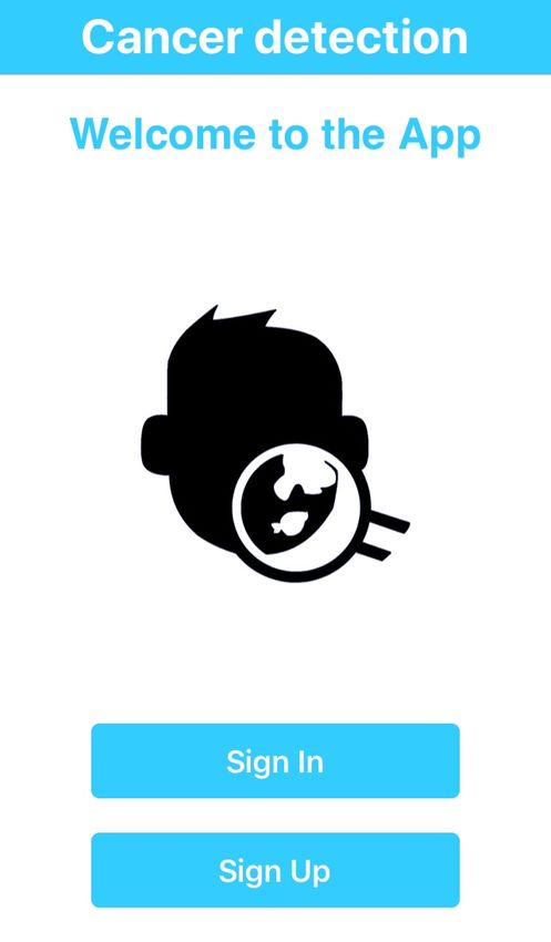
in 2020 International Joint Conference on Neural Networks (IJCNN).
       IEEE, 2020, pp. 1–8, under review.
[37]   N. C. Codella, Q.-B. Nguyen, S. Pankanti, D. Gutman, B. Helba,
       A. Halpern, and J. R. Smith, “Deep learning ensembles for melanoma
       recognition in dermoscopy images,” IBM Journal of Research and
       Development, vol. 61, no. 4, pp. 5–1, 2017.
[38]   B. Harangi, A. Baran, and A. Hajdu, “Classification of skin lesions using
       an ensemble of deep neural networks,” in 40th Annual International
       Conference of the IEEE Engineering in Medicine and Biology Society
       (EMBC), July 2018, pp. 2575–2578.
[39]   G. G. De Angelo, A. G. Pacheco, and R. A. Krohling, “Skin lesion seg-
       mentation using deep learning for images acquired from smartphones,”
       in 2019 International Joint Conference on Neural Networks (IJCNN).
       IEEE, 2019, pp. 1–8.
[40]   A. Ali, J. Li, S. J. O’Shea, G. Yang, T. Trappenberg, and X. Ye, “A
       deep learning based approach to skin lesion border extraction with a
       novel edge detector in dermoscopy images,” in 2019 International Joint
       Conference on Neural Networks (IJCNN), July 2019, pp. 1–7.
[41]   O. Ronneberger, P. Fischer, and T. Brox, “U-net: Convolutional networks
       for biomedical image segmentation,” in International Conference on
       Medical Image Computing and Computer-Assisted Intervention, 2015,
       pp. 234–241.
[42]   P. Krähenbühl and V. Koltun, “Efficient inference in fully connected
       CRFs with Gaussian edge potentials,” in Advances in Neural Information
       Processing Systems 24, J. Shawe-Taylor, R. S. Zemel, P. L. Bartlett,
       F. Pereira, and K. Q. Weinberger, Eds. Curran Associates, Inc., 2011,
       pp. 109–117.
[43]   A. Namozov, D. Ergashev, and Y. I. Cho, “Adaptive activation functions
       for skin lesion classification using deep neural networks,” in 2018 Joint
       10th International Conference on Soft Computing and Intelligent Sys-
       tems (SCIS) and 19th International Symposium on Advanced Intelligent
       Systems (ISIS), Dec 2018, pp. 232–235.
[44]   C. Barata and J. S. Marques, “Deep learning for skin cancer diagnosis
       with hierarchical architectures,” in IEEE 16th International Symposium
       on Biomedical Imaging (ISBI 2019), April 2019, pp. 841–845.
[45]   A. A. Adegun and S. Viriri, “Deep learning-based system for automatic
       melanoma detection,” IEEE Access, vol. 8, pp. 7160–7172, 2020.
[46]   F. Pereira dos Santos and M. Antonelli Ponti, “Robust feature spaces
       from pre-trained deep network layers for skin lesion classification,” in
       31st SIBGRAPI Conference on Graphics, Patterns and Images (SIB-
       GRAPI), Oct 2018, pp. 189–196.
[47]   A. Aggarwal, N. Das, and I. Sreedevi, “Attention-guided deep convolu-
       tional neural networks for skin cancer classification,” in Ninth Interna-
       tional Conference on Image Processing Theory, Tools and Applications
       (IPTA), Nov 2019, pp. 1–6.
[48]   R. Akbani, S. Kwek, and . Japkowicz, “Applying support vector ma-
       chines to imbalanced datasets,” in Machine Learning: ECML 2004.
       Berlin, Heidelberg: Springer Berlin Heidelberg, 2004, pp. 39–50.
[49]   R. Storn and K. Price, “Differential evolution - a simple and efficient
       heuristic for global optimization over continuous spaces,” Journal of
       Global Optimization, vol. 11, no. 4, pp. 341–359, 1997.
[50]   F. Perez and et.al., “Data augmentation for skin lesion analysis,” in
       OR 2.0 Context-Aware Operating Theaters, Computer Assisted Robotic
       Endoscopy, Clinical Image-Based Procedures, and Skin Image Analysis.
       Springer International Publishing, 2018, pp. 303–311.
[51]   L. v. d. Maaten and G. Hinton, “Visualizing data using t-SNE,” Journal
       of Machine Learning Rsesearch, vol. 9, pp. 2579–2605, 2008.

You can also read