Dragon fruit calli development and fungal contamination as influenced by activated charcoal
←
→
Page content transcription
If your browser does not render page correctly, please read the page content below
Balendres & Cruz., 2021

     Dragon fruit calli development and fungal contamination as
                   influenced by activated charcoal

                     María Angela Cruz1 and Mark Angelo Balendres1*

1
    Institute of Plant Breeding, College of Agriculture and Food Science, University of the
                                     Philippines Los Baños

                     *
                         Corresponding author: mobalendres@up.edu.ph

                                        ABSTRACT

      Activated charcoal is often used in plant tissue culture systems to reduce phenolic
oxidation and improve cell growth and development. This study investigated activated
charcoal's effect on the somatic embryogenesis of dragon fruit (Hylocereus species).
Nine-month-old seed-derived dragon fruit stems were placed in basal Murashige and
Skoog (MS) plates supplemented with 2, 4-D (1.5 mg/L) and activated charcoal (1g/L).
Cultures were incubated in 14 h continuous light or under dark condition. MS medium
without activated charcoal served as the control. Percent fungal contamination, degree
of tissue browning, and callus growth (length, width, and total area) were assessed. A
significant decrease in contamination was observed four days after inoculation (dai) in a
medium containing activated charcoal regardless of the light condition used (p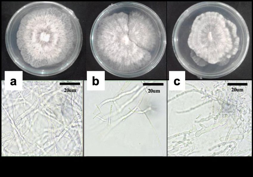
Balendres & Cruz., 2021

activities (Bakar et al., 2011; Luo et al., 2014). Some of the commonly cultivated species
of dragon fruit are Hylocereus undatus (pink-skinned, white-fleshed), Hylocereus
megalanthus (yellow-skinned, white-fleshed), and Hylocereus polyrhizus or
monacanthus (red-skinned, red-fleshed) (Ortiz-Hernandez and Carrillo-Salazar, 2012;
Mercado-Silva, 2018). Dragon fruits are propagated by seeds, by micropropagation
(Mohamed-Yassen, 2002), and, more commonly, through vegetative (stem) cuttings
(Ortiz-Hernandez, 1999). However, successful crop production is hampered by many
factors. Among these factors are plant diseases. These diseases can negatively affect
the yield and fruit quality of dragon fruits (Balendres and Bengoa, 2019). Recently,
dragon fruit diseases have been reported in the Philippines (Taguiam et al. 2020a; 2020b;
2020c).

Dragon fruits are mainly propagated by using stem cuttings. These stem cuttings are
transported from one region to another and can quickly be established in an area.
Nevertheless, vegetative (stem) cuttings could harbor disease causing microorganisms
or pathogens. Hence, while large-scale propagation is possible, stem cuttings must be
diseased-free to avoid disease epidemics in the field. One of the best methods for large-
scale and disease-free crop production is through in-vitro tissue culture (Bozkurt et al.,
2020; Dahanayake and Ranawake, 2011). Several studies have examined various
propagation methods of dragon fruit (Bozkurt et al., 2020). However, currently, little
information exists on the protocols for producing high-quality crop planting material via
the tissue culture system (Dahayanake and Ranawake, 2011). Moreover, problems such
as explant browning remain a constraint in producing the crop via the tissue-culture
system.

Activated charcoal has been found to reduce the oxidation of phenolic compounds, which
leads to tissue browning and eventually cell death (Thomas, 2008). Several studies
found activated charcoal improves plant growth by adsorption of toxic metabolites
released by the wounded plant (Liang et al., 2019; Olah, 2017; Pan and Staden, 1998;
Shimelis et al., 2015; Thomas, 2008; Wang and Huang, 1976) and increases uptake of
growth hormones (Wei et al., 2006; Pan and Staden, 2001). Explant health is important
for callus induction. Tissue or explant browning is a challenge in an in-vitro culture
system. This study provides evidence of the applicability of activated charcoal in
decreasing tissue or explant browning in dragon fruit explants. Establishing an excellent
in-vitro micropropagation technique will be useful for large-scale and disease-free
production of dragon fruit. Using stem cuttings became a more popular way in
propagating dragon fruit, but pathogen-infested stem cuttings could lead to disease
epidemic in the field (Balendres and Bengoa 2019). Although, propagation is quite time-
consuming and laborious, especially when using seeds (Trivelleni et al., 2020), a
successful in-vitro culture system of dragon fruit could lead to the development of
potentially disease resistant or high-yielding clones as a result of somaclonal variation.

                             MATERIALS AND METHODS

Explant source and preparation
The explants were taken from the stems nine-month-old, seed-derived H. monacanthus
(red-skinned, red-fleshed) and H. undatus (red-skinned, white-fleshed) plants. These
plants were grown in pots containing sterilized soils and were maintained at the screen
JPACD (2021) 23:58-73                                                                  59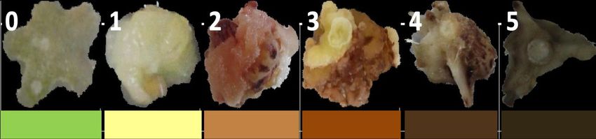
Balendres & Cruz., 2021

house of the Plant Pathology Laboratory, Institute of Plant Breeding, College of
Agriculture and Food Science, University of the Philippines Los Baños. Newly-emerged
stems (from the main stem) of dragon fruits were cut and placed in tissue paper. In the
lab, the stems were transferred in a 125 mL beaker, covered with gauze, and washed in
running water for at 30 min. Tissue samples were sterilized in 50% sodium hypochlorite
solution (Zonrox, Philipines) for 5 min and were rinsed three times with sterile distilled
water. Additional sterilization was done by soaking tissue samples in 70% ethanol for 3
min, and finally, rinsed with sterile distilled water three times. The sterilized tissues were
air dried for at 30 min under the laminar flow hood.

Effect of activated charcoal and light on callus induction and tissue browning
The sterilized 2-mm cut explants were inoculated in basal Murashige and Skoog (MS)
plates supplemented with 1.5 mg/L 2,4-D and activated charcoal (1g/L). All media had
3% (w/v) sucrose, adjusted to pH 5.7 before adding 0.8% (w/v) agar, and autoclaved at
121℃ for 20 min. Explants that were cultured on basal MS media without activated
charcoal served as the control check. The experiment was performed twice and each
experiment consists of 4 replicate plates. Callus color changes were observed visually
and measured using an arbitrary 9-point and 5-point scales. For the light effect trial,
cultures were maintained under 14 hours of cycled fluorescent light and in no light
condition (dark) with temperature ranging from 20±5℃. For dark conditions, cultures
were covered with foil. Percent contamination was recorded four days and eight days
after inoculation in the medium. The type of callus formed, degree of explant browning,
and callus growth [measured by length (mm), width (mm), and total area (mm2)] were
collected 28 days after incubation. A paired sample (Independent) T-test was performed
using Statistical Tool for Agricultural Research (STAR Nebula) with a 95% confidence
level.

Isolation, morpho-cultural, and molecular characterization of fungal contaminants
A 3 mm2 agar block of hyphae growing from the stem of the dragon fruit explants was
transferred onto a potato dextrose agar (PDA) medium. Plates were stored at room
temperature (28-30°C) for three days (with 14 hours light in 24 hours cycle). The fungus
was then purified and further characterized. Five-mm of the fungal mycelial plug from a
seven-day-old culture was transferred to a new PDA medium and incubated (same
condition as above). Fungal morphology was assessed using a light microscope
(Olympus CX23, Japan and colony characteristics were recorded. For molecular
analyses, the fungal genomic DNA was extracted using the procedure of Talbot (2017).
The fungal genomic DNA was normalized to 30 ng/μL and used as a template for the
succeeding polymerase chain reaction (PCR) assay to amplify the partial sequence of
the internal transcribed spacer (ITS) gene region. The PCR assay was performed in
MyCycler™ Thermal Cycler (Bio-Rad, USA) in a 15-μL reaction volume. The reaction
mix contained 1x PCR Buffer (Invitrogen), 2.0 mM MgCl2 (Invitrogen), 0.2 mM dNTPs
(Invitrogen), 0.2 μM each of the forward (ITS4, 5’-TCCTCCGCTTATTGATATGC3’) and
reverse (ITS5, 5’-GGAAGTAAAAGTCGTAACAAGG-3’) primers (White et al., 1990), one
U Taq DNA Polymerase (Invitrogen), 1 μL of the fungal genomic DNA, and DEPC-water
to volume. The thermal cycling conditions were as follows; initial denaturation at 94 °C
for five min, followed by 28 cycles of denaturation at 94 °C for 45 sec, annealing at 55 °C
for 45 s and extension at 72 °C for one min and a final extension at 72 °C for seven min.
The PCR product was resolved by gel electrophoresis [1.0% Agarose (Vivantis) 0.5X
JPACD (2021) 23:58-73                                                                      60
Balendres & Cruz., 2021

Tris-Acetate-EDTA buffer containing one μL GelRed solution (Biotium) (PowerPac™ and
Sub-Cell GT, (Bio-Rad Laboratories)]. The PCR product was sent to Apical Scientific
Sdn. Bhd. (Malaysia) for DNA sequencing.

Molecular characterization and phylogenetic analysis
A consensus DNA sequence was derived from the forward and reverse sequences using
the sequence editing software Geneious. Sequence similarity analysis was performed
using the BLASTN program (Zhang and Madden, 1997), based on the highest percent
similarity, e-value, and highest query cover. The authentic DNA sequences of the partial
sequence of the ITS gene region of 10 Diaporthe species (Table 1) were used for
comparison of the sequences from the three fungi (MDF1a, MDF1b MDF1c) isolated in
this study. Septoria steviae (CBS 120132) was used as the outgroup (Jiang et al., 2020).
Sequences were aligned using CLUSTALW (Kearse et al., 2012). The phylogenetic tree
was constructed using the Maximum Likelihood method based on the Tamura-Nei model
(Tamura and Nei, 1993) with 1,000 bootstrap replicates. The analysis was conducted in
MEGA X software (Kumar et al., 2018).

JPACD (2021) 23:58-73                                                                61
Balendres & Cruz., 2021

Table 1. Diaporthe species and their corresponding isolates, host, and ITS gene sequences used in this study.
        CPC= Culture collection of Pedro Crous, housed at CBS; ATCC=American Type Culture Collection, Historic District, 10801 University
 Species                  Isolate                 Host                             Locality        ITS Genbank Reference
                                                                                                   Accession
 D. passifloricola        CPC 27480               Passiflora foetida               Malaysia        NR_147595.1   Crous and Groenewald (2016)
 D. ueckerae                  CBS 139283                  Cucumis melo                          USA                 NR_147543            Udayanga et al. (2015)

 D. miriciae                  BRIP 54736j                 Helianthus annuus                     Australia           NR_147535            Thompson et al. (2015)
 D. tectonae                  MFLUCC 12-0777              Tectona grandis                       Thailand            NR_147590            Doilom et al. (2017)
 D. tulliensis                BRIP 62248a                 Theobroma cacao                       Australia           NR_147574            Crous et al. (2015)
 D. endophytica               CBS 133811                  Schinus terebinthifolius (leaf)       Brazil              NR_111847.1          Gomes et al. (2013)

 D. longicolla                ATCC 60325                  Glycine max                           USA                 NR_144924            Udayanga et al. (2015)
 D. tectonendophytica         MFLUCC 13-0471              Tectona grandis                       Thailand            NR_147591            Doilom et al. (2017)
 D. yunnanensis               CGMCC 3.18289               Coffea sp.                            China               NR_152472.1          Gao et al. (2017)
 D. melonis                   CBS 507.78                  Cucumis melo                          USA                 NR_103700.1          Gomes et al. (2013)
 Septoria steviae             CBS 120132                  Stevia rebaudiana                     China               NR_163307            Koehler et al. (2019)
        Boulevard, Manassas (VA), USA; CBS= Culture Collection of the Westerdijk Fungal Biodiversity Institute, Utrecht, The Netherlands; BRIP= The Plant Pathology
    Herbarium, Department of Agriculture, Fisheries, and Forestry, Mareeba Queensland, Australia; MFLUCC= Mae Fah Luang Culture Collection, Mae Fah Luang University,
    Center of Excellence in Fungal Research, School of Science, Mae Fah Luang University, 333 Moo 1 Muan, Chiang-Rai, Chiang-Rai 57100, Thailand; CGMCC= China
    General Microbiological Culture Collection, Institute of Microbiology, Chinese Academy of Sciences, Beijing, China. ITS= internal transcribed spacers

JPACD (2021) 23:58-73                                                                                                                                             62
Balendres & Cruz., 2021
RESULTS
Culture Establishment
Successful production of dragon fruit calli were observed in MS supplemented with 2,4-D
(1.5mg/l) with (Figure 1A) or without the addition of activated charcoal (Figure 1B). Ideal callus
with green to pale and friable callus characteristics were produced in cultures without activated
charcoal (Figure 1a). White, non-friable to no callus formation were observed in explants
placed to activated charcoal (Figure 1b; 1c; 1d).
Figure 1. Dragon fruit callus formation in MS with 2,4-D (1.5ppm) supplemented with
activated charcoal (A) and without activated charcoal added (B) 28 days after
inoculation. Friable calli which are green to pale in color (a) were observed in
medium without activated charcoal, while white, non-friable calli were formed (b)
in medium exposed to activated charcoal. Dragon fruit explants showing
different callus reactions in 2,4-D (1.5mg/l) with (a-b) and without
supplementation of activated charcoal (1 g/l) (c-e).
Growth and reduction of browning using activated charcoal.
Callus growth was similar between explants grown medium with and without activated
charcoal, but the browning rate was significantly different. A nine (Figure 2) and five-point
(Figure 3) visual hedonic scale was developed based on the varying degree of explant
browning observed. A significant decrease in browning of dragon fruit calli was observed in
media with activated charcoal (Figure 4). Degree of tissue browning in H. undatus calli
JPACD (2021) 23:58-73 63Balendres & Cruz., 2021
exposed to activated charcoal scored 2 or 3 using a 9-point scale (Figure 4a) and 1 or 2 using
a 5-point system (Figure 4b). Meanwhile, callus cultured in medium without activated charcoal
scored 6 or 7 (9-point scale) (Figure 4a) and 4 (5-point scale) (Figure 4b) on average. Light
conditions did not affect the degree of tissue browning (Figure 4). Further, the addition of
activated charcoal did not significantly increase the length (Figure 5a), width (Figure 5b), and
the total area (Figure 5c) of the calli produced. The average callus length and width values for
both treatments ranged from 13.9-16.3 mm and 11.5-12.5 mm, respectively. On the other
hand, the callis total area ranges from 380.2 to 550.6 mm2. Similarly, no significant effect of
light condition used on callus growth was observed (Figure 5). Due to the high contamination
rate in H. monacanthus, data for callus induction and growth were only collected from H.
undatus explants (see next section).
Figure 2. Nine-point (0-9) visual hedonic scale for measuring the degree of tissue
browning of dragon fruit explants.
JPACD (2021) 23:58-73 64Balendres & Cruz., 2021
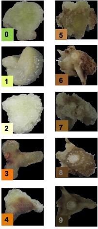
Figure 3. Five-point (0-5) visual hedonic scale for measuring the degree of tissue browning
of dragon fruit explants.
Figure 4. Effect of activated charcoal on the degree of explant browning of dragon fruit calli
using 9-point (A) and 5-point (B) arbitrary visual hedonic scale. Bars, within a
figure, with different letter indicates means were significant at pBalendres & Cruz., 2021
Figure 5. Length (a), width (b), and total area (c) of H. undatus calli 28 days after
inoculation (dai) in media with and without activated charcoal. No significant
difference was found (p>0.05) based on T-test analysis. Values represent the
mean of the two trials performed.
Fungal Contamination
Significant reduction of contamination of H. monacanthus cultures with activated charcoal
under 14-hour light (25%) and dark condition (50%) was observed four days after inoculation,
compared with untreated cultures (62.5 and 100%) (Figure 6a). The same results were
obtained for H. undatus. The contamination values for activated charcoal under 14-hour light
and dark conditions were 3.1% and 6.3%, respectively, while without activated charcoal were
15.6 18.8%, respectively (Figure 6b). In contrast, an increased contamination rate of both H.
JPACD (2021) 23:58-73 66Balendres & Cruz., 2021
monacanthus (87.5-100%) and H. undatus (50-75%) explants were observed eight days post-
inoculation in both media (Figure 6).
Figure 6. Percent fungal contamination in H. monacanthus (A) and H. undatus (b)
explants four and 8 days after incubation (dai). Means with different letters are
significantly different at p < 0.05 based on T-test. Values represent the mean of
the two trials performed.
Identities of fungal contaminants
Fungal mycelia with white to grayish growth were isolated (Figure 7) from all infected dragon
fruit cultures. The fungi were fast-growing, with mycelial growth reaching the edge of the plate
on the 7th-day post-incubation (Figure 7A). Thick mycelia with no spores formed were
observed (Figure 7B) from the seven-day-old cultures. Culture and morphology characteristics
resemble those of Diaporthe species. Three isolates (MDF1a, MDF1b, and MDF1c) were
chosen for sequence analysis based on their unique morphocultural characters. Analysis of
the DNA sequence of the partial sequence of the ITS-rDNA gene region using the BLASTN
software showed high similarities (99-100%) of the isolates to D. passifloricola and D. tectonae
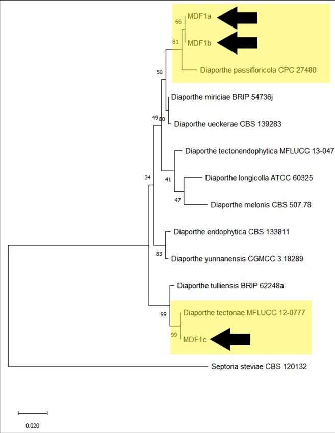
(Table 2). The phylogenetic tree (Figure 8) further supported close similarity of isolate MDF1a
and MDF1b to D. passifloricola and isolate MDF1c to D. tectonae.
JPACD (2021) 23:58-73 67Balendres & Cruz., 2021
Figure 7. Cultural (A) and morphological (B) characteristics of seven-day-old D.
passifloricola (a and b) and D. tectonae (c) in potato dextrose agar medium
(PDA).
Figure 8. Phylogenetic tree based on the maximum likelihood analysis of the partial
sequence of the ITS gene of the three fungal isolates (MDF1a, MDF1b, and
MDF1c) from this study and other Diaporthe species. Septoria steviae CBS
120132 served as an outgroup.
JPACD (2021) 23:58-73 68Balendres & Cruz., 2021
Table 2. Percent similarities of the fungal isolates associated with the tissue-cultured
dragon fruit.
Isolate/Sample Species ITS (Accession)
MDF1a D. passifloricola CPC 27480 99.20% (NR_1475.95.1)
MDF1b D. passifloricola CPC 27480 99.20% (NR_147595.1)
MDF1c D. tectonae MFLUCC 12-0777 100% (NR_147574.1)
DISCUSSION
This study showed activated charcoal (AC) reduces tissue browning of dragon fruit explant.
The findings were consistent with the previous results obtained in other plants, such as potato
(Buckseth et al., 2018), sugarcane (Shimelis et al., 2015), carrots (Pan and Staden, 2001),
and grapevine (Olah, 2017). The arbitrary visual hedonic scales used in this study could be
used in future status to assess callus browning intensity. Both the 5- and 9-point scale were
able to discern the browning variation. Some calli were formed, but the presence of AC in the
media affected the friability (ideal) of the calli. Activated charcoal did not effect on the length,
width, and total area of the calli produced compared to the control check. Activated charcoal
has been shown to adsorb auxin and cytokinin from culture media, thus rendering them
inactive (Weatherhead et al., 1979). Activated charcoal can also absorb α-naphthyl acetic acid
(NAA), up to 300 mg/l, (Weatherhead, 1979), dichlorophenoxyacetic acid (2, 4-D) (Ebert and
Taylor, 1990), and 6- benzyl amino purine (BA) (Ebert et al., 1993). Three cytokinins, 6-
furfuryl-aminopurine, 6-benzyl amino purine, and 6-(γ,γ-dimethylallylamino) purine, were
likewise adsorbed at a concentration of 10 mg/l (Weatherhead et al., 1979) by AC. Thus, the
addition of activated charcoal in the media possibly interfered with the growth and callus
development of dragon fruit explants through adsorption of the hormone and nutrients
available in the medium.
A significant decrease in the dragon fruit cultures' contamination was also documented four
days after inoculation (dai) using activated charcoal. Aside from reducing phenolic oxidation,
activated charcoal (AC) is known to adsorb undesirable/inhibitory substances and nutrients
found in the medium (Pan and Staden, 2001; Thomas, 2008). Activated charcoal in the media
might have influenced the environment necessary for the growth of specific microorganisms
(Weatherhead et al., 1978). However, no significant effect of AC in contamination was
observed eight dai. A higher contamination rate was observed in H. monacanthus (87.5-100%)
than in H. undatus (50-75%) callus cultures. The relatively higher rate of contamination in H.
monacanthus might be associated with the rich microflora present in the initial explant, which
was not effectively removed during the sterilization process. AC's activity is still unclear, but
some workers believe that AC gradually releases certain adsorbed products, such as nutrients
and growth regulators, which become available to plants (Thomas, 2008). This gradual release
of adsorbed nutrients by activated charcoal might have supported microorganisms' growth in
the cultures. Hence, activated charcoal for reducing contamination rate in tissue culture is
ineffective in the long run.
Molecular analyses of the ITS region of the fungal contaminants found in the dragon fruit
cultures revealed that the isolates were highly similar to two Diaporthe species: D.
passifloricola and D. tectonae. Although these fungi were not associated with any disease of
dragon fruit stem explants used in this study, Diaporthe species are known to cause diseases
on a wide range of host plants. Diaporthe species cause root and fruit rots, dieback, cankers,
JPACD (2021) 23:58-73 69Balendres & Cruz., 2021
leaf spots, blights, decay, and wilt (Mostert et al. 2001; van Rensburg et al. 2006; Thompson
et al. 2011; Udayanga et al. 2014; Dissanayake et al. 2015). For example, D. ampelina is one
of the pathogenic species best known as the causal agent of Phomopsis cane, leaf spot, and
grape yield losses in temperate regions (Erincik and Madden 2001). At the same time, D.
phaseolorum and D. longicolla are reported to be pathogenic to soybean (Santos et al., 2011).
The occurrence of fruit rot disease in dragon fruit caused by Diaporthe sp. was also reported
in China (Tsai et al., 2001). Recently, stem rot disease in H. undatus caused by D.
phaseolorum has been reported (Karim et al., 2019). However, the two Diaporthe species
found in this study was not associated to any symptom in the explant. These two species are
considered as an endophyte in dragon fruit, unless proven pathogenic to dragon fruits in future
studies.
CONCLUSION
The addition of AC in the tissue culture media reduced callus browning regardless of presence
or absence of light. Both the 5- and 9-point visual hedonic scale can be useful for future callus
development studies. Activated charcoal does not inhibit fungal contamination. However,
although callus were formed, AC (at 1 g/L) negatively affects callus friability. Therefore,
determining the optimum concentration of activated charcoal that can significantly reduce
tissue browning without hindering callus friability would be worthwhile. Diaporthe passifloricola
and D. tectonae were also identified as fungal contaminants in dragon fruit tissue culture and
are considered endophytes in dragon fruit stem. This is the first report of the association of
these two Diaporthe species in stems of dragon fruits. Nevertheless, the two species’
pathogenicity to dragon fruit plants would be tested to further elucidate their interaction with
dragon fruits.
ACKNOWLEDGMENTS
We thank Fe Dela Cueva, Edzel Evallo, John Darby Taguiam, Mari Neila Seco, Diane Biglete,
Loida Pascual, Ryan Tiongco, and Mr. Yron Retuta for the technical assistance.
FUNDING
This study was supported by the Department of Agriculture Bureau of Agricultural Research.
The Institute of Plant Breeding, College of Agriculture and Food Science, University of the
Philippines Los Baños has provided in kind support.
CONFLICT OF INTEREST
The authors declare that they have no conflict of interests.
ETHICAL APPROVAL
This article does not contain any studies with human participants or animals performed by any
of the authors. The research did not contain any experiment that uses animals or humans.
JPACD (2021) 23:58-73 70Balendres & Cruz., 2021
REFERENCES
Bakar, J, Shu CE, Muhammad S, Kharidah S and Hashim DM. 2011. Physico-chemical
characteristics of red pitaya (Hylocereus polyrhizus) peel. Int. Food Res. J.,18: 279-
286.6.
Balendres MA, Bengoa JC. 2019. Diseases of dragon fruit (Hylocereus species): etiology and
current management options. Crop Protection 126, 104920.
Bozkurt T, İnan S, Dündar I. 2020. Micropropagation of different pitaya varieties. International
Journal of Agricultural and Natural Sciences 13(1): 39-46.
Buckseth TS, Singh RK, Sharma AK, Sharma S, Moudgil V, Saraswati A. 2018. Optimization
of activated charcoal on in vitro growth and development of potato (Solanum tuberosum
L.). Int. J. Curr. Microbiol. App.Sci 7(10): 3543-3548.
Casas, A., Barbera, G., 2002. Mesoamerican domestication and diffusion. In: Nobel, P.S. (Ed.),
Cacti: Biology and Uses. University of California Press, Los Angeles, California, pp.
143–162.
Crous PW and Groenewald JZ. 2016. Fungal Planet description sheets: 400-468. Persoonia
36:316-458.
Crous PW, Wingfield MJ, Le Roux JJ, Richardson DM et al. 2015. Fungal Planet description
sheets: 371-399. Persoonia 35:264-327.
Dahanayake N and Ranawake AL. 2011. Regeneration of dragon fruit (Hylecereus undatus)
plantlets from leaf and stem explants. Tropical Agricultural Research & Extension 14(4):
85-89.
Dissanayake AJ, Liu M, Zhang W, Chen Z, Udayanga D, Chukeatirote E, Li X, Yan J, Hyde
KD. 2015. Morphological and molecular characterisation of Diaporthe species
associated with grapevine trunk disease in China. Fungal Biol. 119:283–294.
Doilom M, Dissanayake AJ, Wanasinghe DN, Boonmee S, Liu J, Bhat DJ, Taylor J, Bahkali
AH, Mckenzie EH, Hyde K. 2016. Microfungi on Tectona grandis (teak) in Northern
Thailand. Fungal Diversity, 82, 107-182.
Ebert A, Taylor HF. 1990. Assessment of the changes of 2, 4-dichlorophenoxyacetic acid
concentrations in plant tissue culture media in the presence of activated charcoal. Plant
Cell Tissue Organ Cult 20:165–72.
Ebert A, Taylor F, Blake J. 1993. Changes of 6-benzylaminopurine and 2, 4-
dichiorophenoxyacetic acid concentrations in plant tissue culture media in the presence
of activated charcoal. Plant Cell Tissue Organ Cult 33:157–63.
Erincik O, Madden LV (2001) Effect of growth stage on susceptibility of grape berry and rachis
tissues to infection by Phomopsis viticola. Plant Dis 85:517–520.
Gao Y, Liu F, Duan W, Crous PW, Cai L. 2017. Diaporthe is paraphyletic. IMA Fungus
8(1):153-187. doi:10.5598/imafungus.2017.08.01.11.
Gomes RR, Glienke C, Videira SI, Lombard L, Groenewald JZ, Crous PW. 2013. Diaporthe:
a genus of endophytic, saprobic and plant pathogenic fungi. Persoonia 31:1-41.
Jiang N, Fan X, Tian C, Crous PW. 2020. Reevaluating Cryphonectriaceae and allied families
in Diaporthales. Mycologia 112 (2): 267-292.
Karim MM, Rahman ME, Islam MN, Akhter MS, Khatun F, Rahman ML, Goswami BK. 2019.
Occurrence of stem rot disease of Hylocereus undatus in Bangladesh. Indian
Phytopathology (Short Communication).
Kearse, M., Moir, R., Wilson, A., Stones-Hava, S., Cheung, M., Sturrock, S., Buxton, S.,
Cooper, A., Markowitz, S., Duran, C., Thierer, T., Ashton, B., Mentjies, P., Drummond,
JPACD (2021) 23:58-73 71Balendres & Cruz., 2021
A. (2012). Geneious Basic: an integrated and extendable desktop software platform for
the organization and analysis of sequence data. Bioinformatics, 28(12), 1647-1649.
Koehler AM, Larkin MT, Rogers LW, Carbone I, Cubeta MA, Shew HD. 2019. Identification
and characterization of Septoria steviae as the causal agent of Septoria leaf spot
disease of stevia in North Carolina. Mycologia 111(3):456-465.
Kumar S., Stecher G., Li M., Knyaz C., and Tamura K. (2018). MEGA X: Molecular
Evolutionary Genetics Analysis across computing platforms. Molecular Biology and
Evolution 35:1547-1549.
Liang S, He Y, Zheng H, Yuan Q, Zhang F, Sun B. 2019. Effect of sucrose and browning
inhibitors on callus proliferation and anti-browning of chinese kale. IOP Conf. Ser.: Earth
Environ. Sci. 252 022018.
Luo H, Cai Y, Peng Z, Liu T and Yang S. 2014. Chemical composition and in vitro evaluation
of the cytotoxic and antioxidant activities of supercritical carbon dioxide extracts
of pitaya (dragon fruit) peel. Chem. Central J. 8 (1):1.
Mercado-Silva EM. 2018. Pitaya- Hylocereus undatus (Haw). Exotic Fruits: pp. 339-349.
Mohamed-Yasseen Y., 2002. Micropropagation of pitaya (Hylocereus undatus Britton et Rose).
In 492 Vitro Cell. Dev. Biol.—Plant. 38, 427–429.
Mostert L, Crous PW, Kang JC, Phillips AJL. 2001. Species of Phomopsis and a Libertella sp.
occurring on grapevines with specific reference to South Africa: morphological, cultural,
molecular and pathological characterization. Mycologia 93:146–167.
Olah R. 2017. The use of activated charcoal in grapevine tissue culture. Vitis 56: 161–171.
Ortíz-Hernandez, YD. 1999. Pitahaya. Un nuevo cultivo para México. Editorial Limusa, México
City.
Ortiz-Hernandez, YD and Carrillo-Salazar JA. 2012. Pitahaya (Hylocereus spp.): A short
review. Commun. Sci., 3: 220-237.
Pan M, Staden J. 1998. The use of activated charcoal in in vitro culture- A review. Plant Growth
Regulation 26: 155–163.
Pan M, Staden J. 2001. The effect of activated charcoal on the production and development
of somatic embryos in cultures of carrot Daucus carota. South African Journal of Botany
67: 629-635.
Rebecca OPS, Boyce AN and Chandran S. 2010. Pigment identification and antioxidant
properties of red dragon fruit (Hylocereus polyrhizus). Afri. J. Biotechnol., 9: 1450-
1454.5.
Rodeo AJD, Castro AC, Esguerra EB. 2018. Postharvest handling of dragon fruit
(Hylocereus spp.) in the Philippines. Dragon Fruit Regional Network Initiation Workshop:
pp 125-131.
Santos JM, Vrandečić K, Ćosić J, Duvnjak T, Phillips AJL (2011) Resolving the Diaporthe
species occurring on soybean in Croatia. Persoonia 27:9–19.
Shimelis D, Bantte K, Feyissa T. 2015. Effects of polyvinyl pyrrolidone and activated charcoal
to control effect of phenolic oxidation on in vitro culture establishment stage of
micropropagation of sugarcane (Saccharum officinarum L.). Adv Crop Sci Tech 3: 184.
Taguiam JD, Evallo E, Bengoa J, Maghirang R, Balendres MA. 2020a. Detection of Nigrospora
sphaerica in the Philippines and the susceptibility of three Hylocereus species to
reddish-brown spot disease. J Prof Assoc Cactus Devt. 22, 49-61.
Taguiam JD, Evallo E, Bengoa J, Maghirang R, Balendres MA. 2020b. Susceptibility of the
three dragon fruit species to stem canker and growth inhibition of Neoscytalidium
dimidiatum by chemicals. Journal of Plant Pathology 102, 1077–1084.
JPACD (2021) 23:58-73 72Balendres & Cruz., 2021
Taguiam JD, Evallo E, Bengoa JC, Maghirang RM, Balendres MA (2020c) Pathogenicity of
Epicoccum sorghinum towards dragon fruits (Hylocereus species) and in vitro evaluation
of chemicals with antifungal activity. Journal of Phytopathology, 168(6), 303-310.
Talbot N. 2017. DNA Extraction for fungi. University of Exeter. Retrieved 11 February 2019
from http://www.exeter.ac.uk/nicktalbot/protocols/.
Tamura K. and Nei M. (1993). Estimation of the number of nucleotide substitutions in the
control region of mitochondrial DNA in humans and chimpanzees. Molecular Biology
and Evolution 10:512-526.
Tan YP, Edwards J, Grice KRE, Shivas RG. 2013. Molecular phylogenetic analysis reveals
six new species of Diaporthe from Australia. Fungal Diversity 61 (1):
Thomas TD. 2008. The role of activated charcoal in plant tissue culture. Biotechnol Adv.
26:618–631.
Thompson SM, Tan YP, Young AJ, Neate SM, Aitken EA, Shivas RG. 2011. Stem cankers on
sunflower (Helianthus annuus) in Australia reveal a complex of pathogenic Diaporthe
(Phomopsis) species. Persoonia 27:80–89.
Thompson SM, Tan YP, Shivas RG, Neate SM, Morin L, Bissett A, Aitken EA. 2015. Green
and brown bridges between weeds and crops reveal novel Diaporthe species in
Australia. Persoonia 35:39-49. doi: 10.3767/003158515X687506.
Tsai JN, Hsu TH, Cheng SF, Ann PJ. 2010. Preliminary surveying report of fungal diseases
on postharvest dragon fruit. Plant Path. Bull. Taiwan 19: 293. (Abstract in Chinese).
Udayanga D, Castlebury LA, Rossman AY, Chukeatirote E, Hyde KD. 2014. Insights into the
genus Diaporthe: phylogenetic species delimitation in the D. eres species complex.
Fungal Divers 67:203–229.
Udayanga D, Castlebury LA, Rossman AY, Chukeatirote E, Hyde KD. 2015. The Diaporthe
sojae species complex: Phylogenetic re-assessment of pathogens associated with
soybean, cucurbits and other field crops. Fungal Biol. 119, 383-407
Wang and Huang. 1976. Beneficial effects of activated charcoal on plant tissue and organ
cultures. In Vitro 12, 260–262 (1976).
Wei X, Gou X, Yuan T, et al. 2006. A highly efficient in vitro plant regeneration system and
Agrobacterium-mediated transformation in Plumbago zeylanica. Plant Cell Rep.
25:513–521.
Weatherhead MA, Burdon J, Henshaw GG. 1978. Some effects of activated charcoal as an
additive to plant tissue culture media. Zeitschrift für Pflanzenphysiologie Volume 89 (2):
141-147.
Weatherhead MA, Burdona J, Henshaw GG. 1979. Effects of Activated Charcoal as an
Additive Plant Tissue Culture Media: Part 2. Zeitschrift für Pflanzenphysiologie 94,
399-405.
van Rensburg JCJ, Lamprecht SC, Groenewald JZ, Castlebury LA, Crous PW. 2006.
Characterization of Phomopsis spp. associated with dieback of rooibos (Aspalathus
linearis) in South Africa. Stud Mycol 55:65–74.
Zhang J, Madden TL. 1997. PowerBLAST: A new network BLAST application for interactive
or automated sequence analysis and annotation. Genome Research, 7:649-656.
JPACD (2021) 23:58-73 73You can also read