Increasing Lignification in Translucent Disorder Aril of Mangosteen Related to the ROS Defensive Function - Hindawi.com
←
→
Page content transcription
If your browser does not render page correctly, please read the page content below
Hindawi
Journal of Food Quality
Volume 2021, Article ID 6674208, 10 pages
https://doi.org/10.1155/2021/6674208

Research Article
Increasing Lignification in Translucent Disorder Aril of
Mangosteen Related to the ROS Defensive Function

 Chalermchai Wongs-Aree ,1 Patchayapon Siripirom,2 Apinya Satitpongchai,2
 Kitti Bodhipadma ,2 and Sompoch Noichinda 2
 1
 Division of Postharvest Technology, School of Bioresources and Technology, King Mongkut’s University of Technology Thonburi,
 Bangkhuntien, Bangkok 10150, Thailand
 2
 Division of Agro-Industrial Technology, Faculty of Applied Science, King Mongkut’s University of Technology North Bangkok,
 Bang-sue, Bangkok 10800, Thailand

 Correspondence should be addressed to Sompoch Noichinda; sompoch.n@sci.kmutnb.ac.th

 Received 27 November 2020; Revised 22 January 2021; Accepted 27 January 2021; Published 15 February 2021

 Academic Editor: Giorgia Liguori

 Copyright © 2021 Chalermchai Wongs-Aree et al. This is an open access article distributed under the Creative Commons
 Attribution License, which permits unrestricted use, distribution, and reproduction in any medium, provided the original work is
 properly cited.

 Mangosteen fruit has a high potential on the global fruit market, but some disorders, including translucent flesh, are major
 problems of fruit quality, limiting the marketability. The present study was conducted to compare physiological changes of
 reactive oxygen species (ROS), cellular lignification between translucent and normal aril, and elucidate the relation. Mangosteen
 fruits at purple peel color were collected from eastern Thailand during the middle of the rainy season of 2019. Translucent aril
 accumulated higher lignin content in the tissues, expressing firmer texture ten times higher than normal aril. Lignification was
 increased in translucent aril by 740% and 25% higher coniferyl alcohol dehydrogenase (CAD) and peroxidase (POD) activity,
 respectively, induced by high H2O2. Healthy aril performed higher activities of superoxide dismutase (SOD) (8.5 times) and
 ascorbate peroxidase (APX) (1.3 times) to those in translucent aril. Furthermore, the higher flavonoid content, ascorbic acid
 content, and antioxidant capacities detected in normal aril could significantly reduce oxidative stress. Although containing high
 antioxidant systems, healthy aril was found to accumulate higher malonaldehyde content (MDA). This study provides intensive
 evidence of oxidative stress and the defensive systems between normal and translucent tissues.

1. Introduction shows uneven ripening among aril segments in the fruit [4].
 Furthermore, abnormal symptoms, including aril translu-
Mangosteen (Garcinia mangostana) belongs to the Gutti- cency and stiff texture of the aril, could be operated during
ferae family. It is a unique tropical fruit, expressing ripening maturation. These disorders are severe obstacles in
prominent calyx on top of the fruit looking like a crown and mangosteen marketability.
so-called the ‘Queen of fruit.’ Mangosteen fruit consists of a Translucent flesh disorder usually occurs during on-tree
thick pericarp derived from the floral ovary wall and 5–7 aril fruit ripening during the rainy season. The previous study
segments. The edible part (aril) is developed from the seed showed that the number of fruits generating flesh translu-
integument. The periodical cycle of fruit growth from flower cency increased when the tree was applied by water
opening until maturity takes about 11–12 weeks [1]. The sprinkling over the canopy for 2–3 h [5]. On the other hand,
fruit ripening starts by the pericarp color turning from pale a high level of underground water did not stimulate the
green to purple followed by aril softening [2]. When mature induction of translucent flesh disorder [6]. Moreover, an
green fruit starts ripening with the peel showing a red pad application of water supplied at the harvested fruit peduncle
sign, mangosteen is classified as a climacteric fruit according did not induce translucent flesh disorder in ripe fruit [3].
to its respiratory pattern [3]. However, mangosteen fruit Since capillary water (lenticel-occupied water) in the
2 Journal of Food Quality

pericarp of on-tree mature green fruit induced a hypoxic plunger with 5 mm distance depth and 1.0 mm·s−1 test speed.
condition, this incident enhanced translucent flesh disorder The maximum force was recorded in Newton unit.
which found a remarkable accumulation of lignin in the ripe
aril [7]. Furthermore, our previous study found a signifi-
 2.3. Cross Section and Staining. A thin piece of free-hand
cantly high proportion of Na2CO3-SP between differential
 cross section (40 microns) from normal and translucent
pectin fractions of translucent disorder aril [7]. A com-
 disorder arils was stained with 0.1 M potassium phosphate
parison of respiratory patterns indicates that translucent
 buffer (pH 6.8) containing 0.05% (w/v) Toluidine Blue
disorder aril conducted a higher respiration rate than
 O-dye for 2 min and then washed with distilled water [16].
healthy aril [8]. Typically, translucent disorder initially
 The stained tissues were observed under a light microscope
occurs in the largest aril segment, which showed high vig-
 (EMZ, Meiji Techno, Japan).
orous energies in both aril and seed than healthy arils [9]. As
hypoxic cellular conditions are stimulated by alternative
catabolic pathways to produce more energy for maintaining 2.4. Lignin Determination. Lignin content in the flesh was
systematic survival, these stress phenomena could be en- investigated according to the method of Bruce and West [17].
couraged by the overemission of reactive oxygen species Four g of aril flesh was homogenized in 16 mL of methanol by
(ROS) in several plant parts [10, 11]. The ROS directly at- using a homogenizer (IKA Ultrarax T 25, Germany). Ho-
tacks the cell membrane or is transformed into a harmful mogenate was filtered through Whatman GF/Ag filter No. 1.
hydroxyl radical (OH_), enhancing the lipid membrane’s The remaining residual was washed twice by methanol and
peroxidation. However, ROS can be subsequently reduced dried in an oven at 60°C for 24 h. Fifty mg of dried residual was
by some biological pathways, including cellular defensive mixed in 5 mL of 2 M hydrochloric acid and 0.5 mL of thio-
mechanisms. Plants under stress drive their defensive glycolic acid. The mixture was boiled at 100°C for 4 h. After
resilience through the alternative antioxidant systems, both cooling, the suspension was centrifuged at 12,000 × g for
enzymatic and nonenzymatic shuttles. The enzymatic system 30 min. The pallet was rinsed with 5 mL of distilled water. The
includes superoxide dismutase (SOD), catalase (CAT), and pallet was mixed in 5 mL of 0.5 M sodium hydroxide for lignin
ascorbate peroxidase (APX). On the other hand, phenolics, thioglycolate extraction and left for 18 h. The mixture was
flavonoids, and ascorbates are involved in preventing stress. centrifuged at 12,000 × g for 30 min. The supernatant (1 mL)
Some abiotic stresses inducing the defensive mechanisms in was added with 1 mL of concentrated hydrochloric acid and
plants through ROS metabolisms have been recently re- incubated for 4 min. The reaction was centrifuged at 10,000 × g
ported [12–15]. Thus, the purposes of this research were to for 10 min. The radish-brown residual was collected and dis-
investigate ROS generations, phenolic accumulation, and solved in 25 mL of 0.5 M sodium hydroxide. The dissolved
lignification and to explore the relation of enzymes asso- lignin solution’s absorbance was measured by using a spec-
ciated with ROS removal in ripe mangosteen aril between trophotometer (Shimadzu UV-1800, Japan) at 280 nm. The
the healthy and translucent tissues. content was reported in Abs280 nm/50 mg DW.

2. Materials and Methods 2.5. ROS Generation and.−
 Its Transformation Determination.
 Superoxide anion (O2 ) was extracted and measured
2.1. Plant Materials. Fresh ripe mangosteen fruits at the peel according to Chaitanya and Naithani [18]. Two g of aril flesh
purple stage were obtained from a commercial orchard in was homogenized in 5 mL of 0.05 M potassium phosphate
Chanthaburi Province, eastern Thailand, in July 2019, and buffer (pH 7.8) including 1 mM diethyl-dithiocarbamate (a
were transported to the Applied Science Laboratory at King SOD inhibitor). The homogenate was centrifuged at
Mongkut’s University of North Bangkok (KMUTNB) within 12,000 × g at 4°C for 15 min. The supernatant (1 mL) was
a day. Fruits were then sorted by the uniformity of size (ca. added with 3 mL of a reaction mixture containing 0.1 M
70–90 g/fruit), peel color, and absence of defects. For a phosphate (pH 7.8), 1 mM diethyl-dithiocarbamate, and
preliminary classification of translucent flesh disorder, fruits 0.25 mM nitro blue tetrazolium (NBT). The absorbance of
were floated onto water containing 1.0% sodium chloride. the reaction solution was spectrophotometrically measured
The fruit that sank in the solution was presumed as having at 540 nm within a min.
translucent flesh disorder, comprising higher gravity. After .−
 Hydrogen peroxide (H2O2), an O2 transformation, was
air-drying at the ambient room temperature (23–28°C), the measured according to the work of Zouari et al. [19]. One g
fruit pericarp was carefully removed by a sharp knife without of aril was homogenized in 5 mL of 0.1% trichloroacetic acid
aril damage. In the present study, the fruit largest aril (TCA) and centrifuged at 12,000 × g at 4°C for 15 min. The
segment of normal white (Supplementary Figure 1(a)) and supernatant (1 mL) was then added to 2 mL of a reaction
translucent disorder flesh (Supplementary Figure 1(b); mixture containing 0.01 M potassium phosphate buffer (pH
pointing arrow) was collected from one hundred mango- 7.0) and 1 M potassium iodide. The reaction absorbance was
steen fruits for investigating the ROS stress and lignification. spectrophotometrically measured at 390 nm.

2.2. Firmness Measurement. Aril firmness was measured in 2.6. ROS Defensive Enzyme Assays. Extracts of superoxide
the middle of the segment by using a TA-XT 21 texture dismutase (SOD), catalase (CAT), peroxidase (POD), and
analyzer (Stable Microsystems, UK) using a 2 mm spherical ascorbate peroxidase (APX) were performed according to
Journal of Food Quality 3

the methods of Dhindsa et al. [20] and Jiménez et al. [21]. for 10 min and then cooled down on ice. The absorbances
For SOD, POD, and CAT, 1 g of aril flesh was homogenized were spectrophotometrically measured at 532 and 600 nm,
in 10 mL of 0.05 M potassium phosphate buffer (pH 7) respectively. MDA content was calculated from the following
containing 1% (w/v) polyvinylpolypyrrolidone (PVPP). For formula with 155 mM−1 cm−1 extinction coefficient value:
APX extraction, 1 g of aril flesh was homogenized in 10 mL
of 0.05 M potassium phosphate buffer (pH 7.8) containing μmol Abs532nm − Abs600nm
 MDA � . (2)
0.1% (v/v) Triton X-100. The homogenate was centrifuged at gFW 155 mM−1 1cm−1 × 1000
12,000 × g at 4°C for 15 min, and the supernatant was used
for activity assay.
 The extraction and assay of CAD were performed with 2.8. Phenolics Accumulation and Phenylalanine Ammonia
the slightly modified Goffner et al. [22]. Briefly, 2 g of aril was Lyase (PAL) Activity Assay. Determination of aril phenolics
homogenized in 10 mL of 100 mM phosphate buffer (pH 7.5) was performed according to Recuenco et al.’s method [25]. One
containing 1 mM EDTA, 5 mM magnesium chloride, 0.05% g of flesh was homogenized in 10 mL of methanol. After
(v/v) Tween 20, and 2.5 mM 2-mercaptoethanol. The ho- centrifugation at 10,000 × g for 15 min, the supernatant
mogenate was high-speed centrifuged at 18,000 × g at 4°C for (100 µL) was mixed in 3 mL of distilled water and 0.5 mL of
30 min. The supernatant was used for enzyme activity assay. Folin–Ciocalteu phenol reagent for 3 min. The mixture was
One mL of the crude enzyme was incubated in 3 mL of added with 2 mL of 20% (w/v) of sodium carbonate. The
100 mM phosphate buffer (pH 7.5) containing 0.2 mM absorbance of the reaction was recorded at 750 nm. Phenolic
NADP and 0.1 mM coniferyl alcohol for 1 min. The activity acid content was compared to the absorbance of gallic acid
of the enzyme was measured by using a spectrophotometer standard. For flavonoid determination, 2 mL of supernatant
at 400 nm. was mixed in 5 mL of distilled water and 0.15 mL of 5% (w/v)
 SOD activity assay was perfprmed according to the work sodium nitrate for 5 min, and then, 0.15 mL of 10% (w/v)
of Dhindsa et al. [20]. The crude (100 µL) was mixed in 3 mL aluminium chloride was added. The reaction absorbance was
of 0.05 M potassium phosphate buffer (pH 7.8) containing measured at 510 nm. Flavonoid content was compared with
13 mM methionine, 75 mM NBT, 4 mM riboflavin, and catechin standard.
100 µM ethylene diamine tetra-acetic acid. The solution PAL assay was performed according to the work of Camm
mixture was vortexed for 1 min and then left under fluo- and Towers [26]. Five g of flesh was homogenized in 60 mL of
rescent light (15 watts) for 30 min. The absorbance was cold acetone and filtered through Whatman filter paper No. 2.
spectrophotometrically measured at 560 nm. SOD activity The remaining brown residual was rewashed with cold acetone
was calculated from the reaction mixture’s absorbency with until the residual color turned white. The residual was blended
the enzyme (sample) and without enzyme (control). One in 50 mL of cold 95% ethanol and filtered through Whatman
unit of SOD activity was represented by 50% NBT inhibition filter paper No. 2. The residual was dried at room temperature
calculated from the following formula: (25°C) and then kept in a desiccant chamber. For PAL, 0.5 mg
 of the acetone powder was mildly stirred in 50 mL of 0.1 M
 unit AbsControl − AbsSample /AsbControl × 100
 SOD � . sodium borate buffer (pH 8.8) at 4°C for 30 min. After cen-
 gFW 2 trifugation at 12,000 × g at 4°C for 20 min, the supernatant
 (1) (1 mL) was mixed in 1.5 mL of 0.1 M sodium borate buffer (pH
 8.8) and 1 mL of 10 mg/mL phenylalanine and incubated in a
 POD and CAT activity assays were performed according hot bath at 30°C for 1 h. Five N HCl (0.5 mL) was added into
to Song et al.’s method [23]. One hundred µL of the crude the reaction to stop the enzyme activity. The reaction absor-
extract was mixed in 3 mL of 0.05 M potassium phosphate bance with and without phenylalanine was measured at
buffer (pH 7.0) containing 200 mM hydrogen peroxide and 290 nm. One unit of PAL activity was represented by an in-
20 mM guaiacol. The absorbance of POD was measured at crease in 1 µmol cinnamic acid per 1 h.
470 nm. One POD activity unit was represented by an in-
crease in the absorbance (0.01) in 1 min. For CAT activity,
100 µL of the crude was mixed in 3 mL of 0.05 M potassium 2.9. Ascorbic Acid Determination. An ascorbic acid deter-
phosphate buffer (pH 7.0) containing 200 mM hydrogen mination was performed, according to Klein and Perry’s
peroxide. The absorbency was measured at 240 nm. One unit method [27]. One g of aril flesh was homogenized in 10 mL of
of CAT activity was represented by a decrease in the ab- 5% (w/v) metaphosphoric acid and centrifuged at 12,000 × g
sorbance (0.01) for 1 min. for 10 min. The supernatant (0.5 mL) was mixed in 4.5 mL of
 0.1 mM 2, 6-DCIP. The absorbance of ascorbic acid in the
 mixture was then spectrophotometrically measured at 515 nm
2.7. Malondialdehyde (MDA) Determination. Determination and compared to the ascorbic acid standard curve.
of MDA was performed according to Dipierro and Leonardis
[24]. Two g of aril flesh was homogenized in 10 mL of 0.1% 2.10. Antioxidant Ability Assays
(w/v) trichloroacetic acid (TCA) and centrifuged at
12,000° × °g for 10 min. The supernatant (1 mL) was reacted 2.10.1. Reducing power Assay. One g of aril flesh was ho-
with 2 mL of 20% (v/v) TCA containing 0.5% (v/v) of thi- mogenized in 9 mL of methanol with a homogenizer. The
obarbituric acid (TBA) and incubated in a hot bath at 100°C homogenate was made to stand at ambient temperature
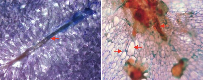
4 Journal of Food Quality

 Healthy aril flesh Translucent aril flesh

 Toluidine Blue O staining (100X)
 Fibrous integument
 coated lignin
 Fibrous
 integument Thicken wall
 (Lignin) Aerenchyma

 (a)
 1.8 0.8

 1.6 0.7

 Lignin (Abs280 nm/50 mgDW)
 1.4 a a
 0.6
 Firmness (Newton)

 1.2
 0.5
 1
 0.4 b
 0.8
 0.3
 0.6
 0.2
 0.4

 0.2 b 0.1

 0 0
 Healthy aril Healthy aril
 Translucent aril Translucent aril
 (b) (c)
 5 1.6
 4.5 a 1.4
 4
 1.2
 CAD (unit/mg protein)

 POD (unit/µg protein)

 3.5
 3 1

 2.5 0.8 a

 2 0.6 b
 1.5
 0.4
 1
 b
 0.5 0.2

 0 0
 Healthy aril Healthy aril
 Translucent aril Translucent aril
 (d) (e)

Figure 1: Toluidine Blue O-stained tissues (a), firmness (b), lignin content (c), coniferyl alcohol dehydrogenase activity (d), and peroxidase
activity (e) in healthy and translucent arils. Means (n � 4) with different lower-case letters on the bars are significantly different.
Journal of Food Quality 5

(24–28°C) for 3 h and then suctioned through Whatman higher lignin content detected in the translucent tissues,
filter paper No. 1. The methanolic supernatant (5 mL) was double the amount compared to healthy arils
mixed with 1.25 mL of 0.2 M potassium phosphate buffer (Figure 1(c)). The translucent aril conducted high ligni-
(pH 6.6) and 1.25 mL of 1.0% potassium ferricyanide (w/v). fication, which was increased by the activity of two key
The mixture was then incubated in a hot water bath at 50°C enzymes in lignin biosynthesis, namely, CAD and POD.
for 20 min. The mixture was cooled down on ice before When the former increased by 740% (Figure 1(d)), the
adding 1.25 mL of 10% (w/v) trichloroacetic acid. The latter was 25% up (Figure 1(e)) in the translucent aril.
cleared zone fraction (2.5 mL) was mixed with 2.5 mL of From these two enzymes directly involved in the accu-
distilled water and 0.5 mL of 0.1% (w/v) ferric chloride and mulation of lignins, CAD could be the rate-limiting step
incubated at ambient temperature for 10 min. The absor- of lignification in mangosteen aril under the stress con-
bance of the solution mixture was spectrophotometrically ditions. Translucent flesh disorder is usually generated in
measured at 700 nm [24]. mangosteen fruit during fruit ripening developed on tree
 in the rainy season. Water covering the fruit could induce
 hypoxic and oxidative stress in the fruit [5]. Lignification
2.10.2. DPPH Scavenging Inhibition Assay. DPPH scav-
 induction was found in wheat under waterlogging con-
enging ability was measured, according to Chang et al.
 ditions related to increasing CAD and POD activities [10].
[28]. One g of aril flesh was homogenized in 10 mL of
 The modification of lignins in the cell wall, simultaneously
methanol and centrifuged at 10,000 × g for 15 min. The
 induced by abiotic stress, could modify the cell-wall
supernatant (0.1 mL) was added into 2.9 mL of 1 mM 1,1-
 matrix and the properties. Schopfer [30] reported that
diphenyl-2-picrylhydrazyl (DPPH) and kept in the dark
 cell-wall stiffening of maize coleoptile was related to an
condition for 30 min. Then, mixture absorbance was
 intercellular coupling of monolignol residues by POD, a
monitored at 517 nm. The reaction without the methanol-
 lignin bound-membrane enzyme, using H2O2 as an
extraction solution was demonstrated as a control. The
 electron acceptor. As a result, changes in a high pro-
percentage of scavenging inhibition was calculated from
 portion of Na2CO3-SP in the pectin [7] and the high
the following formula:
 lignification could transform the white soft aril tissues
 AbsControl − AbsSample into translucent crispy tissues under stress conditions.
 DPPH inhibition (%) � × 100.
 AbsControl
 (3) 3.2. ROS Generation and Its Transformation. Oxidative in-
 termediates were investigated in both healthy and translu-
 .−
 cent arils of mangosteen fruit. Superoxide anion (O2 ) was
2.11. Protein Determination. Protein content in the enzyme dramatically higher in translucent aril, which was 8.5-fold
reaction was measured according to Bradford’s method [29]. compared to a healthy one (Figure 2(a)). This harmful ROS
The crude at 0.5 mL was mixed in a protein reagent com- was suddenly dismutated to hydrogen peroxide (H2O2) by
posed of 100 mg of Coomassie Brilliant Blue G-250 (CBB) in superoxide dismutase (SOD). When H2O2 content was 125%
50 mL of ethanol and 100 mL of 85% (w/v) phosphoric acid. higher accumulated in translucent aril (Figure 2(b)), SOD
The absorbance was recorded at 595 nm and then compared activities were, however, equal in both healthy and trans-
to Bovine Serum Albumin (BSA) standard. lucent arils (Figure 2(c)). Levels of lipid peroxidation could
 imply oxidative stress and damage. Interestingly, malo-
2.12. Statistical Analysis. Independent Sample t-test analysis naldehyde (MDA), a membrane-damaged end-product by
was performed to compare the variation of the parameter lipid peroxidation, was slightly higher in healthy aril
mean values between both treatments (4 replicates each; 4 (Figure 2(d)). MDA is widely used as a marker of oxidative
fruit/replicates) at p < 0.05 using an SPSS software version lipid injury in plant tissues under biotic and abiotic stress.
26 (IBM, Chicago, IL, USA). However, this reaction in plant tissues could be potentially
 interfered by many biological compounds which vary
3. Results and Discussion according to the tissue types and stress conditions [31]. For
 oxidative stress in mangosteen fruit, H2O2 in healthy aril
3.1. Lignification and Firmness. Translucent aril, behaving could be transformed into hydroxyl radical (OH·) via Fenton
as a stiffening structure, exhibits firm-crispy texture and a reaction in the presence of Fe2+ than translucent aril. Al-
translucent tissue character. The microscopic images of ternatively, H2O2 was contributed to be a cosubstrate of
Toluidine Blue O-stained aril structure show that lignin POD to produce lignins in translucent aril, which is higher .−
was localized in the parenchyma cell wall and high in the than that in healthy aril. Oxidative intermediates such as O2
protruding fibrous-chain of the seed coat of translucent and H2O2 were highly induced in okra pods [12] and as-
flesh disorder (Figure 1(a)). Moreover, we found that paragus shoots [32] during storage under abiotic stress.
some parenchyma cells in translucent tissues were either Furthermore, Jia et al. [11] found that cherry rootstock
transformed into collenchyma cells or collapsed to form released a high ROS amount under waterlogging conditions.
aerenchyma. The firmness of healthy aril was only 0.21 For survival, the harmful radical was discriminated by a
Newtons, which was 10 times lower than that of trans- scavenging mechanism using several enzymes and
lucent aril (Figure 1(b)). This evidence was related to the cosubstrates.
6 Journal of Food Quality

 0.035 14
 a
 0.03 12 a

 O2·-(Abs 540nm/min/gFW)
 0.025 10

 H2O2 (µM/gFW)
 0.02 8

 0.015 6
 b
 0.01 4

 0.005 b 2

 0 0
 Healthy aril Healthy aril
 Translucent aril Translucent aril
 (a) (b)
 1.5 0.18

 0.16
 1.2 0.14
 a
 SOD (unit/µg protein)

 MDA (µmol/gFW)

 0.12
 0.9 a
 a 0.1
 b
 0.08
 0.6
 0.06

 0.3 0.04

 0.02

 0 0
 Healthy aril Healthy aril
 Translucent aril Translucent aril
 (c) (d)
 .−
Figure 2: O2 (a), H2O2 content (b), superoxide dismutase activity (c), and malonaldehyde content (d) in healthy and translucent arils.
Means (n � 4) with different lower-case letters on the bars are significantly different.

3.3. The Differential ROS Defensive Mechanisms. The phe- defense in the healthy mangosteen aril by utilizing ascorbic
nolics content was 54 mg GAE/g FW in translucent aril and acid and H2O2 into water via the ascorbate-glutathione cycle
1.7 times higher than in healthy aril (Figure 3(a)). Since PAL [33]. When the lower content of ascorbate by 24% was
activity was significantly 100% higher in translucent aril detected in translucent aril (Figure 4(a)), the higher range of
(Figure 3(b)), the phenolics were contributed to form lignin flavonoids by 35% was measured in healthy aril
along with the lignified enzyme series. Phenolic biosynthesis (Figure 4(b)). Flavonoids could be an effective antioxidant
could be stimulated by some abiotic stresses such as hypoxia supplement to reduce the activity of ROS. The supportive
and physical damage to produce phenylpropanoid inter- evidence was shown by approximately 100% higher of the
mediates as defensive mechanisms of plant cells [32]. high reducing power ability (Figure 4(c)) and potent inhi-
Translucent flesh disorder is developed during on-tree fruit bition of DPPH scavenging in healthy aril (Figure 4(d)).
ripening in rainy season or when a fruit was covered with Abiotic stresses inducing antioxidant systems’ defensive
water for several hours, which could induce hypoxic con- mechanisms through redox-reaction via ROS metabolism
ditions in the fruit [5]. Furthermore, the level of transformed were reported in some field crops [14, 15] and horticultural
H2O2 can alternatively be reduced by other plant defensive crops [12, 34].
reactions. Although CAT activity, catalyzing H2O2 into We propose the comparative routes of ROS generation
water in peroxisome, was almost equal in both healthy and and the defensive mechanism, taking place in the healthy
translucent arils (Figure 3(c)), APX activity in healthy aril and translucent disorder of ripe mangosteen arils in
was significantly higher than that in translucent aril Figure 5. Translucent aril is an abnormal symptom oc-
(Figure 3(d)). Thus, APX could play a key role in ROS curring during fruit ripening on the tree. The initial
Journal of Food Quality 7

 80 50

 70 45

 a 40
 a

 Phenolics (mg GAE/gFW)
 60

 PAL (unit/mg protein)
 35
 50 30
 40 25
 b b
 30 20
 15
 20
 10
 10 5
 0 0
 Healthy aril Healthy aril
 Translucent aril Translucent aril
 (a) (b)
 0.045 0.5

 0.04 0.45

 0.035 0.4 a
 a
 0.35
 APX (unit/µg protein)
 CAT (unit/µg protein)

 0.03 b
 0.3
 0.025 a
 0.25
 0.02
 0.2
 0.015
 0.15
 0.01 0.1
 0.005 0.05
 0 0
 Healthy aril Healthy aril
 Translucent aril Translucent aril
 (c) (d)

Figure 3: Phenolic content (a), phenylalanine ammonia lyase activity (b), catalase (c), and ascorbate peroxidase (d) in healthy and
translucent arils. Means (n � 4) with different lower-case letters on the bars are significantly different.

 20 30
 18
 25
 16
 Flavonoid (mg catechin/gFW)

 14 a
 Ascorbate (µM/gFW)

 a 20
 12
 b
 10 15 b
 8
 10
 6
 4
 5
 2
 0 0
 Healthy aril Healthy aril
 Translucent aril Translucent aril
 (a) (b)
 Figure 4: Continued.
8 Journal of Food Quality

 0.14 20
 18
 0.12 a

 Reducing power (Abs700 nm/gFW)
 16
 0.1 14

 DPPH (% inhibition)
 a
 12
 0.08 b
 10
 0.06 b
 8

 0.04 6
 4
 0.02
 2
 0 0
 Healthy aril Healthy aril
 Translucent aril Translucent aril
 (c) (d)

Figure 4: Ascorbic acid (a), flavonoid content (b), reducing power activity (c), and DPPH inhibition (d) in healthy and translucent arils.
Means (n � 4) with different lower-case letters on the bars are significantly different.

 Glycolysis

 Abnormal ripe
 Normal ripe
 Anaerobic respiration
 Aerobic respiration
 Mitochondria
 (Redox reaction)
 Fermentation
 .-
 O2
 [+]
 Pentose phosphate
 pathway High content of
 Ascorbate-defensive

 capillary water
 in healthy flesh

 Ascorbate in peel during
 O2.- DPPH fruit ripening
 Pool
 ROS

 scavenging (Noichinda et al. [7])

 SOD
 Phenolics-defensive

 in healthy flesh

 Ascorbate
 CAT
 ROS

 APX H2O2 H2O
 Dehydrascorbate
 H2O
 Fe3+ Fe2+ Flavonoid
 Fenton
 [+]
 HO. + H+ reaction
 Lipid
 PAL
 peroxidation
 Phenylalanine Cinnamic acid
 Phenylpropanoid CAD
 pathway
 MDA
 Monolignols

 POD
 Lignino-cell wall

 Normal ripe Major route
 Abnormal ripe Minor route

Figure 5: Putative pathways of lignin formation in translucent mangosteen aril related to an increase in phenolics accumulation and the
failure of ROS defensive mechanism.
Journal of Food Quality 9

mechanism is induced under hypoxia by water by ap- Supplementary Materials
plying either rainfalls or artificial water supply over the
fruits [7]. Under a hypoxic condition in fruit by full
capillary water in the mangosteen pericarp, the living cells Supplementary Figure 1. Mangosteen fruit with healthy aril
of aril lack metabolite-driving energy and so called ‘an (a) and translucent aril (arrow point) (b). (Supplementary
energy crisis’ stress [35]. As a result of survival, cells must Materials)
produce alternative energy via the fermentation route
instead of the typical oxidative phosphorylation. Alter- References
natively, glucose could be dehydrogenated to form ribose
via the pentose phosphate pathway. This phenomenal [1] K. Wanichkul and S. Kosiyachinda, “Fruit development of
stress is dramatically released of ROS content through the mangosteen (Garcinia mangostana Linn.),” Agricultural Sci-
 ence Journal, vol. 13, pp. 63–72, 1979.
redox-reaction process. Since ROS could further react to
 [2] S. Noichinda, K. Bodhipadma, S. Singkhornart, and S. Ketsa,
lipid peroxidation in the cell membrane, plant cells “Changes in pectic substances and cell wall hydrolase enzymes
comprise their defensive
 .−
 mechanism to defend the ROS. of mangosteen (Garcinia mangostana) fruit during storage,”
SOD can catalyze O2 into H2O2 (a nonactive ROS form), New Zealand Journal of Crop and Horticultural Science,
and H2O2 is then detoxified into water by CAT. However, vol. 35, no. 2, pp. 229–233, 2007.
due to the low activity of CAT in both healthy and [3] S. Noichinda, Effect of Modified Atmosphere Condition on
translucent arils instead, APX was important to oxidize Quality and Storage Life Mangosteen (Garcinia mangostana
H2O2 and ascorbate into the water in healthy aril, but not L.) Fruit, MS Thesis, Department of Horticulture, Kasetsart
in translucent aril. Consequently, a high concentration of University, Bangkok, Thailand, 1992, in Thai.
remaining H2O2 could induce the PAL activity to produce [4] C. Wongs-Aree and S. Noichinda, “Postharvest physiology
 and quality maintenance of tropical fruits,” in Postharvest
cinnamic acid via a translucent aril’s phenylpropanoid
 Handling A Systems Approach, W. J. Florkowski,
pathway. This phenolic (cinnamic acid) was derived into R. L. Shewfelt, B. Brueckner, and S. E. Prussia, Eds., Academic
two substances. The first was to produce flavonoids, which Press is an imprint of Elsevier, Cambridge, MA, USA, 3rd
were potent inhibitors of DPPH in healthy aril. Secondly, edition, 2014.
cinnamate is dehydrogenated into monolignol by CAD, [5] S. Noichinda, K. Bodhipadma, and C. Wongs-Aree, “Man-
and the phenolic is then bound to the cell wall by POD. gosteen,” in Postharvest Physiological Disorders in Fruits and
This evidence plays an outstanding role in defense ROS in Vegetables, S. Tonetto de Freitas and S. Pareek, Eds., CRC
translucent aril by increasing the cell wall’s lignin. In Press, Boca Raton, FL, USA, 2019.
normal aril, high activities of CAT and APX and high [6] P. Chuennakorn, P. Paiboon, and S. Yingjajaval, Rate of Water
content of flavonoids and ascorbate could effectively re- Flow and the Occurrence of Gamboges and Translucent Flesh
duce H2O2 in the cells. However, the remaining H2O2 in Disorders in Mangosteen Fruit, Center for Agricultural Bio-
 technology, Kasetsart University, Kamphaeng Saen Campus,
healthy aril could be further reacted to a vigorous lipid Nakhon Pathom, Nakhon, Thailand, in Thai, 2011.
peroxidation form such as OH· when presenting of Fe2+ [7] S. Noichinda, K. Bodhipadma, and S. Kong, “Capillary water
via Fenton reaction that damaged membrane lipid as in pericarp enhances hypoxic condition during on-tree fruit
indicated by MDA concentration. maturation that induces lignification and triggers translucent
 flesh disorder in Mangosteen (Garcinia mangostana L.),”
4. Conclusions Journal of Food Quality, vol. 2017, Article ID 7428959, 7 pages,
 2017.
Translucent aril of mangosteen contained high oxidative [8] V. Luckanatinvong, The study on chemical composition, cell
supplements, whereas normal tissues comprised the better viability and influence of water on flesh translucent disorder
defensive mechanism. Normal aril conducted not only high in mangosteen (Garcinia mangostana L.), MS Thesis, De-
 partment of Horticulture, Kasetsart University, Bangkok,
activities of antioxidant enzymes of superoxide dismutase
 Thailand, 1996, in Thai.
and ascorbate peroxidase but also high contents of anti- [9] J. Siritikul, S. Noichinda, K. Bodhippadma, and C. Wongs-
oxidants of flavonoids and ascorbic acid. Translucent aril Aree, “Aril and seed viabilities related to the developmental
reduced a high amount of H2O2 by inducing lignification of stage of translucent pulp in mangosteen,” Agricultural Science
the cell wall through cinnamyl alcohol dehydrogenase and Journal, vol. 45, no. 2, pp. 561–564, 2014.
peroxidase, resulting in higher firmness with stiffening [10] T. Nguyen, S. Son, M. C. Jordan, D. B. Levin, and B. T. Ayele,
structure and crispy texture. “Lignin biosynthesis in wheat (Triticum aestivum L.): its re-
 sponse to waterlogging and association with hormonal levels,”
 MBC Plant Biology, vol. 16, no. 28, 2016.
Data Availability [11] L. Jia, X. Qin, D. Lyu, S. Qin, and P. Zhang, “ROS production
 and scavenging in three cherry rootstocks under short-term
The supplementary figure data used to support the findings
 waterlogging conditions,” Scientia Horticulturae, vol. 257,
of this study are included within the supplementary infor- Article ID 108647, 2019.
mation file. [12] S. Phornvillay, N. Prongprasert, C. Wongs-Aree,
 A. Uthairatanakij, and V. Srilaong, “Physio-biochemical re-
Conflicts of Interest sponses of okra (Abelmoschus esculentus) to oxidative stress
 under low temperature storage,” The Horticulture Journal,
The authors declare that they have no conflicts of interest. vol. 89, no. 1, pp. 69–77, 2020.
10 Journal of Food Quality

[13] M. H. Siddiqui, S. Alamri, M. Nasir Khan et al., “Melatonin [28] C.-H. Chang, H.-Y. Lin, C.-Y. Chang, and Y.-C. Liu,
 and calcium function synergistically to promote the resilience “Comparisons on the antioxidant properties of fresh, freeze-
 through ROS metabolism under arsenic-induced stress,” dried and hot-air-dried tomatoes,” Journal of Food Engi-
 Journal of Hazardous Materials, vol. 398, Article ID 122882, neering, vol. 77, no. 3, pp. 478–485, 2006.
 2020. [29] M. M. Bradford, “A rapid and sensitive method for the
[14] A. A. Alsahli, J. A. Bhat, M. N. Alyemeni, M. Ashraf, and quantitation of microgram quantities of protein utilizing the
 P. Ahmad, “Hydrogen sulfide (H2S) mitigates arsenic (As)- principle of protein-dye binding,” Analytical Biochemistry,
 Induced toxicity in pea (Pisum sativum L.) plants by regu- vol. 72, no. 1-2, pp. 248–254, 1976.
 lating osmoregulation, antioxidant defense system, ascorbate [30] P. Schopfer, “Hydrogen peroxide-mediated cell-wall stiffen-
 glutathione cycle and glyoxalase system,” Journal of Plant ing in vitro in maize coleoptiles,” Planta, vol. 199, pp. 43–49,
 Growth Regulation, vol. 166, 2020. 1996.
[15] C. Kaya, M. Ashraf, M. N. Alyemeni, F. J. Corpas, and [31] M. W. Davey, E. Stals, B. Panis, J. Keulemans, and
 P. Ahmad, “Salicylic acid-induced nitric oxide enhances ar- R. L. Swennen, “High-throughput determination of malon-
 senic toxicity tolerance in maize plants by upregulating the dialdehyde in plant tissues,” Analytical Biochemistry, vol. 347,
 ascorbate-glutathione cycle and glyoxalase system,” Journal of no. 2, pp. 201–207, 2005.
 Hazardous Materials, vol. 399, Article ID 123020, 2020. [32] C. Wongs-Aree and S. Noichinda, “Glycolysis fermentative
[16] T. P. O’Brien, N. Feder, and M. E. McCully, “Polychromatic by-products and secondary metabolites involved in plant
 staining of plant cell walls by Toluidine-blue O,” Phytoplasma, adaptation under hypoxia during pre- and postharvest,” in
 vol. 59, pp. 367–373, 1964. Hypoxia and Anoxia, K. Das and M. S. Biradar, Eds., Inte-
[17] R. J. Bruce and C. A. West, “Elicitation of lignin biosynthesis chOpen, London, UK, 2018.
 and isoperoxidase activity by pectic fragments in suspension [33] T. Ushimaru, Y. Maki, S. Sano, K. Koshiba, K. Asada, and
 cultures of Castor bean,” Plant Physiology, vol. 91, no. 3, H. Tsuji, “Induction of enzymes involved in the ascorbate-
 pp. 889–897, 1989. dependent antioxidative system, namely, ascorbate peroxi-
[18] K. S. K. Chaitanya and S. C. Naithani, “Role of superoxide, dase, monodehydroascorbate reductase and dehy-
 lipid peroxidation and superoxide dismutase in membrane droascorbate reductase, after exposure to air of rice (Oryza
 perturbation during loss of viability in seeds of Shorea robusta sativa) seedlings germinated under water,” Plant and Cell
 Gaertn.f.” New Phytologist, vol. 126, no. 4, pp. 623–627, 1994. Physiology, vol. 38, no. 5, pp. 541–549, 1997.
[19] M. Zouari, C. Ben Ahmed, W. Zorrig et al., “Exogenous [34] W. W. Lwin, V. Srilaong, P. Boonyaritthongchai, C. Wongs-
 proline mediates alleviation of cadmium stress by promoting Aree, and N. Pongprasert, “Electrostatic atomized water
 photosynthetic activity, water status and antioxidative en- particles reduces postharvest lignification and maintain as-
 zymes activities of young date palm (Phoenix dactylifera L.),” paragus quality,” Scientia Horticulturae, vol. 271, Article ID
 Ecotoxicology and Environmental Safety, vol. 128, pp. 100–108, 10948, 2020.
 2016. [35] T. Fukao and J. Bailey-Serres, “Plant responses to hypoxia - is
[20] R. S. Dhindsa, P. Plumb-Dhindsa, and T. A. Thorpe, “Leaf survival a balancing act?” Trends in Plant Science, vol. 9, no. 9,
 senescence: correlated with increased levels of membrane pp. 449–456, 2004.
 permeability and lipid peroxidation, and decreased levels of
 superoxide dismutase and catalase,” Journal of Experimental
 Botany, vol. 32, no. 1, pp. 93–101, 1981.
[21] A. Jiménez, J. A. Hernández, L. A. del Rio, and F. Sevilla,
 “Evidence for the presence of the ascorbate-glutathione cycle
 in mitochondria and peroxisomes of Pea leaves,” Plant
 Physiology, vol. 114, no. 1, pp. 275–284, 1997.
[22] D. Goffner, I. Joffroy, J. Grima-Pettenati et al., “Purification
 and characterization of isoforms of cinnamyl alcohol dehy-
 drogenase fromEucalyptus xylem,” Planta, vol. 188, no. 1,
 pp. 48–53, 1992.
[23] H. Song, X. Gao, B. Feng et al., “Leaf senescence and phys-
 iological characters in different Adzuki bean (Vigna angularis)
 cultivars (lines),” African Journal of Agricultural Research,
 vol. 8, pp. 4025–4032, 2013.
[24] S. Dipierro and S. De Leonardis, “The ascorbate system and
 lipid peroxidation in stored potato (Solanum tuberosum L.)
 tubers,” Journal of Experimental Botany, vol. 48, no. 3,
 pp. 779–783, 1997.
[25] M. C. Recuenco, M. S. Lacsamana, W. A. Hurtada, and
 V. C. Sabularse, “Total phenolic and total flavonoid contents
 of selected fruits in the Philippines,” Philippine Journal of
 Science, vol. 145, pp. 275–281, 2016.
[26] E. L. Camm and G. H. N. Towers, “Phenylalanine ammonia
 lyase,” Phytochemistry, vol. 12, no. 5, pp. 961–973, 1973.
[27] B. P. Klein and A. K. Perry, “Ascorbic acid and vitamin A
 activity in selected vegetables from different geographical
 areas of the United States,” Journal of Food Science, vol. 47,
 no. 3, pp. 941–945, 1982.
You can also read