BREATHE
EASY
HOW RADIOLOGY
HELPS TO FIND AND
FIGHT LUNG DISEASES
←
→
Page content transcription
If your browser does not render page correctly, please read the page content below
BREATHE EASY HOW RADIOLOGY HELPS TO FIND AND FIGHT LUNG DISEASES
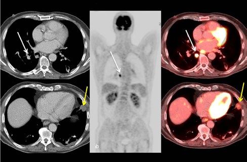
TABLE OF CONTENTS

                                                                          3
                                                                 breathe easy

BREATHE
EASY
TABLE OF CONTENTS

4     INTRODUCTION: AN OVERVIEW OF CHEST IMAGING
9     CHAPTER 1: Lung imaging: the techniques
21    CHAPTER 2: L
                  ung cancer: diagnosis, staging,
                 radiological treatment options, follow-up
33    CHAPTER 3: Lung cancer: screening
41    CHAPTER 4: Diffuse lung diseases
53    CHAPTER 5: Pulmonary Embolism
65    CHAPTER 6: CT Lung Cancer Screening: A Powerful
                  Example of How Research Advances Radiology
85    CHAPTER 7: Imaging patients with lung disease:
                  a roundtable interview
99    CHAPTER 8: IDoR and patients’ organisations:
                  new collaborations to benefit all
105   Authors & Interviewees
116   Photocredits
INTRODUCTION INTRODUCTION
4 5
breathe easy breathe easy
AN OVERVIEW OF
CHEST IMAGING
By Cornelia Schaefer-Prokop
C
hest imaging, and imaging in general, serves important diagnostic tool for suggesting the correct require intravenous injection of a contrast agent and recommend screening, there has been no such recom-
many goals. It is initially used to diagnose or diagnosis and monitoring the course of treatment. rapid imaging while the contrast agent passes the vessel mendation in Europe.
exclude a disease. During this process, imaging territory of interest. CT angiography, for example, is the
is frequently only one of many components and is The most frequently used primary imaging technique technique of choice for evaluating the lung arteries in Radiologists are the doctors responsible for imaging
combined with information provided by the physical to examine the chest is the chest radiograph. It is widely suspected pulmonary embolism. and image interpretation. They are trained to recog-
examination, patient history, laboratory results and available, fast and relatively cheap. Many diseases can nise the normal appearance of a chest radiograph,
pathological findings, in order to come to the right be diagnosed or excluded with a chest radiograph, for In some countries, screening programmes for lung a CT or MR scan. This normal appearance includes
diagnosis. Once a diagnosis has been made, imaging example pneumonia, pneumothorax or fluid overload cancer have been put in place or are currently being changes that may occur with increasing age or as
can be used to show the response to treatment, such due to heart problems. Tumours are also frequently studied. A number of trials have been carried out over residuals of past disease, for example pneumonia,
as chemotherapy for tumours or antibiotic treatment diagnosed using a chest x-ray. Many suspicious the past few decades to assess whether screening of but that do not represent acute disease requesting
for pneumonia. This is done to see whether response to abnormalities on the chest x-ray will trigger a further a large part of the population can detect lung cancers treatment. Like a detective, a radiologist looks for any
treatment is adequate, or to see if a change of therapy is work-up, most of the time using a computed tomogra- early enough to make successful treatment possible differences from that appearance and then works on
required in cases of inadequate or insufficient response. phy (CT) examination. CT is superior to chest radiogra- and reduce the chances of dying from lung cancer (lung finding potential reasons. Sometimes the changes are
Similarly, imaging is used to look for complications or phy at detecting small lesions and characterising them. cancer-specific mortality). Chest radiography alone, or so typical that only one underlying disease is possi-
progression of disease if the clinical symptoms worsen That’s because CT has a higher spatial resolution and in conjunction with sputum analysis, was not found to ble. Often, however, there are two or more possible
or new symptoms arise. it does not produce a shadow image like a chest radio- be able to decrease mortality significantly; CT is much underlying diseases. There are abnormalities that, for
graph. It creates a true cross-sectional image that does better suited to this purpose. CT screening has been example, resemble pneumonia or a tumour. In these
Imaging can also be used for screening in order to not suffer from superimposition of various structures. endorsed in Japan, Korea, the United States and some cases, following up over time, studying the effect of
detect diseases that are already present but do not Certain diseases such as pulmonary embolism (a clot European countries since the 1990s. However, no real specific treatments, or imaging again can clarify the
yet cause symptoms. By doing this, the disease, such obstructing a pulmonary artery) can not be seen by scientific proof for the effectiveness of screening was situation. The radiologist chooses the most appro-
as lung cancer, can often be detected at such an early radiography but require CT, MR or scintigraphy. Ultra- available then. This changed two years ago, when the priate test that provides the highest likelihood of a
stage that it can be treated more successfully. Imaging sound is mainly used for pleural diseases (e.g. character- largest clinical study, the National Lung Screening Trial correct diagnosis. If imaging alone is insufficient, a
can be helpful for detecting asymptomatic diseases isation of pleural fluid) or diseases that are located close (NLST trial), involving around 50,000 smokers in the biopsy might be needed to determine the underlying
that have nothing to do with cancer. Indications for to the chest wall. Magnetic resonance (MR) is used for U.S., found a statistically significant reduction in lung cause of disease.
such screening exams include an increased risk of emerging indications such as further analysis of chest cancer-specific mortality within the screening group.
inherited disease; contact with patients who have a wall lesions or lesions located in the mediastinum. X-ray The results were published in 2011 in the prestigious To avoid mistakes in image interpretation, interdis-
highly infectious disease, such as tuberculosis; known angiography uses a catheter that is introduced into a New England Journal of Medicine (NEJM). A number ciplinary conferences are held, in which all informa-
diseases located outside the region that might involve vessel and pushed to the region that needs to be exam- of European trials, all of them much smaller, were tion regarding an individual patient is reviewed and
the lung without creating symptoms. Radiologic imag- ined. The technique is invasive and has a small but sig- unable to find similarly positive results. The results of discussed with various disciplines (surgery, oncology,
ing relies on the change of morphology of any of the nificant risk of bleeding or other serious complications. the largest European screening trial, the Dutch/Belgian pathology and radiology). Such conferences are very
anatomic structures in the chest to detect and char- For the chest, it is normally substituted by CT angiogra- NELSON trial, are expected to be published in 2016. important to making the best diagnostic and therapeu-
acterise disease. For this reason, it is often the most phy or MR angiography, which are much safer and only While many professional medical societies in the U.S. tic decisions in cancer patients, but are also held forINTRODUCTION INTRODUCTION
6 7
breathe easy breathe easy
diseases of the lung tissues (interstitial lung diseases), potential, or known, allergic reactions or limited renal women have a higher risk associated with radia- necessary to gain the information needed for diagnosis
which require the specific skills of the radiologists and function. tion than men, and risk decreases with old age and or treatment decisions.
lung physicians. Such conferences may also include increases in children.
experts from different institutions. Radiological procedures very rarely have side effects. The future will bring new possibilities, as tech-
Most potential side effects stem from the medication The benefits of imaging tests are their ability to help nological improvements continue. The technical
Modern picture archive and communication systems applied to optimise image quality such as contrast make the correct diagnosis, decide the most promis- performance of CT technology has doubled every
(PACS) store all image data digitally, which means that agents. The strong magnetic field used in MRI exam- ing treatment and monitor the effect of treatment. two years over the past 20 years. This has led to a
images can be transferred to different institutions if inations may influence pacemakers or other electronic Since the risks are so low, the benefit of an imaging tremendous increase in diagnostic opportunities and
a patient changes clinic, and they can also be used to implants. Thus anyone undergoing CT or MR examina- test normally vastly outweighs the potential radiation has made it possible to avoid using more invasive and
consult external specialists in particularly difficult tions is asked about factors that may influence his or risks. Radiologists and radiographers, who are specially risky imaging techniques such as angiography. CT is,
situations. Specific image interpretation workstations her individual risk of experiencing such side effects. In trained operators of the imaging equipment, use the and remains, the most important imaging technique
allow for interactive evaluation of the datasets, which in patients with an increased risk of side effects, imaging ALARA principle in their daily practice. This means for the lung. Technological development in CT has
the case of chest CT may comprise hundreds of images. can be an option but may have to be adapted to their that radiation exposure is kept ‘as low as reasonably recently switched from improving performance to
They can display current and previous images side-by- individual risk profile. achievable’. Modern equipment for CT or chest radi- decreasing radiation exposure. MRI techniques have
side, thus ensuring optimum evaluation of changes over ography uses automated exposure control techniques been continuously evolving over the last few decades.
time or after treatment. Some imaging modalities use ionising radiation in and advanced processing to use only as high a dose as MRI remains problematic for imaging of the lungs but
very low doses. Patients should keep in mind that is required to gain a diagnostic image. Over the past new techniques look promising, although their diag-
Undergoing any kind of radiological exam involves their physicians have thoroughly weighed the benefits decade, a significant reduction in radiation exposure nostic performance is still inferior to CT. Both CT and
little or no discomfort. The imaging process itself and risks of any diagnostic test; this is also true for has been achieved thanks to technical advances. MRI will move from morphological imaging to func-
cannot be felt at all. Patients need to follow breathing imaging. In general the radiation exposure associated tional imaging. Morphological imaging serves to eval-
instructions to make sure that images are not blurred, with x-rays is considerably lower than the exposure In general imaging techniques that involve no radia- uate anatomic and pathological details in the chest,
and they should try to lie still on the examination associated with CT. For both techniques, however, a tion at all, such as ultrasound or MRI, are preferred in while functional imaging focuses on evaluating and
table. Patients with difficulties holding their breath single examination or even several examinations do children and used whenever possible. Air in the lungs, quantifying natural or disease processes. We will learn
can often continue breathing shallowly, although not produce dose levels that put the individual patient however, makes ultrasound and MRI examination dif- to quantify tissue perfusion, which can be expected
image quality will be somewhat reduced in this case. at significant risk. Most risk estimates are derived from ficult or even impossible. For this reason, CT will still to help differentiate various disease processes, predict
Many CT or MRI examinations require injection of atomic bomb data, which can only provide a rough be required. All examinations are specifically adapted response to treatment and select the most promising
an intravenous contrast agent, a dye that improves risk estimate for imaging procedures. The life-time for children: depending on their size and age, the med- treatment. Currently computer programmes are being
the display of vessels and various organs. These con- risk for developing cancer in the general population ication and the radiation dose will be reduced. In very developed that can aid radiologists in better detecting
trast agents often cause a warm sensation within the varies between countries and is in the order of 30 per- small children, who do not understand the instructions or quantifying disease. These programmes will make
body that ceases after about 15–30 seconds. Patients cent. A single CT of the chest will lead to an estimated (breath hold and lying still) required for obtaining image interpretation more effective and less expen-
may temporarily experience an unusual taste in this increase in cancer risk by 0.01–0.05%, depending on images of diagnostic quality, medication will be required sive, contributing to the sustainability of high-quality
period. Precautions have to be taken in patients with the imaging technique used, patient age and gender: to sedate them. In some cases, even anaesthesia will be healthcare services for future generations.LUNG IMAGING: TECHNIQUES
9
breathe easy
CHAPTER 1
LUNG IMAGING:
THE TECHNIQUES
CHAPTER 1LUNG IMAGING: TECHNIQUES LUNG IMAGING: TECHNIQUES
10 11
breathe easy breathe easy
1 CHEST
RADIOGRAPHY
By José Vilar
Chest radiograph reveals a nodular
lesion in the left lung (arrow).
T
he chest radiograph (CXR) is the oldest radio- It is important that radiologists review and report the
graphic technique and remains the most com- chest radiograph findings. This report will also indicate
mon radiological examination performed in the whether the patient needs further work-up such as a
world today. Approximately 25 percent of all radio- chest CT. In most cases the report will inform the refer-
graphic examinations are CXR. The enduring use of ring physician that the patient has a pulmonary disease
CXR can be explained by the advantages it offers. It is (e.g. pneumonia), triggering appropriate treatment or
easy to perform and widely available. The radiograph allowing the physician to rule out a suspected pulmo-
provides instant information about the lung, the heart, nary disease.
the large vessels that bring blood to and from the heart
(great vessels) and the chest wall. It also involves a low In bed-ridden patients, CXR can be obtained using
radiation dose and is relatively inexpensive. portable machines, which can be transferred to inten-
sive care units. Portable CXR is essential in the evalua-
Despite these apparent advantages, the technique tion of critically ill patients who frequently suffer from
also has some significant limitations. These limitations serious pulmonary diseases caused by pneumonia,
mainly relate to its limited spatial resolution and the heart failure or respiratory distress, requiring mechani-
fact that all structures, within the chest, lying in the cal ventilation.
path of the x-ray are projected over one another. These
effects of ‘overprojection’ mean that some pulmonary Recent advances in chest radiology include the tran-
lesions are difficult to see or analyse on a chest radio- sition from analogue to digital techniques, as in regular
graph, and in these cases the usual response is to obtain photography. The advantages of digital radiographic
a CT scan for further analysis. Conditions that are techniques include a more consistent and optimised
usually easy to diagnose with a chest radiograph are image quality. Image data can be transferred anywhere
pneumonia, pneumothorax, symptomatic pleural effu- and made instantly available in multiple locations at
sion, cardiac enlargement with vascular congestion, and the same time. Elaborate computerised analysis of the
symptomatic tumours. Small nodules, a lung fibrosis data is becoming more available and has the potential
and complex diseases affecting the mediastinum and to support radiologists in the detection of pathology, e.g.
the lung are often not entirely visible and require CT pulmonary nodules.
imaging.
In conclusion it can be stated that chest radiography
CXR is usually performed with the patient in a stand- is still in everyday use. The technique has been con-
ing position. Normally two views are obtained: one fron- stantly improved over the last few decades and it serves
tal and one lateral in order to improve the ability of the as a baseline examination for lung diseases worldwide
radiologist to localise pathology within the chest. If only and, as such – if wisely used by radiologists – is of great
one view is available, then visualisation is even more value.
limited by the described projection effects.
CHAPTER 1 CHAPTER 1LUNG IMAGING: TECHNIQUES LUNG IMAGING: TECHNIQUES
12 13
breathe easy breathe easy
2
Ultrasound is part of the diagnostic
ULTRASOUND work-up; Transudate is often anechoic
Exudate varies in echogenicity; Strands
and septae are suggestive of exudate,
By Fergus Gleeson
pus is rarely anechoic; 40% have effusion,
10% empyema
T
he most common method of imaging the chest is examination and to be more reliable than the CXR in
the CXR; in fact it is the most common radiologi- detecting fluid. It has also been shown that ultrasound
cal examination performed worldwide. Like CXR, guidance is of substantial benefit when inserting chest
ultrasound is also becoming more widely accessible, drains. Unnecessary procedures can be prevented when
along with the ensuing benefits in diagnosis and treat- there is only limited fluid present, or complications can
ment. As computing power increases and both software be avoided, e.g. inadvertent puncture of organs, such as
and hardware become cheaper and more readily availa- of the liver or spleen.
ble, ultrasound, previously a specialised and expensive
technology, has become more widely available. Initially, thoracic ultrasound was performed by
radiologists, but it is now routinely used by physicians
Thoracic ultrasound uses high-frequency sound and surgeons in clinics and on the wards. It has become
waves, above the audible range of the human ear. These a standard of care in emergency departments, and many
sound waves are passed from an ultrasound probe professional medical societies around the world have set
(transducer) into the body and their reflections, caused up ultrasound training programmes for junior staff.
by differences in tissue density, are detected and conver-
ted into images for visual interpretation using dedicated Although its primary use is in the diagnosis of pleu-
computer algorithms. Because it has to pass through ral effusions and their drainage, it may also be used to
structures and be reflected back, ultrasound is unable assess and biopsy masses abutting the chest wall, and
to detect disease in an aerated lung, and requires either for the diagnosis of pneumothorax, especially in critical-
fluid or a solid mass abutting the chest wall to produce ly ill patients.
an image.
As computing power increases and sophisticated
The main use of ultrasound in the chest is the detec- hardware becomes cheaper, ultrasound probes will
tion and characterisation of pleural effusions. Multiple become smaller and more portable, and may eventually
studies have shown ultrasound to be better than clinical become the ‘stethoscope of the 21st century’.
CHAPTER 1 CHAPTER 1LUNG IMAGING: TECHNIQUES LUNG IMAGING: TECHNIQUES
14 15
breathe easy breathe easy
3 CT, HRCT AND MDCT: Three CT images showing
a
the complete chest based
COMPUTED on a volumetric scan in
coronal (A and b) and
TOMOGRAPHY sagittal reconstruction (c):
OF THE LUNG
By eva castaÑer
(a) is optimised to show
the lung parenchyma,
(B) the soft tissues of the
C
B
T (computed tomography) uses x-rays
to produce detailed images of the
for further analysis of findings that have
been obtained with a chest radiograph. CT is
are so fast, provide new insights into imaging
of moving organs (cardiac imaging). This mediastinum with the
inside of the body. As opposed to chest
radiography, CT does not suffer from over-
the main tool for the diagnosis and staging of
lung cancer.
continuous volumetric assessment allows for
the precise assessment of volumetric data of heart and the large
projection and it offers a spatial resolution in
the submillimetre range. CT is by far the best HRCT is the method of choice for asses-
a whole lung, nodule or tumour.
vessels and
method for evaluating very small lesions wit-
hin the lungs. The abbreviation HRCT stands
sing lung tissue. This technique is very useful
for analysing diffuse lung diseases such as
The main drawback of CT is that it invol-
ves radiation. A CT examination should never
(C) to illustrate the bone
for high resolution CT, which is based on thin
(1–1.5mm) sections. The abbreviation MDCT
pulmonary fibrosis, emphysema or diseases
affecting the airways. For diseases affec-
be undertaken without very clear indications
to do so and alternative techniques should
structures of the
stands for Multidetector CT, which uses
multiple detectors and provides a 3D volume-
ting the airways CT images are sometimes
obtained during full inspiration and full
always be considered, especially if children
and pregnant women are involved. Over the
thoracic spine.
tric examination of the whole thorax within expiration, in order to gain information about last few years in particular, the industry has
seconds; it is the most modern type of CT the functional aspects of ventilation. CT can developed very elaborate techniques to dra-
scanner. MDCT also allows for the producti- also help locate the abnormality and suggest stically reduce the dose of radiation involved
on of cross sections (slices) through the chest the most suitable location for a histological in an examination and to allow for the dose
C
in any direction (axial, sagittal or coronal) and biopsy, if needed. to be adapted to suit the indication, the body
the production of 3D images. part to be examined and the individual body
MDCT scanners have been available since weight. This ensures that only the amount of
CT is equally well-suited to examining the the nineties. The development of MDCT after radiation absolutely necessary to answer the
lungs, the thoracic vessels, the chest wall or the introduction of the first spiral CT scanner diagnostic question is used.
the mediastinum. Depending on the clinical at the end of the eighties was very fast: star-
question and the type of suspected disease, a ting with four detector rings, then going up In the future CT scanners will provide
contrast agent may be injected intravenously to 16 then 40, 64 and now a maximum of 256 more detailed images, quicker and with less
to help enhance the visual contrast between to 320 detector rings, using one or two x-ray radiation. CT scanners are an essential tool in
vascularised and non-vascularised, or less tubes at the same time, depending on the modern medicine and will certainly continue
vascularised, structures. CT can be used as scanner type. These modern scanners can ex- to play an important role for many years to
the first imaging test (e.g. in emergency situa- amine a chest within three seconds, acquire come.
tions when a small and very subtle pathology continuous volumetric data, assess perfusion
is suspected), but most of the time it serves and functional ventilation and, because they
CHAPTER 1 CHAPTER 1LUNG IMAGING: TECHNIQUES LUNG IMAGING: TECHNIQUES
16 17
breathe easy breathe easy
4
Patient with colon carcinoma and lung nodules:
PET AND PET/CT: On CT (a) two lesions can be seen: a small lesion
in the right lower lung (white arrow) and a
IMPROVING larger lesion in the left lung (yellow arrow).
FDG-PET (b) only shows FDG uptake (hotspot) in
DETECTION AND the lesion in the right lung (white arrow).
PET/CT (c) localised hotspot on right lung (white
ASSESSMENT OF arrow) suggestive of lung metastasis but not on
the left lung (yellow arrow). Therefore not
LUNG CANCER
By Walter De Wever
suggestive of lung metastasis.
a B C
I
maging provided by computed tomography (CT) and with CT or PET. Improved diagnostic accuracy (Figure 3)
magnetic resonance imaging (MRI) does not always allows for the detection of lesions not initially seen on
give us all the information we need to diagnose and CT or PET images; more precise lesion localisation and
stage patients with lung cancer. Tumours or tumour better delineation of the surrounding structures; and
relapses (reappearance of the tumour) can be missed or better characterisation of lesions as benign or malig-
diagnosed too late. Tumours or other alterations may nant.
appear similar on CT or MRI images before and after
treatment because functional or metabolic changes The radiologist should be aware that FDG not only
may occur, even in the absence of a noticeable change in enhances most malignant tumours, but can also
appearance. enhance other non-malignant areas that are metabol-
ically active, like inflammation or brown fatty tissue.
Positron emission tomography (PET) can image Some tumours, like slowly growing adenocarcinoma or
these functional processes by using radioactive tracers carcinoid tumours, often show little or no FDG-uptake,
and photon detectors. PET is based on the injection of which may lead to misinterpretation.
radioactive-labelled biomolecules (tracers), which are
then followed and detected (enhancement). In oncol- The patient radiation dose from PET/CT is clearly an
ogy, 18F-fluorodeoxyglucose (FDG), which is a glucose issue today. However, as long as a disease like cancer
analogue, is the most widely used PET and PET/CT remains primarily a disease of the elderly and presents
tracer. The disadvantages of PET are that small lesions a life-threatening disease if not treated appropriately,
(less than five millimetres) are difficult to detect and it then the benefits of nuclear and x-ray imaging will
can be hard to accurately pinpoint the location of the largely outweigh the risks. The fact that PET frequently
abnormality. But by combining PET and CT (PET/CT) provides indispensable information, with an impact on
functional and structural imaging are available in one patient management in cases of malignant tumours,
machine. This ‘anatomo-metabolic’ imaging technique has meant that it increasingly represents an integral
improves diagnostic accuracy for staging compared part of patient management, especially in oncology.
CHAPTER 1 CHAPTER 1LUNG IMAGING: TECHNIQUES LUNG IMAGING: TECHNIQUES
18 19
breathe easy breathe easy
5 LUNG MRI
By JÜRGEN BIEDERER
M Young man with
agnetic resonance imaging (MRI) is the latest lung imaging. Being a non-radiation alternative, lung
technique for lung examinations. It uses the MRI is particularly attractive for use in children, young
subtle resonant signal that can be obtained
from hydrogen nuclei (protons) of water or organic
patients, and pregnant women.
Pneumonia a large tumour
substances when they are exposed to a strong magnet-
ic field and excited by precise radio frequency pulses.
Furthermore, beyond its excellent morphological
imaging capacities, MRI provides more functional
(white spots of the chest
Since the human body is made of proteins and fat, and
contains a large amount of water, anatomic structures,
information than any other technology. Blood circu-
lation and air exchange inside the lung, as well as the
and areas) in wall originating
as well as changes caused by diseases, can be easily
visualised with MRI. In contrast to x-ray and computed
movement of the lung and the breathing muscles (dia-
phragm, chest wall), can be studied with a routine exam-
a young man from a rib.
tomography, images are acquired without any radiation ination. This makes MRI a preferred modality in specific
exposure. clinical conditions such as cystic fibrosis (when the
air flow inside the lung is blocked by large amounts of
However, MRI of the lung is particularly challeng- viscous mucus) and acute pulmonary embolism (when
ing, since the lung contains a large volume of air with blood clots are blocking the pulmonary arteries). In
no signal and only small amounts of liquid and tissue, other situations, e.g. tumours or pneumonia in children,
generating a low signal. This, in addition to a number lung MRI may be considered an alternative or adjunct
of artefact sources (factors that lead to distortions in to other modalities with similar diagnostic value.
images), makes MRI of the lung a challenging endeav-
our that is less widely available and for which there is Overall, MRI is more complex and more expensive
relatively little experience among the radiological com- than x-ray or CT. When resources were limited, it was
munity. This and many other issues, however, have been important to define standardised protocols and clarify
addressed by recent technological advances. the indications in which MRI is preferred. This was a
crucial step in introducing lung MRI into clinical use.
Today, modern MRI scanners produce images with This information is now widely available and makes
great soft tissue contrast and they are well suited to it more likely that MRI will play a bigger role in lung
neurological, musculoskeletal, abdominal, heart, and imaging in the future.
CHAPTER 1 CHAPTER 1LUNG IMAGING: TECHNIQUES LUNG CANCER: DIAGNOSIS, STAGING, RADIOLOGICAL TREATMENT OPTIONS, FOLLOW-UP
20 21
breathe easy breathe easy
CHAPTER 2
LUNG CANCER:
DIAGNOSIS,
STAGING,
RADIOLOGICAL
TREATMENT
OPTIONS,
FOLLOW-UP
CHAPTER 1 CHAPTER 2LUNG CANCER: DIAGNOSIS, STAGING, RADIOLOGICAL TREATMENT OPTIONS, FOLLOW-UP LUNG CANCER: DIAGNOSIS, STAGING, RADIOLOGICAL TREATMENT OPTIONS, FOLLOW-UP
22 23
breathe easy breathe easy
1 LUNG CANCER:
HOW LUNG CANCER
IS DIAGNOSED
Ach, dass der
Mensch doch
By Cornelia Schaefer-Prokop and Nigel Howarth
durchsichtig wäre
P wie eine Qualle
atients with pulmonary symptoms, such as tumour, as well as the presence of distant metastases, is
a cough or increasing dyspnoea, normally called ‘staging’ and determines whether the patient will
undergo a chest radiograph first, in order to undergo surgery, chemotherapy, radiotherapy, or some
diagnose or rule out any abnormal pulmonary opaci- combination of the three.
und dass man den
fication. Depending on the symptoms of the patient,
e.g. increased temperature or sputum production, such Malignant cells show a pathologically increased
an opacification may be caused by pneumonia, and glucose metabolism compared to non-malignant cells.
so a control image following treatment will demon- This process is exploited by combining a PET scan with
Sitz seiner Leiden
strate adequate regression of the opacification. Certain CT. While the CT provides the anatomic information
morphological findings or a lack of therapy response, (where the lesion is), the PET scan shows the patholog-
however, are indicative of lung cancer and will trigger ical metabolism indicative of malignancy. Many studies
immediate further diagnostic work-up. This is usually a have demonstrated the increased sensitivity of PET/CT
CT examination with intravenous contrast injection. for the detection of metastases compared to CT alone.
Because of its 3D information and lack of overprojec-
tion, CT is superior to CXR in showing the exact loca-
tion and size of a tumour. For tumours located in the
centre of the lung it is important to analyse how exten-
sively the tumour has grown into the central structures
Nevertheless, any suspicious finding that determines
the therapeutic management has to be histopathologi-
cally confirmed. Therefore, patients frequently have to
undergo a biopsy, e.g. of a bone lesion or a liver lesion.
There are several options for acquiring tissue from
schauen könnte.
Wilhelm Conrad Röntgen (1845 - 1923)
of the thorax, called the mediastinum, where the large mediastinal lymph nodes for histological examination:
vessels, the oesophagus and the central tracheobron- via direct surgical access to the mediastinum under
chial system are located. The invasion of the tumour anaesthesia (mediastinoscopy), the tracheobronchial
into the chest wall, the infiltration of lymph nodes system (EBUS) or the oesophagus (EUS). Similarly, tis-
or the presence of metastases in the bones, adrenals sue from the tumour itself has to be examined by the
or liver are other important findings. The diagnostic pathologist in order to determine the best therapy for
process of determining the exact local extent of the the patient, depending on tumour type and stage.
CHAPTER 2 CHAPTER 2LUNG CANCER: DIAGNOSIS, STAGING, RADIOLOGICAL TREATMENT OPTIONS, FOLLOW-UP LUNG CANCER: DIAGNOSIS, STAGING, RADIOLOGICAL TREATMENT OPTIONS, FOLLOW-UP
24 25
breathe easy breathe easy
2
FIgure 1a FIgure 1B FIgure 2a FIgure 2b
LUNG CANCER:
VARIOUS
RADIOLOGICAL
APPEARANCES
By Paul Flechsig, Claus Peter Heussel and Hans-Ulrich Kauczor
FIgure 3a FIgure 3B FIgure 3C
L
ung cancer can appear with a huge glands and bones are signs of more advanced with the irregularly shaped nodules seen in
variety of shapes and sizes. These disease. the chest x-ray (orange arrows).
appearances differ between tumour
entities, types and stages, as well as between Figures 1 and 2 show two lung cancer To analyse chest wall infiltration, an addi-
different imaging modalities. patients with two very different radiological tional MRI examination would be helpful.
appearances. In the first patient, the chest Figure 3 shows a mass in the upper right lung.
One of the most common ways to examine x-ray (Figure 1a) shows a solid mass in the In the x-ray (Figure 3a) and CT (Figure 3b), the
lung diseases is with chest x-ray. In cases upper left lung (red arrow). The CT image tumour borders cannot be clearly assessed
of doubtful findings from a chest x-ray, an (Figure 1b) reveals the chest wall as tumour and infiltration into the ribs and chest wall
additional CT examination is considered free (blue arrows) on one side, but with close muscles needs to be ruled out (Figure 3b,
helpful. CT also captures the adjacent ana- contact between the tumour and mediastinal green arrows, question mark). The tumour
tomic structures, i.e. the mediastinum, chest structures (green arrows). borders are more clearly demarcated in the
wall and heart. This helps to assess the local MRI image (Figure 3c, red line): the lung can-
tumour burden and infiltration in patients In the second patient, the chest x-ray cer is limited to the lung tissue while the ribs FIgure 4a FIgure 4B FIgure 4c
suffering from lung cancer. (Figure 2a) shows many irregularly shaped and chest wall muscles are unaffected.
nodules widely distributed in the lung
In the early stages, lung cancer appears as (orange arrows). The mediastinum is wid- Figure 4 shows chest x-ray (Figure 4a), CT
a small lung nodule; the larger the nodule or ened, indicating enlarged lymph nodes (green (Figure 4b) and MR images (Figure 4c) of a
mass, the higher the stage. More advanced arrows). The CT image (Figure 2b) shows a lung cancer patient with a large mass in the
tumour stages are often accompanied by tumour consisting of a large cavitation (yel- upper left lobe (blue arrow). Local chest wall
central necrosis, infiltration of the chest low arrow). The blue arrows indicate chest infiltration, including the destruction of the
wall, ribs, or mediastinal structures, as well wall infiltration; green arrows show enlarged first and second rib can be seen on CT (Figure
as metastases to the lymph nodes. Moreover, metastatic lymph nodes. The orange arrows 4b, yellow arrows) as well as on the MR image
metastases in the lung, liver, brain, adrenal indicate multiple lung metastases, correlating (Figure 4c, yellow arrows).
CHAPTER 2 CHAPTER 2LUNG CANCER: DIAGNOSIS, STAGING, RADIOLOGICAL TREATMENT OPTIONS, FOLLOW-UP LUNG CANCER: DIAGNOSIS, STAGING, RADIOLOGICAL TREATMENT OPTIONS, FOLLOW-UP
26 27
breathe easy breathe easy
3 BIOPSY IN
CHEST DISEASE
By Katerina Malagari and Dimitrios Filippiadis
The patient is lying on his side: A needle
Is placed from the back, crossing the
chestwall to approach the intrapulmonary
nodule. The needle is placed in such way
to bypass the bones.
T
he purpose of a biopsy procedure is adenopathy, chest wall masses and lytic bony Complications are usually minor (some
to obtain a sample of tissue or cells cage lesions. Contraindications include a pain, or mild bleeding from the puncture
from a diseased organ. A fine-needle patient’s refusal (the sole absolute contrain- site). The most serious, and also the most
aspiration is a procedure whereby cells are dication), while other potentially correctable common, complication in lung biopsies is the
obtained using a very thin needle. Diagnostic contraindications include bleeding diathesis; insertion of air in the pleura room leading
interpretation of the cellular specimens or severe emphysema, especially if there is previ- to a collapsed lung of varying severity (the
sample tissue from the lung, pleura, chest ous contralateral pneumonectomy; intractable latter is called pneumothorax). Pneumotho-
wall and other organs of the thorax is now cough; suspected echinococcal cyst (hydatid); rax occurs in 20–25 percent of lung biopsies,
practiced in virtually every major medical possible arteriovenous malformation; and but only one in four of them require treat-
institution. These two procedures are per- severe pulmonary hypertension. ment by inserting a chest tube. Usually, the
formed in most patients suspected of having collapse is so minor that it can be managed
lung cancer, in order to confirm the final diag- The patient is told about possible compli- conservatively with the administration of
nosis and determine the histological type of cations prior to the procedure and is given oxygen and a few hours of follow-up. Bleed-
the cancer, which is necessary for appropriate instructions to discontinue certain medi- ing and haemoptysis occurs in less than 5–10
treatment planning. cation (e.g., aspirin or other non-steroidal percent of cases and is self-limited. Fatal hae-
anti-inflammatory drugs). Patients on oral morrhaging occurs in less than one case in a
These procedures are guided by suitable anticoagulants should consult their physi- thousand.
imaging modalities to ensure the needle cians. A couple of hours before the procedure,
is accurately inserted into the tissue, and clotting tests are performed to ensure proper A biopsy is a highly accurate procedure
to decrease the risk of side-effects such as coagulation and reduce the risk of bleeding. yielding a definite (histological) diagnosis in
bleeding or pneumothorax. In most cases, The patient should fast for six to eight hours more than 90 percent of cases. Diagnosis can
CT is used but other common alternatives prior to the procedure. be more difficult in benign lesions, especially
include ultrasound or fluoroscopy. By using since a negative result always needs to be
imaging guidance the physician can make After placing the patient in the most suitable considered critically to safely rule out a false
sure that the sample is taken exactly from position, to access the tumour/lesion, the pro- negative result. In a certain percentage of
the suspicious mass, nodule or lesion and not cedure is performed under local anaesthesia. cases, it might happen that the material aspi-
from the surrounding organs, while avoiding Special needles are used to obtain the sample rated was insufficient for the pathologist to
injury to the neighbouring organs. (cytological or actual tissue fragment in biopsy) make a diagnosis and it might be necessary
and the sample is subsequently handled by to repeat the procedure, perhaps modifying
The indications for these procedures include pathology or cytology experts. After complet- the type of access or the type of needle.
the evaluation of lung nodules or masses, ing the procedure, additional views may be
pleural masses, mediastinal lesions, lymph taken to rule out possible complications.
CHAPTER 2 CHAPTER 2LUNG CANCER: DIAGNOSIS, STAGING, RADIOLOGICAL TREATMENT OPTIONS, FOLLOW-UP LUNG CANCER: DIAGNOSIS, STAGING, RADIOLOGICAL TREATMENT OPTIONS, FOLLOW-UP
28 29
breathe easy breathe easy
4
New lung
LUNG CANCER cancer in the
left lung of
TREATMENT OPTIONS: a patient
RADIOFREQUENCY who under-
went surgical
AND MICROWAVE resection of a
right-sided lung
ABLATION
By Benoît Ghaye
cancer a few
years earlier.
A
bout one-third of patients with lung cancer are is inserted into the tumour under CT guidance. The
actually inoperable, even though the tumour type of energy and anaesthesia (general anaesthesia
itself is localised. This is usually because the or conscious sedation) depend on the patient, tumour
patients present with poor overall condition, i.e. they location, nature of the tumour, treatment goal, and
have low cardiopulmonary function, which makes sur- operator experience or preference. The tumour should
gery too risky. In such cases, alternative therapies like be no larger than two or three centimetres in diameter
radiation therapy or chemotherapy are applied, often to be suitable for this type of treatment. The duration
accompanied with significant toxicity to the patient. of the procedure varies from 30 minutes to three hours,
Recently, minimally invasive treatments, including per- depending on the number and type of lesions.
cutaneous thermal ablation, have been developed and
appear to offer a valuable alternative. Indeed, as most patients treated with thermal abla-
tion have contraindications to other treatments, the
Thermal ablation is currently used as a substitute, results of ablative therapy look very encouraging. Nev-
or adjunct, to other therapeutic modalities for treat- ertheless, studies are still needed to accurately assess
ing focal tumours in the liver, kidney, breast, thyroid, the role of ablation compared with other emerging
head and neck, bones and more recently, the lungs. The techniques, like stereotactic radiotherapy, as well as its
advantages of thermal ablation include reduced mor- potential synergy with other treatments.
bidity and mortality; faster recovery; earlier discharge
from hospital; more outpatient treatment; lower costs; Complications are few and basically the same as for
and a relative sparing of healthy peritumoural tissue, percutaneous lung biopsy. They mainly concern pneu-
which is especially important for treating patients with mothorax, which is easily treated during intervention.
reduced cardiopulmonary reserve. After intervention, patients are followed up with PET,
and CT or MRI. When necessary, thermal ablations can
Thermal ablation is performed by delivering either be repeated to complete treatment in patients showing
extreme heat (radiofrequency, microwave or laser) a persistence of viable tumour tissue.
or extreme cold (cryotherapy) through a needle that
CHAPTER 2 CHAPTER 2LUNG CANCER: DIAGNOSIS, STAGING, RADIOLOGICAL TREATMENT OPTIONS, FOLLOW-UP LUNG CANCER: DIAGNOSIS, STAGING, RADIOLOGICAL TREATMENT OPTIONS, FOLLOW-UP
30 31
breathe easy breathe easy
5
FIgure 1 FIgure 2
LUNG CANCER:
FOLLOW-UP
By Anna Rita Larici, Lucio Calandriello and Lorenzo Bonomo
S
urgery remains the mainstay of treatment with therapy, selected patients may subsequently undergo
curative intent for patients with lung cancer in surgery.
the early stages (with specific reference to non-
small cell lung cancer = NSCLC). However, most patients PET/CT is a highly accurate means of detecting
are not eligible for surgery at the time of diagnosis, due residual disease after treatment (Figure 1) and for deter-
to the advanced tumour stage or the coexistence of mining further treatment. It is known that metabol-
cardiopulmonary diseases limiting the indications for ic-functional alterations precede morphological changes
surgery. In these patients possible treatment options and, therefore, a reduction in the uptake of 18F-FDG by
are combined (neoadjuvant) radio-chemotherapy, che- tumour cells after treatment is indicative of a reduction
motherapy alone or local treatments, such as radiother- in the number of viable tumour cells. PET/CT is also
apy or tumour ablation, according to the extent of the useful for distinguishing metabolically active tumours
disease. from inactive scarring (fibrosis), which can occur after
radiation therapy in the lung parenchyma surrounding
Imaging plays an important role in the assessment the treated lesion.
of treatment response after radio-chemotherapy and Recurrence of NSCLC. A 70-year-old man underwent right
in the follow-up of patients surgically treated for lung Even when treated with curative intent, lung cancer upper lobe lobectomy for a large mass (A), with ipsilateral
cancer. Computed tomography (CT) and positron emis- can recur, depending mainly on the pathological stage. mediastinal and subcarinal lymphadenectomy. One month
sion tomography with computed tomography (PET/CT) Most recurrences occur within the first two years fol- after surgery, the baseline CT exam showed a pleural effu-
with 18F-fluorodeoxyglucose (18F-FDG) are the imaging lowing completion of treatment. Therefore, it is import- sion and metal clips in the subcarinal station (B). Two years
modalities commonly used in this context. ant to schedule a tighter follow-up for the patient later a rounded soft tissue mass is seen close to the clips
during that period. Radiology is essential for investigat- on CT (C) highly suspicious for loco-regional recurrence
CT provides information on morphological changes ing both loco-regional (within the treated hemithorax) Good response to treatment of NSCLC. A 66-year-old and confirmed by PET/CT scan (D).
affecting the tumour after treatment (Figure 1). Accord- and distant recurrences. In particular, the integration woman with a large right upper lobe mass infiltrating the
ing to dimensional criteria and following internationally between morphological and metabolic information mediastinal fat and closely adjacent to the right innomi-
accepted rules, radiologists and clinicians define the obtained with PET/CT is useful for confirming a tumour nate vein and superior vena cava. Vessels show mildly irre-
presence of ‘complete’ or ‘partial response’ to treatment recurrence (Figure 2), distinguishing lung abnormalities gular margins (A, B). After three months of neoadjuvant
as well as ‘progression’ or ‘stability’ of the disease. When from tumour recurrence after treatment, and identify- combined chemotherapy and radiotherapy (C), the mass
a good response is seen after neoadjuvant radio-chemo- ing distant metastases. showed evident volume shrinkage (D) with residual meta-
bolic activity in the PET/CT scan (E). Patient subsequently
underwent surgery.
CHAPTER 2 CHAPTER 2LUNG CANCER: DIAGNOSIS, STAGING, RADIOLOGICAL TREATMENT OPTIONS, FOLLOW-UP LUNG CANCER: SCREENING
32 33
breathe easy breathe easy
CHAPTER 3
LUNG CANCER:
SCREENING
CHAPTER 2 CHAPTER 3LUNG CANCER: SCREENING LUNG CANCER: SCREENING
34 35
breathe easy breathe easy
1
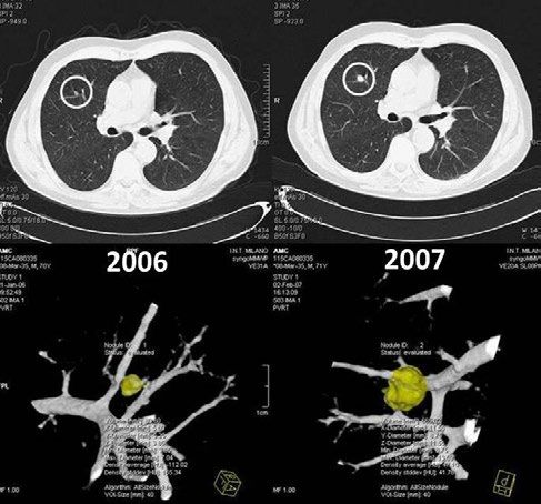
TWO CTs and automatic volumetry
LUNG CANCER showing significant growth of the
SCREENING: WHAT IS nodule within one year indicative of
an early lung cancer.
IT AND WHAT HAVE
WE LEARNT SO FAR?
By Anand Devaraj
L
ung cancer is still one of the deadliest diseases in reported 87 fewer deaths due to lung cancer among
the world today. It is the leading cause of death those individuals who were screened with CT, which
in many countries and is responsible for the larg- corresponds to a 20 percent reduction in lung can-
est number of cancer-related deaths worldwide. When cer-specific mortality.
detected early, however, there is a good chance of curing
lung cancer through surgery. Unfortunately, lung cancer Like in many trials before, the Prostate, Lung, Colorec-
only has symptoms in more advanced stages, and most tal and Ovarian Randomized Cancer Screening Trial
people suffering from lung cancer present at their doc- (PLCO), which was published at about the same time as
tor’s office when their chances of being cured are slim. the NLST, confirmed that screening with chest x-rays
alone has no effect on lung cancer survival. No differ-
Computed tomography (CT) scanning of the chest is ence in mortality was identified between those receiv-
a medical imaging technique that can detect early-stage ing chest radiography and those who were not screened
lung cancer before symptoms become apparent (Figure at all.
1b). Over the past decade, numerous investigations have
studied whether CT can be used as an effective tool to The results from the NLST have led many American
detect these early stages of lung cancer in asymptom- national organisations to recommend the introduction
atic individuals. Many randomised controlled trials of lung cancer screening in clinical practice. In Europe,
(either completed or in progress) have addressed the however, smaller trials (MILD, DANTE and DLST) with
question of whether CT screening can reduce mortality approximately 2,500–4,000 patients each did not show
in those who are at high risk of developing lung cancer. any benefit from CT screening. In fact, these trials even
suggested an increased mortality in those who under-
In 2002, the National Lung Screening Trial (NLST) went annual CT screening. The larger Dutch-Belgian
embarked upon recruiting 53,454 former and current NELSON lung cancer screening trial, with more than
smokers to participate in a trial where participants 15,000 participants, will publish its results in the next
were randomised to undergo annual screening with few years, and will help clarify whether CT screening
either CT or chest x-rays for three years. In 2011, six and should also be introduced in Europe.
a half years after the end of the trial, the investigators
CHAPTER 3 CHAPTER 3LUNG CANCER: SCREENING LUNG CANCER: SCREENING
36 37
breathe easy breathe easy
2
Minimally invasive lung
LUNG CANCER adenocarcinoma in a
SCREENING: WHO 65-year-old female lung
cancer screening participant.
SHOULD TAKE PART Baseline CT image (left) shows
a ground-glass nodule with
AND WHAT ARE THE a barely solid component on
CONCERNS?
By Nicola Sverzellati
the left lower lobe. CT images
repeated annually show an
increase in the size of a small,
T central and solid component.
he American National Lung Screening Invasive procedures such as biopsies or The discussion continues as to how best
Trial (NLST) demonstrated a 20 per- surgery are required to distinguish cancers to identify those who are likely to benefit
cent reduction in death due to lung
cancer among those who were screened with
from benign lesions. Participants with a
benign lesion (false positive) will still have to
from screening. Lung cancer risk increases
with age and with the number of pack-years
Choosing the best work-up for
CT. Even the total number of deaths within
the six-and-a-half year follow-up period was
undergo such procedures, along with their
costs and potential complications. Anxiety
smoked. The number of pack-years is calcu-
lated by multiplying the number of years a
this type of slow-growing lesion
reduced by more than six percent. While
these results are spectacular for any screen-
is also an important consideration, partic-
ularly while waiting for screening results.
person has smoked by the average number
of packets of cigarettes smoked per day. The
can be difficult due to the risk
ing programme, the screening procedure
itself has its risks. Careful selection is there-
False-positive results have been associated
with depression and changes in overall per-
NLST targeted high-risk smokers and for-
mer smokers between 50 and 75 years of age
of overdiagnosis.
fore necessary to identify those for whom the ception of one’s health4. with at least 30 pack-years. Most European
benefits outweigh the potential risks. studies, which did not show a benefit from
Other risks are overdiagnosis and the CT screening, also included individuals who
The risks are mainly due to false-positive potential risk of radiation-induced cancer had smoked less. It is also not clear whether
results, suspicious lung lesions that are from the radiation used by CT1. Overdiagno- screening might benefit individuals with
thought to represent lung cancer on screen- sis refers to the detection of slow-growing other risk factors for lung cancer, such as var-
ing CT, but are not actually malignant. Small non-fatal cancers, which if detected in a ious lung diseases, occupational exposure to
lung nodules are common: in almost 20 per- screening programme will cause unnecessary asbestos or other carcinogens, or lung cancer
cent of lung screening participants a lesion treatment because they would not have lim- in a first-degree relative.
within the size range of 5–10 mm is detected. ited the patient’s life expectancy nor effect
The vast majority of these lesions are in his/her quality of life. The risk of cancer Current research focuses on how to best
fact benign; less than 10 percent are cancer- being induced by the low dose from lung select screening participants, how to reduce
ous. These lesions are usually followed up cancer CT screening is unlikely, but cannot be the number of false positives and cases of
using CT to see whether they have grown. If completely ruled out. The average effective overdiagnosis, and how to keep radiation
lesions grow, their likelihood of being cancer dose is comparable to the annual radiation exposure to a minimum.
is much greater, but many are still benign. dose from natural sources3.
References
1. Aberle DR, Abtin F, Brown K. Computed Tomography screening for sung Cancer: has it finally arrived? Implications of the National Lung Screening Trial. J Clin Oncol 2013; 31:1002-1008.
2. White CS. National Lung Screening trial. A breakthrough in lung cancer screening? J Thorac Imaging 2011; 26: 86-87
3. Larke FJ, Kruger RL, Cagnon CH, et al. Estimated radiation dose associated with low-dose chest CT of average-size participants in the National Lung Screening Trial. AJR Am J Roentgenol 2011; 197: 1165-1169.
4. van den Bergh KA, Essink-Bot ML, Bunge EM, et al. Impact of computed tomography screening for lung cancer on participants in a randomized controlled trial (NELSON trial). Cancer. 2008;113:396-404.
CHAPTER 3 CHAPTER 3LUNG CANCER: SCREENING LUNG CANCER: SCREENING
38 39
breathe easy breathe easy
3 LUNG CANCER
SCREENING:
AUTOMATED
DETECTION &
FOLLOW-UP
By Bram van Ginneken
W
idespread implementation of CT lung screen- This reading bottleneck in CT lung screening could resent slow-growing cancers, which could otherwise be Researchers foresee that in the future computer algo-
ing would require millions of CT scans to be be solved by letting specially trained non-radiologists easily missed. It is important to verify that no lesions rithms will use computerised quantitative features of
evaluated for the detection of potential lung evaluate the images, but the effort involved would still requiring direct work-up or short term follow-up CT are the lesion for this purpose. Ultimately, this software will
cancers. Evaluation is normally carried out by radiol- be vast. A more promising solution would be to use missed by this procedure. become good enough to determine which nodule can be
ogists, medical specialists who have been trained for computerised nodule detection software. Such software left alone, which one requires follow-up with CT scans,
many years to read such medical images. The demand has been commercially available for about a decade, Workflow has to be optimised to increase throughput. and which one has such a high likelihood of being can-
for evaluations that would be brought about by wide- but literature reports that the sensitivity of these algo- For example, nodules in prior and current scans have to cer that immediate resection is required. At this time,
spread CT lung screening would create a major bottle- rithms is too low. be automatically linked to ensure correct comparison. however, more knowledge about the natural evolution
neck: radiologists require five to fifteen minutes reading Automated measurement of their volume and their of pulmonary nodules and shared databases is needed
time to locate pulmonary nodules on a 3D CT dataset. Recently, it was shown that combining multiple algo- growth rate also needs to be optimised. For subsolid to develop and validate such algorithms.
This excludes the interaction time needed to locate the rithms for nodule detection in CT can substantially lesions in particular, automated assessment of lesion
same nodule in prior exams and measure the size of improve overall performance, far exceeding that of volume remains a challenge. Size and growth help
the nodule. These tasks are fundamentally different the best single algorithm1. Current research focuses on in assessing the probability that a nodule is a cancer,
from those required for mammography screening, the developing algorithms that are better than most human which determines work-up.
only widespread image-based screening implemented readers for detecting the locations of potential cancers,
in the western world. Mammography screening uses 2D so that a human reader would only inspect locations References
1. van Ginneken, B.; Armato, S. G.; de Hoop, B.; van de Vorst, S.; Duindam, T.; Niemeijer, M.; Murphy, K.; Schilham, A. M. R.; Retico, A.; Fantacci, M. E.; Camarlinghi, N.;
images and requires, on average, substantially less read- selected by the computer. Specific software has to be Bagagli, F.; Gori, I.; Hara, T.; Fujita, H.; Gargano, G.; Belloti, R.; Carlo, F. D.; Megna, R.; Tangaro, S.; Bolanos, L.; Cerello, P.; Cheran, S. C.; Torres, E. L. & Prokop, M. Compar-
ing time for radiologists. developed for so-called ‘subsolid’ lesions that may rep- ing and combining algorithms for computer-aided detection of pulmonary nodules in computed tomography scans: the ANODE09 study, Medical Image Analysis,
2010, 14, 707-722.
CHAPTER 3 CHAPTER 3You can also read