Amine oxidase, copper containing 3 exerts anti mesenchymal transformation and enhances CD4+ T cell recruitment to prolong survival in lung cancer
←
→
Page content transcription
If your browser does not render page correctly, please read the page content below
ONCOLOGY REPORTS 46: 203, 2021

                   Amine oxidase, copper containing 3 exerts
             anti‑mesenchymal transformation and enhances CD4+
              T‑cell recruitment to prolong survival in lung cancer
                CHAO‑YUAN CHANG1,2, KUAN‑LI WU1,3,4, YUNG‑YUN CHANG1,3,5, PEI‑HSUN TSAI1,
                  JEN‑YU HUNG1,3,4, WEI‑AN CHANG3,6, SHU‑FANG JIAN1, YUNG‑CHI HUANG1,
                       INN‑WEN CHONG1,3,4, YING‑MING TSAI1,3,4,7 and YA‑LING HSU1,7

                      1
                    Graduate Institute of Medicine, College of Medicine, Kaohsiung Medical University;
         2
         Department of Anatomy, Kaohsiung Medical University; 3Division of Pulmonary and Critical Care Medicine,
     Kaohsiung Medical University Hospital, Kaohsiung Medical University; 4School of Medicine, College of Medicine,
          Kaohsiung Medical University; 5Division of General Medicine, Kaohsiung Medical University Hospital,
Kaohsiung Medical University; 6Graduate Institute of Clinical Medicine, College of Medicine, Kaohsiung Medical University;
  7
    Drug Development and Value Creation Research Center, Kaohsiung Medical University, Kaohsiung 807, Taiwan, R.O.C.

                                        Received November 9, 2020; Accepted June 15, 2021

                                                       DOI: 10.3892/or.2021.8154

Abstract. Lung cancer remains notorious for its poor prog‑             Epigenetic silencing of AOC3 via miR‑3691‑5p caused tumor
nosis. Despite the advent of tyrosine kinase inhibitors and            promotion and progression by increasing migration and epithe‑
immune checkpoint inhibitors, the probability of curing the            lial‑mesenchymal transition (EMT). Furthermore, knockdown
disease in lung cancer patients remains low. Novel mecha‑              of AOC3 caused less CD4+ T‑cell attachment onto lung cancer
nisms and treatment strategies are needed to provide hope to           cells and reduced transendothelial migration in vitro, as well as
patients. Advanced strategies of next generation sequencing            reducing CD4+ T‑cell trafficking to the lung in vivo. In conclu‑
(NGS) and bioinformatics were used to analyze normal and               sion, the present study revealed that downregulation of AOC3
lung cancer tissues from lung cancer patients. Amine oxidases          mediated lung cancer promotion and progression, as well as
have been linked to leukocyte migration and tumorigenesis.             decrease of immune cell recruitment. This novel finding could
However, the roles of amine oxidases in lung cancer are not            expand our understanding of the dysregulation of the tumor
well‑understood. Our results indicated that amine oxidase,             immune microenvironment and could help to develop a novel
copper containing 3 (AOC3) was significantly decreased in              strategy for the treatment of lung cancer.
the tumor tissue compared with the normal tissue, at both the
mRNA and protein level, in the included lung cancer patients           Introduction
and public databases. Lower expression of AOC3 conferred
a poorer survival probability across the different cohorts.            Lung cancer is the leading cause of cancer‑related deaths
                                                                       worldwide with a high annual incidence and a 5‑year survival
                                                                       rate of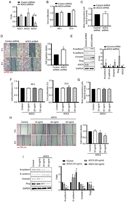
2                              CHANG et al: AOC3 REGULATING EMT AND IMMUNITY IN LUNG CANCER

In addition to the malignant cells, the surrounding micro‑            with 10% fetal bovine serum (FBS), 100 U/ml penicillin
environment is also critical for tumorigenesis (7,8). Amine           and 100 µg/ml streptomycin (Thermo Fisher Scientific,
oxidases refer to a class of enzymes that catalyze the deami‑         Inc.; Waltham, MA, USA) at 37˚C. Recombinant human
nation of amine groups to produce aldehydes, ammonia and              and mouse AOC3 were obtained from R&D Systems, Inc.
hydrogen peroxide (9). There are a variety of amine oxidases          Knockdown of AOC3 in CL1‑5 cells was performed using
consisting of four classes of monoamine oxidases (MAOs),              either pLKO_005 plasmid as a control or AOC3‑shRNA
including MAO‑A and MAO‑B, polyamine oxidases, lysyl                  plasmid (14 µg shRNA plasmid for 5x10⁵ cells in a 6‑well
oxidases, and copper‑containing amine oxidases (CAOs) (10).           plate) obtained from the National RNAi Core Facility
CAOs have been revealed to participate in the regulation of a         (Academia Sinica, Taipei, Taiwan). The plasmid was
variety of pathological and physiological processes, such as          transfected into cells using Lipofectamine 2000™ Thermo
cell proliferation, differentiation, glucose uptake and immune        Fisher Scientific, Inc.) for 2 days, and the stable clone of
regulation (11). Changes in CAO activity are correlated with          AOC3‑knockdown cells were established by puromycin
a variety of human diseases, including diabetes mellitus,             selection (5 µg/ml). All cells were authenticated by short
Alzheimer's disease, and inflammatory disorders (12,13). The          tandem repeat (Promega Corporation) and examined for
four complete genes for CAOs are amine oxidase, copper                mycoplasma contamination using a MycoAlert™ myco‑
containing (AOC)1‑4. AOC1 consists of a homodimeric                   plasma detection kit (Lonza Group, Ltd.) according to the
glycoprotein with an apparent molecular mass of 186 kDa;              manufacturer's protocol every three months.
it is secreted as a diamine oxidase to generate hydrogen
peroxide (14,15). AOC1 is strongly expressed in the kidneys,          Next generation sequencing (NGS) and bioinformatics
placenta, intestine and lungs (14). Little is known about the         analysis. All of the participants selected from January 2018
molecular mechanisms regulating AOC1 gene expression. The             to December 2019, provided written informed consent prior
AOC2 gene encodes retina‑specific amine oxidase (16), which            to inclusion in the present study. The patients who agreed and
was originally identified in ganglion cells. Its functions remain      received surgical intervention were enrolled in this study. The
unclear but it may play a role in hereditary retinal diseases (16).   adjacent non‑tumor lung and tumor tissues of ten patients
The AOC4 gene encodes a soluble plasma amine oxidase in               (7 from LUAD and 3 from LUSC) were obtained from the
cows as bovine AOC4 (17) but not in humans, mice or rats.             Division of Thoracic Surgery and Division of Pulmonary and
The AOC3 gene encodes vascular adhesion protein‑1, which              Critical Care Medicine, Kaohsiung Medical University Hospital
is primarily expressed on the endothelial cell surface but also       (Kaohsiung, Taiwan). The protocol of the present study was
in smooth muscle cells and adipocytes (18). In addition to its        reviewed and approved (approval no. KMUH‑IRB‑20130054
amine oxidase activity, AOC3 functions as a non‑classical             and KMUH‑IRB‑G(II)‑20180021) by the Institutional Review
inflammation‑inducible endothelial molecule which is linked           Board of Kaohsiung Medical University Hospital. The deep
to leukocyte‑subtype specific rolling under physiological             RNA‑seq was carried out at a biotechnology company
shear (18). It has been revealed that the enzymatic activity of       (Welgene, Inc.) using the Solexa platform. RNA and small
AOC3 is functionally important, and leukocyte recruitment is          RNA library construction was carried out using a sample
impaired if its activity is abolished (10).                           preparation kit (Illumina, Inc.) following the protocol of the
    The surrounding microenvironment of cancer contributes            TruSeq RNA or Small RNA Sample Preparation Guide.
to its promotion and progression (19). Due to its unique tumor            The expression of AOCs in lung cancer and normal
microenvironment (TME), cancer promotes and strengthens               specimens (cancer vs. normal) were extracted from the
its own progression as a result of its interactions. The cells        Oncomine® database (http:/www.oncomine.org; Compendia
inside the TME include cancer‑associated fibroblasts, endo‑           Biosciences) (21) and The Cancer Genome Atlas (TCGA)
thelial cells and immune cells (20), which form the tumor             cohort of UALCAN (http://ualcan.path.uab.edu/analysis.
immune microenvironment (TIME). The functions and densi‑              html) (22). The 16 cohorts from Oncomine® database included
ties of different tumor‑infiltrating immune cells in the TIME          Su et al (23), Okayama et al (24), Landi et al (25), Beer et al (26),
are closely associated with prognosis and prediction of the           Stearman et al (27), Selamat et al (28), Garber et al (adeno‑
treatment response (7). Therefore, there is an urgent need for        carcinoma and squamous) (29), Hou et al (adenocarcinoma,
improved understanding of immune dysfunction inside the               squamous and large cell) (30), Wachi et al (squamous) (31),
TIME and the mechanisms by which the tumor modifies its               Bhattacharjee et al (adenocarcinoma, squamous, carcinoid and
environment to remove the functional immunity of the body.            small cell) (32). Criteria in the analysis were fold change >2
The present study aimed to verify the role of AOC3 in lung            and P‑value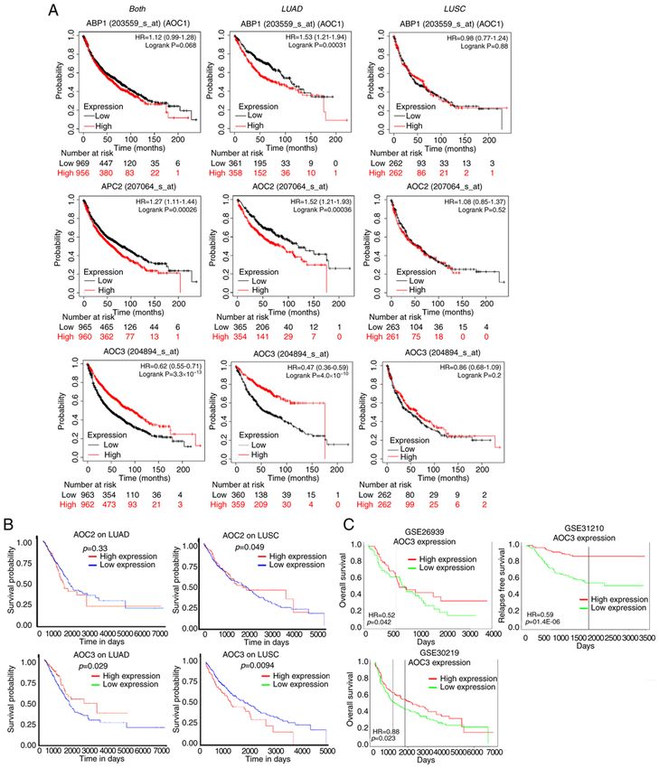
ONCOLOGY REPORTS 46: 203, 2021 3
was predicted using miRWalk (version 3.0) (36), miRanda (37) method according to the manufacturer's protocol (BrdU Cell
and miRDB (38) with restriction of >95% confidence. Proliferation Assay Kit; cat. no. 2750; EMD Millipore).
Measurement of AOC3. All of the participants provided written Wound healing analysis. CL 1‑5 cells were seeded into
informed consent prior to inclusion in the present study. The a 12 well‑pate at 90% confluence and cultured in 1% of
sera of 40 healthy donors and 40 lung cancer patients (healthy FBS‑containing medium for exogenous AOC3 (control,
donors: Age range, 40‑80 years old; M/F 31%/69%; lung 10, 20 and 50 ng/ml) and 10% of FBS‑containing medium
cancer donors: Age range 30‑90 years old; M/F 46/54%) were (since cell proliferation was not affected by AOC3 knock‑
collected from the Division of Thoracic Surgery and Division down, in order to mimic the physiologic conditions, 10% of
of Pulmonary and Critical Care Medicine, Kaohsiung Medical FBS‑containing medium was used and cells were not serum
University Hospital (Kaohsiung, Taiwan) from January 2018 to starved) for AOC3‑shRNA knockdown at 37˚C as previ‑
December 2019. The patients who agreed with written informed ously described by Shao et al (41), and the cell migration
consent were enrolled in this study before starting definite was evaluated by measuring the migration of cells into the
treatment. These samples were assessed using Quantikine acellular region formed by a sterile yellow tip. The wound
Human VAP‑1 Immunoassay (R&D Systems, Inc.). The closure was observed after 8 h. The wound healing assay was
protocol of the present study was reviewed and approved (app closely observed via a Nikon inverted microscope (Nikon
roval no. KMUH‑IRB‑20130054) by the Institutional Review Corporation).
Board of Kaohsiung Medical University Hospital.
CD4+ T‑cell isolation. Peripheral blood mononuclear cells
Reverse transcription‑quantitative (RT‑q) PCR. Total RNAs (PBMCs) of healthy donors (eight healthy donors: Age range,
were extracted from CL 1‑5 lung cancer cells with TRIzol 35‑45 years old; male only) were obtained from the Division
reagent (Thermo Fisher Scientific, Inc.) and reverse tran‑ of Pulmonary and Critical Care Medicine, Kaohsiung Medical
scribed into cDNA using an oligo (dT) primer and reverse University Hospital (Kaohsiung, Taiwan) from January 2020
transcriptase (PrimeScript RT Reagent Kit; Takara Bio, Inc.) to December 2020. The protocol of the present study was
following the manufacturer's protocols. The reaction condi‑ reviewed and approved (approval no. KMUH‑IRB‑20130054)
tions were as follows: Priming for 5 min at 25˚C, reverse by the Institutional Review Board of Kaohsiung Medical
transcription for 20 min at 46˚C, and final inactivation of University Hospital and the donors provided written informed
reverse transcriptase for 1 min at 95˚C (40). The expression consent. PBMCs were isolated using 7.5 ml Ficoll‑Hypaque
levels of specific genes were determined by a StepOne‑Plus gradient reagent (EMD Millipore) in 1 ml blood mixing with
PCR instrument (Applied Biosystems; Thermo Fisher 5 ml PBS, and human CD4+ T cells were isolated form PBMC
Scientific, Inc.), using real‑time analysis with SYBR‑Green using CD4+ T‑cell Isolation Kit (MACS MicroBeads; Miltenyi
(Thermo Fisher Scientific, Inc.). The following primers were Biotec GmbH) according to the manufacturer's instructions.
used: AOC1 forward, 5'‑AOC1_H_F2 GTGATGGAGG CC
AAGATGCA‑3' and reverse, 5'‑AOC1_H_R2 TCTGCAGTG Cell adhesion and transendothelial migration. For transendo‑
TCTGGAAGCTG‑3'; AOC2 forward, 5'‑AOC2_H_F2 GCC thelial migration, HUVECs (5x10⁴) were seeded onto inserts
TTCCACT TCA AGCTGGA‑3' and reverse, 5'‑AOC2_H_R2 with polyester membranes of 3‑µm pore size (EMD Millipore)
GCT C TC AGG T CC T CC T TT C C‑3'; AOC3 for wa rd, and cultured at 37˚C for 48 h to form a 100% confluent
5'‑AOC3_H_F2 gtgggg ccatagaaatac ga‑3' and monolayer. CL1‑5 (1x10⁵) or AOC3‑knockdown CL1‑5 (1x10⁵)
reverse, 5'‑AOC3_H_R2 CAGACCCAGTTCTCCAGTCC‑3'; cells were seeded in the bottom of a 24‑well plate containing
and glyceraldehyde‑3‑phosphate dehydrogenase (GAPDH) RPMI‑1640 culture medium. PKH26‑labeled (EMD Millipore)
forward, 5'‑TTCACCACCATGGAGA AGG C‑3' and reverse, CD4+ T‑cells were seeded onto HUVEC‑coated inserts, which
5'‑GGCATGGACTGTG GTCATGA‑3'. The RT‑qPCR was were placed in the wells of the 24‑well plate and then incu‑
performed at 95˚C for 20 sec, followed by 40 cycles at 95˚C bated for 24 h at 37˚C. The migratory cells were visualized
for 5 sec and 60˚C for 35 sec (39). Relative expression levels of in four randomly selected fields using a Nikon fluorescence
the cellular mRNA were normalized to GAPDH. The relative microscope (Nikon Corporation).
standard method (2‑ΔΔCq) was used to calculate relative RNA
expression (40). Western blot analysis. Total proteins from primary tissues
and cell lines were extracted using RIPA lysis buffer (Thermo
Cell proliferation and 5‑bromo‑2‑deoxyuridine (BrdU) incor‑ Fisher Scientific, Inc.). An equal amount of total protein (2 µg)
poration. Cells (3x10³ cells/well) were seeded in a 96 well was quantitated by bicinchoninic acid (BCA) analysis and
plate, and then cultured for 48 or 72 h. Cell proliferation was separated by SDS‑PAGE (6‑8%). After transferring, the PVDF
determined by cell proliferation reagent WST‑1 proliferation membranes containing bound proteins were blocked at room
assay kit (Takara Bio, Inc.) after 2‑h incubation and measured temperature for 2 h using 5% milk containing TBST buffer
at a 450‑nm wavelength according to the manufacturer's (0.02% Tween‑20) and then incubated overnight at 4˚C with
instructions. Cells were labelled with BrdU (10 µM) at day 2 primary antibodies against a specific target protein. After
after seeding followed by fixation. In the BrdU incorpora‑ incubation with HRP‑coupled secondary antibodies (1:5,000;
tion assay, cells were fixed at room temperature for 30 min anti‑mouse, 7076; anti‑rabbit, 7074; Cell Signaling Technology)
with 200 µl/well of the Fixing Solution (included in the kit at room temperature for 1 h, the protein bands were visualized
undermentioned) and incubated at room temperature for using ECL (EMD Millipore) and detected using a FluorChem
30 min. Integrated BrdU was assessed by ELISA‑based HD2 System (ProteinSimple). The following primary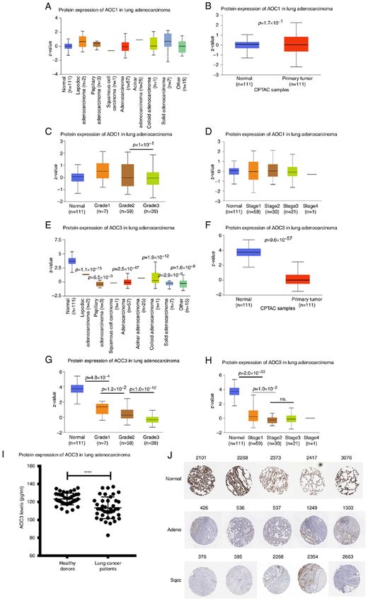
4 CHANG et al: AOC3 REGULATING EMT AND IMMUNITY IN LUNG CANCER
antibodies were used: E‑cadherin (1:500; cat. no. 610182) Results
N‑cadherin (1:500; cat. no. 610921) and vimentin (1:500;
cat. no. 550513; all from BD Biosciences), Slug (1:500; product AOC3 mRNA expression is reduced in lung cancer. The
no. 9585S; Cell Signaling Technology, Inc.), and GAPDH controversial roles of AOCs have been reported in various
(1:5,000; cat. no. MAB374; EMD Millipore). The quantitation cancer types (41‑44), therefore their effect in lung cancer was
of the results of the western blotting was performed using investigated. Tumor tissue and adjacent normal tissue speci‑
AlphaImager software (Version 6.0.0; ProteinSimple). mens from 10 lung cancer patients (7 LUAD and 3 LUSC)
were analyzed via NGS (Table I). The expression of AOC2
miRNA mimics transfection. CL1‑5 cells were transfected (7 out of 10) and AOC3 (8 out of 10) was lower in most of
with microRNA (miR)‑3691‑5p (AGUG GAUGAUGGAGA the tumor tissue of patients compared with their normal
CUCGGUAC; at a concentration of 100 nM; GE Healthcare tissue, however lower AOC1 in tumor tissue was observed in
Dharmacon, Inc.) or scrambled control (negative control 1; only 2 out of 10 patients with lung cancer (Fig. 1A‑C). Using
UCACAACCUCCUAGAA AGAGUAGA; at a concentra‑ Oncomine® datasets, it revealed that AOC3, but not AOC1 or
tion of 100 nM; GE Healthcare Dharmacon, Inc.) by using AOC2, was expressed at lower levels in tumor tissue compared
Dharmafect reagent 4 (GE Healthcare Dharmacon, Inc.) with normal tissue in 16 lung cancer cohorts (Fig. 1D). Further
according to the manufacturer's instructions. The transfection analysis of these 16 cohorts revealed that the expression of
efficacy was monitored by transfecting siGLO fluorescent AOC3 mRNA was lower in the tumor tissue for both the LUAD
oligonucleotides (catalog ID: D‑001630‑02‑05; GE Healthcare and non‑adenocarcinoma patients (Fig. 1E). The expression
Dharmacon, Inc.) concurrently after 24 h of transfection of AOC2 and AOC3 in LUAD and LUSC was also retrieved
at 37˚C according to the manufacturer's protocol. The expres‑ from TCGA cohorts. Overall AOC2 (Fig. 1F) and AOC3
sion of AOC1‑3, cell migration and CD4+ T‑cell migration as (Fig. 1G) expression was significantly lower in the tumor tissue
well as adhesion were assayed after a 48‑h transfection. compared with the adjacent normal tissue, even though this
trend was not observed for all stages. Moreover, the expres‑
Mouse studies. All mice procedures were approved by and sion of AOC3 was significantly lower in the N1 group (with
conducted in accordance with the Institutional Animal lymph node metastasis) compared with the N0 group (without
Care and Use Committee at Kaohsiung Medical University lymph node metastasis), implying that AOC3 may contribute
(IACUC Approval No. 107104; Kaohsiung, Taiwan). C57BL/6 to cancer metastasis (Fig. 1F and G).
mice (12 males in total; weight, 18±2 g; 5 weeks old) were
obtained from the Taiwan National Laboratory Animal AOC3 protein expression is inversely associated with lung
Center (Taipei City, Taiwan). The mice were housed in a cancer grade. AOC protein expression was extracted from the
specific pathogen‑free environment with the room tempera‑ National Cancer Institute Clinical Proteomic Tumor Analysis
ture being maintained at ~20˚C, the humidity at ~45% and a Consortium (CPTAC). AOC1 protein expression did not vary
12‑h light/dark cycle. Each mouse had free access to food and between the tumor and normal tissue for the different types of
water. The mice were subjected to implantation of LLC cells LUAD, grades or stages (Fig. 2A‑D). However, AOC3 protein
(1x10⁶ cells) via tail vein and tumor growth in the lungs was expression was lower in the tumor tissue compared with the
allowed for 7 days. Mice were treated with PBS or recombi‑ normal tissue in every cell type (Fig. 2E and F). Moreover,
nant mouse (rm) AOC3 twice (10 µg/mouse; on days 7 and 14) AOC3 expression was negatively associated with the grades
by intra‑tracheal route. At the end of the experiment, the and stages (early and late) of LUAD (Fig. 2G and H). The
mice were euthanized by CO2 asphyxiation during which the soluble form of AOC3 has been detected in other cancer types
CO2 gas flow rate displaced 10 to 30% of the cage volume such as colorectal cancer (41). To evaluate the role of soluble
per minute. CD4+ T cells of the lungs of mice were isolated AOC3 in lung cancer, serum from lung cancer patients was
by mouse CD4 + T cell isolation kit (MACS MicroBeads; collected. Soluble AOC3 in the sera from lung cancer patients
Miltenyi Biotec GmbH) according to the manufacturer's was lower than in healthy donors (Fig. 2I). In addition, the
instructions and counted after 21 days of LLC implanta‑ public datasets for the expression of AOC3 in lung cancer
tion. Lung tissue was collected and minced and incubated were utilized. Compared with normal tissue, both LUAD
in RPMI‑1640 medium with collagenase type 1 (400 U/ml; and LUSC expressed lower levels of AOC3 from The Human
Worthington Biochemical Corporation) at 37˚C for 1 h. The Protein Atlas (Fig. 2J). The combination of these results and
digested tissues were filtered through a 70‑µm cell strainer the mRNA expression results indicated that AOC3 could be a
and washed with RPMI‑1640 medium. CD4 + T cells of the promising tumor suppressor in lung cancer.
lung filtered solution were isolated by mouse CD4 isolation
kit and counted after 21 days of LLC implantation. Lower expression of AOC3 confers a poorer survival time.
Since the tumor tissue of lung cancers expressed lower
Statistical analyses. Each experiment was repeated at least levels of AOC3, its prognostic vaule in patients was evalu‑
three times. Data are expressed as the mean ± standard ated by survival analysis. There are several public websites
deviation (SD) using GraphPad Prism version 7.04 (GraphPad that evaluate survival analysis, including the K‑M plotter,
Software, Inc.). Two treatment groups were compared by UALCAN and PROGgeneV2. According to the K‑M plotter,
unpaired Student's t‑test. Multiple group comparisons were low AOC1 expression did not confer a poorer survival time,
performed by two‑way analysis of variance with Tukey's post and it actually conferred a longer survival time in LUAD but
hoc test. P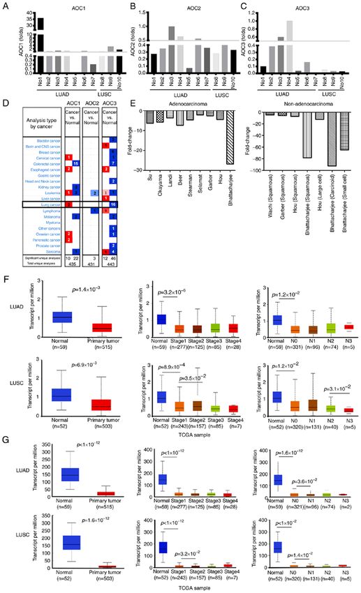
ONCOLOGY REPORTS 46: 203, 2021                                                       5

Table Ι. Characteristics of patients.

Group         Number          Sex       Age               Pathological diagnosis                Stage         T         N          M

I                01            M         70         Adenocarcinoma grade 3                       2B            3         0        0
                 02            M         66         Adenocarcinoma grade 3                       4B           2a         0        1c
                 03            F         51         Adenocarcinoma grade 3                       1B           2a         0        0
                 04            M         53         Adenocarcinoma grade 3                       3A            3         2        0
                 05            F         60         Adenocarcinoma grade 2                       1A           1b         0        0
                 06            M         67         Adenocarcinoma grade 1                       1A           1a         0        0
                 07            M         60         Adenocarcinoma grade 3                       4A            4         1        1a
II               08            M         84         Squamous cell carcinoma grade 2              2B            3         0        0
                 09            F         65         Squamous cell carcinoma grade 2              3A            4         0        b
                 10            M         69         Squamous cell carcinoma grade 2              2B           2b         1        0

panel). However, analysis of AOC3 expression revealed that the    progression. AOC3 expression was knocked down in the
lower the AOC3 expression was, the shorter the survival time      LUAD cell line (CL1‑5) via the shRNA method with >50%
was in LUAD patients but not in LUSC patients (Fig. 3A, lower     efficiency (Fig. 5A). The cells were then studied to evaluate
panel). Moreover, the clinical implication of AOC3 expres‑        the effect of AOC3 knockdown on proliferation. Neither the
sion as detemined by survival rates was validated by cohorts      WST‑1 nor the BrdU assay indicated that AOC3 affected cell
extracted from the UALCAN and PROGgeneV2 websites; low            proliferation in lung cancer (Fig. 5B and C, respectively).
expression of AOC3 conferred a shorter surivival time but this    Cell migration as evaluated via wound healing analysis,
was not observed for AOC1 (ABP1) or AOC2 (Fig. 3B and C).         revealed enhanced healing (increased migration ability) after
These results confirmed that AOC3 was strongly associated         AOC3 knockdown (Fig. 5D). In addition, AOC3 knockdown
with clinical outcomes in lung cancer patients (Fig. 3A and B,    enhanced the mesenchymal characteristics as N‑cadherin,
lower panel; Fig. 3C).                                            vimentin and Slug were increased (Fig. 5E). The rmAOC3
                                                                  protein (rhAOC3) was added to confirm the observed
Mechanism regulating the expression of AOC3. Since AOC3           changes in proliferation and migration. The proliferation
was revealed to be critical in the prognosis of lung cancer       did not change even at a high dose (50 ng/ml) of rhAOC3 as
patients, the dysregulation of AOC3 required investigation.       evaluated by either WST‑1 or BrdU assays (Fig. 5F and G,
Genetic modifications, as DNA copy number variation, DNA          respectively). Cell migration was reduced after the addition
methylation, and post‑transcriptional regulation by miRNAs        of rhAOC3 in a dose‑dependent manner as revealed in the
were utilized. As determined by the TCGA cohort, variation        wound‑healing assay (Fig. 5H). The mesenchymal charac‑
in the DNA copy number of AOC3 was not correlated with the        teristics transitioned to epithelial features as E‑cadherin was
expression of AOC3 mRNA (R=‑0.121; Fig. 4A). In addition,         increased and N‑cadherin, vimentin and Slug were decreased
DNA methylation of AOC3 was not significantly associated          in a dose‑dependent manner (Fig. 5I). The aforementioned
with the expression of AOC3 mRNA (R=‑0.034; Fig. 4B).             results indicated that reduced AOC3 expression played a role
Concerning post‑transcriptional regulation, the miRNAs that       in lung cancer progression by increasing cell migration and
epigenetically regulate AOC3 mRNA were predicted using            EMT but not proliferation.
miRWalk version 3.0 with miRanda and miRDB restrictions
of >95% confidence. There was a total of 27 miRNAs listed as      Lung cancers with low levels of AOC3 fail to recruit CD4+ T
potential regulators of the AOC3 (Fig. 4C). This list of miRNAs   cells to the tumor in vitro and in vivo. CD4+ T‑cell infiltration is
was validated using the TCGA cohort and the ones with the         a critical factor for determining the TIME against cancer (26).
highest probability were miR‑3190 and miR‑3691 since both         The role of AOC3 in the recruitment of CD4+ T cells remains
of them had significantly increased expression in the tumor       unclear in lung cancer. To validate the role of AOC3 in medi‑
tissue compared with the normal tissue (Fig. 4D and E). Both      ating the TIME in lung cancer, in vitro and in vivo studies
of the predicted and highly probable miRNAs were verified in      were performed. CD4+ T‑cell migration and attachment to lung
10 lung cancer patients. The most likely miRNA to contribute      cancer cells were evaluated. As determined by a cell adhesion
to the regulation of AOC3 mRNA was miR‑3691‑5p because            assay, CD4+ T‑cell attachment to lung cancer cells (CL1‑5) was
there was an undetectable read number in most specimens for       decreased after AOC3 knockdown (Fig. 6A). Before CD4+ T
miR‑3190 in our samples (Fig. 4F and G).                          cells arrive at tumor sites, they must traverse the endothelia.
                                                                  A transendothelial migration assay was utilized to reveal the
Low AOC3 expression mediates epithelial‑mesenchymal               transit of CD4+ T cells through the vascular endothelia. When
transition (EMT) in lung cancer. Low levels of AOC3               AOC3 was silenced in cancer cells, CD4+ T‑cell migration
expression conferred poor clinical outcomes in lung cancer        was reduced, (Fig. 6B) indicating that the lower the AOC3
patients. Therefore, the present study set out to verify          expression, the fewer CD4+ T cells were recruited. Conversely,
the mechanisms by which AOC3 mediated lung cancer                 when rhAOC3 was added, more CD4+ T cells attached to the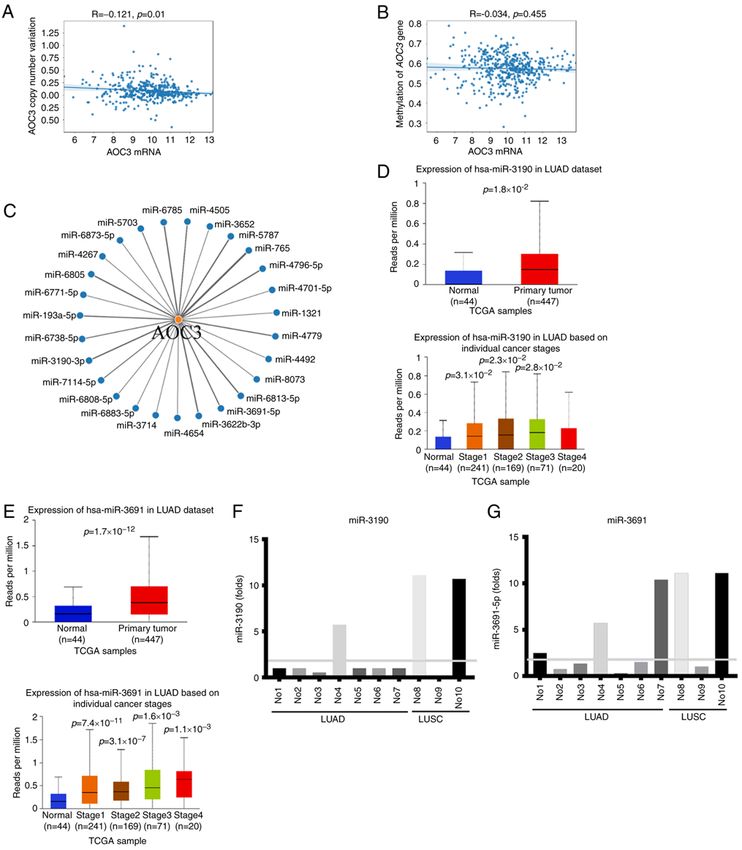
6                                 CHANG et al: AOC3 REGULATING EMT AND IMMUNITY IN LUNG CANCER

Figure 1. AOC mRNA expression in lung cancer. The expression of AOCs in the normal and tumor tissue was obtained from 10 lung cancer patients (7 LUAD
and 3 LUSC). The expression of (A) AOC1, (B) AOC2 and (C) AOC3 between the tumor and normal tissue in lung cancer patients. (D) Based on the Oncomine®
datasets, there was lower expression of AOC3 in the tumor tissue compared with the normal tissue in 16 cohorts. (E) AOC3 expression was lower in the tumor
tissue compared with the normal tissue in the adenocarcinoma and non‑adenocarcinoma cohorts. Furthermore, data from The Cancer Genome Atlas cohort
revealed that there was lower (F) AOC2 and (G) AOC3 expression in the tumor tissue compared with the normal tissue in both LUAD and LUSC cohorts.
Furthermore, there was lower expression of AOC3 in the N1 group compared with the N0 group but not in all stages of lung cancer. AOC, amine oxidase,
copper containing 3; LUAD, lung adenocarcinoma; LUSC, lung squamous cell carcinoma.
ONCOLOGY REPORTS 46: 203, 2021                                                                     7

Figure 2. AOC protein expression in lung cancer. The protein expression of the AOCs was extracted from the National Cancer Institute Clinical Proteomic
Tumor Analysis Consortium. Only AOC1 and AOC3 were available in this dataset. (A) The protein expression of AOC1 in lung cancer tissue vs. normal tissue
was not different in lung cancer (regardless of lung cancer type), (B) lung adenocarcinoma only, (C) different grades of lung adenocarcinoma, or (D) different
stages of lung cancer. (E) Conversely, the expression of AOC3 was lower in tumor tissue compared with the normal tissue in lung cancer (regardless of lung
cancer type), (F) in different types of adenocarcinoma cohorts, (G) different grades of lung adenocarcinoma and (H) different stages of lung cancer. To further
validate the soluble form of AOC3 in lung cancer, the sera from 40 lung cancer patients and 40 normal controls were analyzed. (I) Soluble AOC3 expression
was lower in the lung cancer patients compared with the normal controls. (I) The AOC3 protein expression was also retrieved from the Human Protein Atlas.
(J) The protein expression of AOC3 was attenuated in both adenocarcinoma and squamous cell carcinoma compared with normal lung tissue. ****P
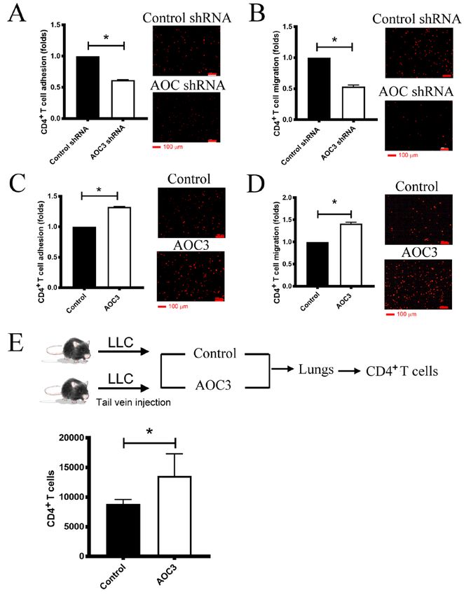
8                                  CHANG et al: AOC3 REGULATING EMT AND IMMUNITY IN LUNG CANCER

Figure 3. Survival analysis of AOCs in lung cancer. The survival time for lung cancer patients from cohorts with different levels of AOC1, 2 and 3 expression
were further analyzed. In the Kaplan‑Meier plotter, survival time was analyzed for ‘both, LUAD and LUSC’, ‘LUAD’ and ‘LUSC’ (from left to right). (A; upper
panel) For AOC1, the high‑expression group was associated with a shorter survival time compared with the low‑expression group. (A; middle panel) For AOC2,
the high‑expression group was not associated with a longer survival time compared with the low‑expression group. (A; lower panel) However, for AOC3, the
low‑expression group was associated with a shorter survival time in lung adenocarcinoma. (B) In addition, UALCAN cohorts also indicated that low expres‑
sion of AOC3 but not AOC2 was associated with a shorter survival time compared with high expression. (C) The low expression of AOC3 also conferred shorter
survival time in different lung adenocarcinoma (GSE26919 and GSE31210) and lung cancer (GSE30219) cohorts in the PROGgeneV2. AOC, amine oxidase,
copper containing 3; LUAD, lung adenocarcinoma; LUSC, lung squamous cell carcinoma.

lung cancer cells (Fig. 6C). The addition of rhAOC3 increased                   the recruitment of CD4+ T cells to lung cancer sites. AOC3
CD4 + T‑cell migration through the vascular endothelial                         facilitation of CD4+ T‑cell recruitment was validated using an
cells (Fig. 6D). These results indicated that AOC3 increased                    animal model. The in vivo study investigated the number of
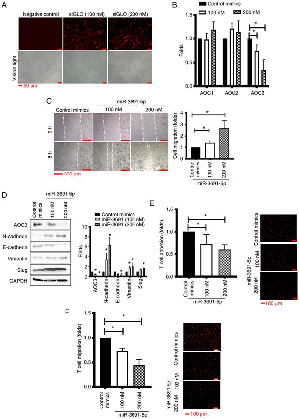
ONCOLOGY REPORTS 46: 203, 2021                                                                 9

Figure 4. Regulation of AOC3 mRNA expression. The expression of AOC3 mRNA was regulated by genetic modifications. The TCGA cohort was used to
investigate these modifications. (A) The DNA copy number of AOC3 was not correlated with AOC3 mRNA expression. (B) Moreover, epigenetic regulation,
such as AOC3 DNA methylation was not associated to AOC3 mRNA expression. (C) MiRNA regulation of AOC3 mRNA was predicted using miRWalk 3.0,
and the 27 possible candidate miRNAs are listed. The present study validated the possibility of each miRNA using the TCGA cohort. The most likely candi‑
date miRNAs included (D) miR‑3190 and (E) miR‑3691. Furthermore, the expression levels of indicated miRs were associated with the stages respectively.
(F and G) These 2 miRNAs were further verified by data from 10 patients (including 7 lung adenocarcinoma and 3 lung squamous cell carcinoma). AOC, amine
oxidase, copper containing 3; TCGA, The Cancer Genome Atlas; miR, microRNA; LUAD, lung adenocarcinoma; LUSC, lung squamous cell carcinoma.

CD4+ T cells in the lungs of mice with tumors and revealed                    miR‑3691‑5 regulates EMT and cancer migration via epigen‑
that the number of CD4+ T cells was increased after rmAOC3                    etic downregulation of AOC3. To further verify the possible
was instilled two times (10 µg/mouse) (Fig. 6E). These results                regulatory role of miR‑3691‑5p in AOC3 expression, miR‑3691
indicated that AOC3 promoted CD4+ T‑cell recruitment into                     mimics were transfected to CL1‑5 cells and then their biological
the TIME.                                                                     functions were assessed. The transfection efficacy of miRNA
10                                CHANG et al: AOC3 REGULATING EMT AND IMMUNITY IN LUNG CANCER

Figure 5. AOC3 mediates EMT in lung cancer. The present study investigated the mechanisms by which low AOC3 expression conferred a poor lung cancer
prognosis. (A) AOC3 shRNA was used to knockdown AOC3 expression in CL1‑5 cells. The knockdown efficiency was >50%. Cell proliferation was not
altered as determined by (B) WST‑1 and (C) BrdU incorporation assays. (D) Migration potential as determined via a wound‑healing assay was enhanced after
AOC3 knockdown. (E) The EMT phenomenon shifted towards the mesenchymal characteristics as indicated by increased N‑cadherin, vimentin and Slug and
decreased epithelial characteristics as indicated by E‑cadherin. Conversely, using rhAOC3, the present study confirmed that AOC3 did not reduce cellular
proliferation as revealed by (F) WST‑1 and (G) BrdU incorporation assays. (H) The wound healing assay also revealed a reduction in migration potential in
a dose‑dependent manner after the addition of rhAOC3. (I) Concurrently, the EMT characteristics were reversed to epithelial characteristics as E‑cadherin
was increased and N‑cadherin, vimentin and Slug were decreased. All experiments were performed independently at least three times. *P
ONCOLOGY REPORTS 46: 203, 2021                                                                      11

Figure 6. AOC3 attracts CD4+ T cells to lung cancer sites. The present study attempted to clarify the role of AOC3 in the recruitment of CD4+ T cells in lung
tumors. The in vitro study used ‘cell adhesion’ of CD4+ T cells on lung cancer cells. After AOC3 knockdown, PKH26‑stained CD4+ T cells were added to the
fully‑recovered lung cancer cells (CL1‑5). (A) When compared with the control, the AOC3‑knockdown lung cancer cells were less likely to be attached to by
CD4+ T cells. (B) Moreover, the transendothelial migration assay revealed that less CD4+ T cells were attracted to AOC3‑knockdown lung cancer cells compared
with the control group. However, rhAOC3 increased CD4+ T‑cell (C) attachment and (D) migration. Furthermore, the in vivo study utilized a mouse model to
verify the in vitro findings. Lewis lung carcinoma cells were injected into mice via their tail vein. Concurrently, rmAOC3 (10 µg/mouse) was instilled into the
trachea of mice twice, 7 days apart (on day 7 and day 14) (intratracheal instillation). After 14 days (day 21), the mice were sacrificed and the lungs were minced.
The CD4+ T cells were then isolated. (E) The CD4+ T cells in the mice instilled with rmAOC3 were increased compared with the controls. All experiments
were performed independently at least three times. *P90% efficacy was                    with miR‑3691‑5p transfection, AOC3 protein expression
achieved (Fig. 7A). miR‑3691‑5p downregulated expression of                        was decreased whereas the expression of the mesenchymal
AOC3 but not of AOC1 and AOC2 (Fig. 7B). The transfected                           markers N‑cadherin, vimentin and Slug were increased
lung cancer cells were then adopted for wound healing analysis                     (Fig. 7D). These results indicated that miR‑3691‑5p affected
and revealed enhanced healing process in a dose‑dependent                          cell migration and EMT through AOC3. Furthermore, to
12                                CHANG et al: AOC3 REGULATING EMT AND IMMUNITY IN LUNG CANCER

Figure 7. miR‑3691 inhibits EMT through downregulation of AOC3. As predicted in Fig. 4E, miR‑3691‑5p was used to evaluate its effect on AOC3. (A) The
transfection efficacy. (B) Low expression of AOC3 but not AOC1 nor AOC2 after miR‑3691‑5p mimics transfection. (C) The miR‑3691‑5p‑transfected CL1‑5
cells were increased as indicated by wound‑healing assay. (D) The EMT phenomenon shifted towards the mesenchymal characteristics as N‑cadherin, vimentin
and Slug were increased and epithelial characteristic indicated by E‑cadherin was decreased after AOC3 knockdown. (E) The in vitro cell adhesion of CD4+
T cells on lung cancer cells was decreased after introduction of miR‑3691‑5p. (F) Moreover, the transendothelial migration assay revealed that less CD4+
T cells were attracted to lung cancer cells compared with the control group. *P
ONCOLOGY REPORTS 46: 203, 2021 13
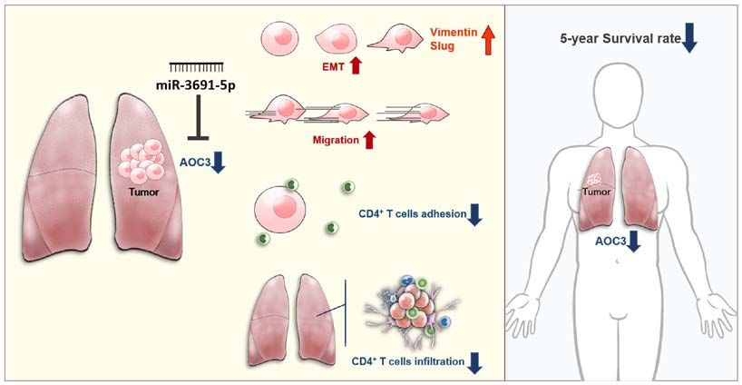
Figure 8. Proposed model of reduced AOC3 mediating lung cancer. The low expression of AOC3 mediated by miR‑3691‑5p in lung cancer conferred the
potential of tumor cells to migrate and mesenchymal characteristics such as vimentin and Slug to increase. Moreover, low expression of AOC3 failed to recruit
CD4+ T cells to the tumor site in vitro and in vivo. These results may explain the reason why loss of AOC3 in tumor tissue leads to shorter 5‑year survival time
in lung cancer patients. AOC, amine oxidase, copper containing 3; miR, microRNA.
evaluate the miR‑3691‑5p‑AOC3 axis in mediating the TIME cancer (47). The present study is the first one, to the best of our
in lung cancer, in vitro studies were performed. CD4+ T‑cell knowledge, which revealed that low‑level AOC3 is the critical
migration and adhesion to lung cancer cells were evaluated. As amine oxidase in lung cancer pathogenesis but not AOC1 or
determined by a cell adhesion assay, CD4+ T‑cell attachment AOC2. Furthermore, miR‑3691‑5p regulation on AOC3 and its
to lung cancer cells (CL1‑5) was decreased after miR‑3691‑5p biological functions were defined. miR‑3691‑5p has been demon‑
transfection in a dose‑dependent manner (Fig. 7E). A tran‑ strated to enhance migration and invasion in hepatocellular
sendothelial migration assay revealed that migration of CD4+ carcinoma (48). Low expression of AOC3 conferred poor clinical
T cells through endothelia was reduced in a dose‑dependent outcomes and lymph node metastasis, supporting the theory that
manner with miR‑3691‑5p transfection (Fig. 7F). These results AOC3 acts as a tumor suppressor in lung cancer. Biological func‑
indicated that miR‑3691‑5p attenuated the recruitment of tion analyses revealed that AOC3 did not affect cell proliferation
CD4+ T cells. Collectively, it was indicated that miR‑3691‑5p but that it did influence cell migration. The process of EMT
regulated AOC3 expression to perform its biological functions. is essential for the enhancement of cell migration (49). AOC3
knockdown enhanced mesenchymal characteristics as revealed
Discussion by an increase in N‑cadherin, vimentin and Slug and attenuated
epithelial characteristics as revealed by a decrease in E‑cadherin.
The present study attempted to identify a novel factor affecting Exogenous rhAOC3 reversed these mesenchymal patterns. This
the different aspects of lung cancer pathogenesis. Through finding revealed that AOC3 is involved in the maintenance of
analysis of lung cancer patients via a high‑throughput NGS epithelial characteristics to decrease the metastasis ability of
tool, and the utilization of genomic data from different cohorts, lung cancer. For the first time, the present study has revealed
the present study determined that AOC3 contributed to lung the pathogenic roles of AOC3 in malignant cells and AOC3
cancer pathogenesis. Different findings at both transcriptional per se providing a useful biomarker and prognostic factor in lung
and translational levels revealed that low levels of AOC3 were cancer patients for clinical diagnosis and treatment.
a critical factor contributing to cancer development. Low‑level AOC3 contributes to both innate and acquired immu‑
AOC3 facilitated mesenchymal transformation and decreased nity (50). Endothelial AOC3 mediates the adhesion of tumor
CD4+ T‑cell recruitment to lung cancer tumors. It was also infiltration lymphocytes, lymphokine‑activated killer cells
revealed that AOC3 expression was under miR3691‑5p epigen‑ and natural killer cells (51) in inflammatory tissue (52) and
etic regulation. The strong negative association between AOC3 tumor tissue. An absence of AOC3 leads to a marked reduction
and the survival rate in patients indicated that it is a key factor in antigen‑specific CD4+ recruitment into the airway bron‑
involved in lung cancer, and that AOC3 could be a valuable target chial lymph nodes (50). In the present study, knockdown of
for drug development (Fig. 8). AOCs have different effects in AOC3 in lung cancer cells caused a reduction in CD4+ T‑cell
different types of cancer (14,42‑44,45,46). High levels of AOCs extravasation through the endothelial layer and attachment to
can act as oncogenes and confer worse clinical outcomes, such cancer cells. On the other hand, exogenous rhAOC3 increased
as AOC1 in gastric cancer (14) and AOC3 in human glioma (45). transendothelial migration and enhanced CD4+ T‑cell attach‑
On the other hand, low levels of AOCs are associated with ment onto lung cancer cells. Furthermore, rmAOC3 facilitated
worse clinical outcomes, such as AOC3 in colorectal (42,47) CD4+ T‑cell recruitment to preexisting lung tumor in a mouse
and gastric cancer (46). Moreover, decreased AOC3 levels are model. These results indicated that AOC3 could modulate the
correlated with lymph node and hepatic metastasis in colorectal TIME in lung cancer cells, and it may be possible to potentiate14 CHANG et al: AOC3 REGULATING EMT AND IMMUNITY IN LUNG CANCER
its effectiveness by immunotherapy. However, before reaching Ethics approval and consent to participate
a definite conclusion, there are some limitations in the present
study. Firstly, CD4+ T cells were utilized as a recruiting immune The protocol of the present study was approved (approval
cell to the lung. However, there are more immune cells such no. KMUH‑IRB‑20130054 and and KMUH-IRB-G(II)-
as dendritic cells and macrophages and consequently, further 20180021) by the Institutional Review Board of Kaohsiung
investigation may be necessary. Secondly, data from immuno‑ Medical University Hospital (Kaohsiung, Taiwan) and written
histochemical staining for membrane‑bound AOC3 in tumor informed consents were acquired from all enrolled patients.
tissues, which would limit the role of AOC3 in lung cancer, are All mice procedures were approved by the Institutional Animal
lacking. Care and Use Committee at Kaohsiung Medical University
Collectively, the results of the present study confirmed the (Kaohsiung, Taiwan).
axis of miR‑3691‑5p‑AOC3 as having a critical role in lung
cancer via inhibiting EMT and migration and a determining Patient consent for publication
factor for the recruitment of CD4+ T cells to restore anticancer
immunity in the TME. AOC3 expression in lung cancer speci‑ Not applicable.
mens may provide valuable information for patient prognosis
and could have valuable applications when determining a Competing interests
therapeutic strategy in immunotherapy/chemotherapy.
The authors declare that they have no competing interests.
Acknowledgements
References
The authors would like to thank the CPTAC of UALCAN and 1. Ferlay J, Colombet M, Soerjomataram I, Mathers C, Parkin DM,
the Human Protein Atlas who generated the data used in this Piñeros M, Znaor A and Bray F: Estimating the global cancer
publication. The authors would also like to thank the Center for incidence and mortality in 2018: GLOBOCAN sources and
methods. Int J Cancer 144: 1941‑1953, 2019.
Research Resources and Development in Kaohsiung Medical 2. Siegel RL, Miller KD and Jemal A: Cancer statistics, 2019.
University for the assistance in Bioinformatics. CA Cancer J Clin 69: 7‑34, 2019.
3. Arbour KC and Riely GJ: Systemic therapy for locally advanced
and metastatic non‑small cell lung cancer: A review. JAMA 322:
Funding 764‑774, 2019.
4. Miller KD, Nogueira L, Mariotto AB, Rowland JH, Yabroff KR,
The present study was supported by the Ministry of Science Alfano CM, Jemal A, Kramer JL and Siegel RL: Cancer treat‑
ment and survivorship statistics, 2019. CA Cancer J Clin 69:
and Technology (grant nos. 110-2314-B-037-124-MY3, 363‑385, 2019.
109‑2314‑B‑037‑091 and 108‑2320‑B‑037‑024‑MY3), the 5. National Lung Screening Trial Research Team: Lung Cancer
Kaohsiung Medical University (grant no. KMU‑DK108008), Incidence and mortality with extended follow‑up in the National
lung screening trial. J Thorac Oncol 14: 1732‑1742, 2019.
the Kaohsiung Medical University Hospital (grant 6. Elmore LW, Greer SF, Daniels EC, Saxe CC, Melner MH,
nos. KMUH108‑8R15, KMUH108‑8R16 and MUH106-6T06) Krawiec GM, Cance WG and Phelps WC: Blueprint for cancer
and the Kaohsiung Municipal Ta‑Tung Hospital (grant research: Critical gaps and opportunities. CA Cancer J Clin 71:
107‑139, 2021.
nos. KMTTH‑103‑019 and KMTTH‑105‑051). 7. Binnewies M, Roberts EW, Kersten K, Chan V, Fearon DF,
Merad M, Coussens LM, Gabrilovich DI, Ostrand‑Rosenberg S,
Availability of data and materials Hedrick CC, et al: Understanding the tumor immune microenvi‑
ronment (TIME) for effective therapy. Nat Med 24: 541‑550, 2018.
8. Duan Q, Zhang H, Zheng J and Zhang L: Turning cold into
The datasets generated and/or analyzed during the present hot: Firing up the tumor microenvironment. Trends Cancer 6:
605‑618, 2020.
study are not publicly available due to ongoing study in our 9. Vakal S, Jalkanen S, Dahlstrom KM and Salminen TA: Human
laboratory but are available from the corresponding author on copper‑containing amine oxidases in drug design and develop‑
reasonable request. ment. Molecules 25: 1293, 2020.
10. Mondovì B and Finazzi Agrò A: Structure and function of amine
oxidase. Adv Exp Med Biol 148: 141‑153, 1982.
Authors' contributions 11. Buffoni F and Ignesti G: The Copper‑containing amine oxidases:
Biochemical aspects and functional role. Mol Genet Metab 71:
559‑564, 2000.
CYC, YMT and YLH conceptualized the present study. SFJ, 12. Boomsma F, Bhaggoe UM, van der Houwen AM and
PHT and YCH provided the technical support, performed van den Meiracker AH: Plasma semicarbazide‑sensitive
the experiments and acquired the data. CYC provided the amine oxidase in human (patho)physiology. Biochim Biophys
Acta 1647: 48‑54, 2003.
software management and analyzed the data. YYC, JYH, 13. Salmi M and Jalkanen S: Vascular adhesion protein‑1: A cell
WAC and IWC validated the results. KLW, YMT and YLH surface amine oxidase in translation. Antioxid Redox Signal 30:
performed the formal analysis. YLH pursued the investigation 314‑332, 2019.
14. Xu F, Xu Y, Xiong JH, Zhang JH, Wu J, Luo J and Xiong JP: AOC1
and provided the resources. YMT, KLW and YLH performed Contributes to tumor progression by promoting the AKT and EMT
data curation and interpreted the data. YMT and KLW wrote pathways in gastric cancer. Cancer Manag Res 12: 1789‑1798, 2020.
original draft. YYC and YLH wrote, reviewed and edited the 15. Lopes de Carvalho L, Bligt‑Linden E, Ramaiah A, Johnson MS
and Salminen TA: Evolution and functional classification of
final manuscript. IWC performed visualization of the imaging mammalian copper amine oxidases. Mol Phylogenet Evol 139:
data. YLH supervised the study, was the project administrator 106571, 2019.
and acquired the funding. YMT and YLH critically revised 16. Imamura Y, Kubota R, Wang Y, Asakawa S, Kudoh J, Mashima Y,
Oguchi Y and Shimizu N: Human retina‑specific amine oxidase
the manuscript for important intellectual content. All authors (RAO): cDNA cloning, tissue expression, and chromosomal
read and approved the final version of the manuscript. mapping. Genomics 40: 277‑283, 1997.ONCOLOGY REPORTS 46: 203, 2021 15
17. Bonaiuto E, Lunelli M, Scarpa M, Vettor R, Milan G and 34. Gyorffy B, Surowiak P, Budczies J and Lanczky A: Online
Di Paolo ML: A structure‑activity study to identify novel and survival analysis software to assess the prognostic value of
efficient substrates of the human semicarbazide‑sensitive amine biomarkers using transcriptomic data in non‑small‑cell lung
oxidase/VAP‑1 enzyme. Biochimie 92: 858‑868, 2010. cancer. PLoS One 8: e82241, 2013.
18. Salmi M and Jalkanen S: A 90‑kilodalton endothelial cell mole‑ 35. Mende DR, Letunic I, Maistrenko OM, Schmidt TS, Milanese A,
cule mediating lymphocyte binding in humans. Science 257: Paoli L, Hernández‑Plaza A, Orakov AN, Forslund SK,
1407‑1409, 1992. Sunagawa S, et al: proGenomes2: An improved database for accu‑
19. Stankovic B, Bjørhovde HAK, Skarshaug R, Aamodt H, rate and consistent habitat, taxonomic and functional annotations
Frafjord A, Müller E, Hammarström C, Beraki K, Bækkevold ES, of prokaryotic genomes. Nucleic Acids Res 48: D621‑D625, 2020.
Woldbæk PR, et al: Immune cell composition in human non‑small 36. Sticht C, De La Torre C, Parveen A and Gretz N: miRWalk: An
cell lung cancer. Front Immunol 9: 3101, 2019. online resource for prediction of microRNA binding sites. PLoS
20. Oja AE, Piet B, van der Zwan D, Blaauwgeers H, Mensink M, One 13: e0206239, 2018.
de Kivit S, Borst J, Nolte MA, van Lier RAW, Stark R and 37. Enright AJ, John B, Gaul U, Tuschl T, Sander C and Marks DS:
Hombrink P: Functional heterogeneity of CD4+ tumor‑infiltrating MicroRNA targets in Drosophila. Genome Biol 5: R1, 2003.
lymphocytes with a resident memory phenotype in NSCLC. 38. Chen Y and Wang X: miRDB: An online database for predic‑
Front Immunol 9: 2654, 2018. tion of functional microRNA targets. Nucleic Acids Res 48:
21. Rhodes DR, Yu J, Shanker K, Deshpande N, Varambally R, D127‑D131, 2020.
Ghosh D, Barrette T, Pandey A and Chinnaiyan AM: ONCOMINE: 39. Ke HL, Li WM, Lin HH, Hsu WC, Hsu YL, Chang LL,
A cancer microarray database and integrated data‑mining plat‑ Huang CN, Li CC, Chang HP, Yeh HC, et al: Hypoxia‑regulated
form. Neoplasia 6: 1‑6, 2004. MicroRNA‑210 overexpression is associated with tumor devel‑
22. Chandrashekar DS, Bashel B, Balasubramanya SAH, opment and progression in upper tract urothelial carcinoma. Int
Creighton CJ, Ponce‑Rodriguez I, Chakravarthi BVSK and J Med Sci 14: 578‑584, 2017.
Varambally S: UALCAN: A portal for facilitating tumor 40. Livak KJ and Schmittgen TD. Analysis of relative gene expres‑
subgroup gene expression and survival analyses. Neoplasia 19: sion data using real‑time quantitative PCR and the 2(‑Delta Delta
649‑658, 2017. C(T)) method. Methods 25: 402‑408, 2001.
23. Su LJ, Chang CW, Wu YC, Chen KC, Lin CJ, Liang SC, Lin CH, 41. Shao L, Li H, Chen J, Song H, Zhang Y, Wu F, Wang W,
Whang‑Peng J, Hsu SL, Chen CH and Huang CY: Selection of Zhang W, Wang F, Li H and Tang D: Irisin suppresses the migra‑
DDX5 as a novel internal control for Q‑RT‑PCR from micro‑ tion, proliferation, and invasion of lung cancer cells via inhibition
array data using a block bootstrap re‑sampling scheme. BMC of epithelial‑to‑mesenchymal transition. Biochem Biophys Res
Genomics 8: 140, 2007. Commun 485: 598‑605, 2017.
24. Okayama H, Kohno T, Ishii Y, Shimada Y, Shiraishi K, 42. Ward ST, Weston CJ, Shepherd EL, Hejmadi R, Ismail T and
Iwakawa R, Furuta K, Tsuta K, Shibata T, Yamamoto S, et al: Adams DH: Evaluation of serum and tissue levels of VAP‑1 in
Identification of genes upregulated in ALK‑positive and colorectal cancer. BMC Cancer 16: 154, 2016.
EGFR/KRAS/ALK‑negative lung adenocarcinomas. Cancer 43. Sun WY, Choi J, Cha YJ and Koo JS: Evaluation of the expres‑
Res 72: 100‑111, 2012. sion of amine oxidase proteins in breast cancer. Int J Mol Sci 18:
25. Landi MT, Dracheva T, Rotunno M, Figueroa JD, Liu H, 2775, 2017.
Dasgupta A, Mann FE, Fukuoka J, Hames M, Bergen AW, et al: 44. Kostoro J, Chang SJ, Clark Lai YC, Wu CC, Chai CY and
Gene expression signature of cigarette smoking and its role in Kwan AL: Overexpression of vascular adhesion protein‑1 is
lung adenocarcinoma development and survival. PLoS One 3: associated with poor prognosis of astrocytomas. APMIS 124:
e1651, 2008. 462‑468, 2016.
26. Beer DG, Kardia SL, Huang CC, Giordano TJ, Levin AM, 45. Chang SJ, Tu HP, Lai YCC, Luo CW, Nejo T, Tanaka S, Chai CY
Misek DE, Lin L, Chen G, Gharib TG, Thomas DG, et al: and Kwan AL: Increased vascular adhesion protein 1 (VAP‑1) levels
Gene‑expression profiles predict survival of patients with lung are associated with alternative M2 macrophage activation and poor
adenocarcinoma. Nat Med 8: 816‑824, 2002. prognosis for human gliomas. Diagnostics (Basel) 10: 256, 2020.
27. Stea r man RS, Dwyer‑Nield L, Zerbe L, Blaine SA, 46. Yasuda H, Toiyama Y, Ohi M, Mohri Y, Miki C and Kusunoki M:
Chan Z, Bunn PA Jr, Johnson GL, Hirsch FR, Merrick DT, Serum soluble vascular adhesion protein‑1 is a valuable prog‑
Franklin WA, et al: Analysis of orthologous gene expres‑ nostic marker in gastric cancer. J Surg Oncol 103: 695‑699, 2011.
sion between human pulmonary adenocarcinoma and a 47. Toiyama Y, Miki C, Inoue Y, Kawamoto A and Kusunoki M:
carcinogen‑induced murine model. Am J Pathol 167: 1763‑1775, Circulating form of human vascular adhesion protein‑1 (VAP‑1):
2005. Decreased serum levels in progression of colorectal cancer and
28. Selamat SA, Chung BS, Girard L, Zhang W, Zhang Y, predictive marker of lymphatic and hepatic metastasis. J Surg
Campan M, Siegmund KD, Koss MN, Hagen JA, Lam WL, et al: Oncol 99: 368‑372, 2009.
Genome‑scale analysis of DNA methylation in lung adenocarci‑ 48. Du W, Zhang X and Wan Z: miR‑3691‑5p promotes hepatocel‑
noma and integration with mRNA expression. Genome Res 22: lular carcinoma cell migration and invasion through activating
1197‑1211, 2012. PI3K/Akt signaling by targeting PTEN. Onco Targets Ther 12:
29. Garber ME, Troyanskaya OG, Schluens K, Petersen S, 4897‑4906, 2019.
Thaesler Z, Pacyna‑Gengelbach M, van de Rijn M, Rosen GD, 49. Leggett SE, Hruska AM, Guo M and Wong IY: The epithelial-
Perou CM, Whyte RI, et al: Diversity of gene expression in mesenchymal transition and the cytoskeleton in bioengineered
adenocarcinoma of the lung. Proc Natl Acad Sci USA 98: systems. Cell Commun Signal 19: 32, 2021.
13784‑13789, 2001. 50. Dun kel J, Aguila r‑Pimentel JA, Oller t M, Fuchs H,
30. Hou J, Aerts J, den Hamer B, van Ijcken W, den Bakker M, Gailus‑Durner V, de Angelis MH, Jalkanen S, Salmi M and
Riegman P, van der Leest C, van der Spek P, Foekens JA, Veres TZ: Endothelial amine oxidase AOC3 transiently
Hoogsteden HC, et al: Gene expression‑based classification of contributes to adaptive immune responses in the airways. Eur
non‑small cell lung carcinomas and survival prediction. PLoS J Immunol 44: 3232‑3239, 2014.
One 5: e10312, 2010. 51. Irjala H, Salmi M, Alanen K, Grénman R and Jalkanen S: Vascular
31. Wachi S, Yoneda K and Wu R: Interactome‑transcriptome anal‑ adhesion protein 1 mediates binding of immunotherapeutic effector
ysis reveals the high centrality of genes differentially expressed cells to tumor endothelium. J Immunol 166: 6937‑6943, 2001.
in lung cancer tissues. Bioinformatics 21: 4205‑4208, 2005. 52. Stolen CM, Marttila‑Ichihara F, Koskinen K, Yegutkin GG,
32. Bhattacharjee A, Richards WG, Staunton J, Li C, Monti S, Vasa P, Turja R, Bono P, Skurnik M, Hänninen A, Jalkanen S and
Ladd C, Beheshti J, Bueno R, Gillette M, et al: Classification of Salmi M: Absence of the endothelial oxidase AOC3 leads to
human lung carcinomas by mRNA expression profiling reveals abnormal leukocyte traffic in vivo. Immunity 22: 105‑115, 2005.
distinct adenocarcinoma subclasses. Proc Natl Acad Sci USA 98:
13790‑13795, 2001. This work is licensed under a Creative Commons
33. Uhlen M, Zhang C, Lee S, Sjöstedt E, Fagerberg L, Bidkhori G, Attribution-NonCommercial-NoDerivatives 4.0
Benfeitas R, Arif M, Liu Z, Edfors F, et al: A pathology atlas of International (CC BY-NC-ND 4.0) License.
the human cancer transcriptome. Science 357: eaan2507, 2017.You can also read