Collagen I triggers directional migration, invasion and matrix remodeling of stroma cells in a 3D spheroid model of endometriosis - Nature
←
→
Page content transcription
If your browser does not render page correctly, please read the page content below
www.nature.com/scientificreports
OPEN Collagen I triggers directional
migration, invasion and matrix
remodeling of stroma cells in a 3D
spheroid model of endometriosis
Anna Stejskalová1*, Victoria Fincke1, Melissa Nowak1,3, Yvonne Schmidt1, Katrin Borrmann2,
Marie‑Kristin von Wahlde1, Sebastian D. Schäfer1, Ludwig Kiesel1, Burkhard Greve2 &
Martin Götte1*
Endometriosis is a painful gynecological condition characterized by ectopic growth of endometrial
cells. Little is known about its pathogenesis, which is partially due to a lack of suitable experimental
models. Here, we use endometrial stromal (St-T1b), primary endometriotic stromal, epithelial
endometriotic (12Z) and co-culture (1:1 St-T1b:12Z) spheroids to mimic the architecture of
endometrium, and either collagen I or Matrigel to model ectopic locations. Stromal spheroids, but not
single cells, assumed coordinated directional migration followed by matrix remodeling of collagen
I on day 5 or 7, resembling ectopic lesions. While generally a higher area fold increase of spheroids
occurred on collagen I compared to Matrigel, directional migration was not observed in co-culture or in
12Z cells. The fold increase in area on collagen I was significantly reduced by MMP inhibition in stromal
but not 12Z cells. Inhibiting ROCK signalling responsible for actomyosin contraction increased the
fold increase of area and metabolic activity compared to untreated controls on Matrigel. The number
of protrusions emanating from 12Z spheroids on Matrigel was decreased by microRNA miR-200b and
increased by miR-145. This study demonstrates that spheroid assay is a promising pre-clinical tool that
can be used to evaluate small molecule drugs and microRNA-based therapeutics for endometriosis.
Endometriosis is a common gynaecological disease in which the uterine lining, the endometrium, grows at
ectopic locations such as the ovaries and peritoneal c avity1. This disease is currently treated using hormonal
therapy and excision surgery. Unfortunately, these treatments are not curative and have high associated side
effects and remission rates1,2. While targeted therapies are urgently needed, their development has been hindered
by the h eterogeneity3,4 and limited mechanistic understanding of the disease.
Based on the widely accepted Sampson’s theory, endometriosis arises when tissue fragments shed during
menstruation implant in the surrounding t issue5 (Fig. 1). To implant, endometrial fragments have to first pen-
etrate either through intact barriers consisting of epithelial cells, basement membranes and collagen or directly
through a damaged tissue (e.g. due to m icrotrauma6 or previous surgical p rocedure7–9) and then s pread10. In
this regard, endometriosis shares many similarities with metastatic cancer11. However, while cancer researchers
have devoted considerable attention to dissecting the invasive processes involved in cancer metastases12, little is
known about invasive processes in endometriosis.
One significant hurdle in studying how endometrial cells invade ectopic tissues has been a lack of suitable
experimental models13. To address this, in vitro models of endometriosis consisting of endometrial e xplants14,
organoids3 or single cells combined with chorioallantoic m embrane15, amniotic m embrane16, peritoneal meso-
thelial cell monolayers17, peritoneal explants18 and h
ydrogels19 have been developed. Nevertheless, each of these
approaches has some inherent limitations. 2D cell culture is the gold standard, but the invasive and migratory
strategies in 2D are markedly different from the coordinated multicellular collective invasion through the extra-
cellular matrix (ECM) that has been observed in vivo20,21. Organoid models typically focus predominantly on
epithelial cells and lack the stromal c omponent22. Explants suffer from high heterogeneity, mixed cell population
1
Department of Gynecology and Obstetrics, Münster University Hospital, Albert‑Schweitzer Campus 1,
D11, 48149 Münster, Germany. 2Department of Radiotherapy‑Radiooncology, Münster University Hospital,
48149 Münster, Germany. 3Present address: Institut für Molekulare Medizin III, Heinrich-Heine-Universität
Düsseldorf, 40225 Düsseldorf, Germany. *email: anna.stejskalova@gmail.com; mgotte@uni‑muenster.de
Scientific Reports | (2021) 11:4115 | https://doi.org/10.1038/s41598-021-83645-8 1
Vol.:(0123456789)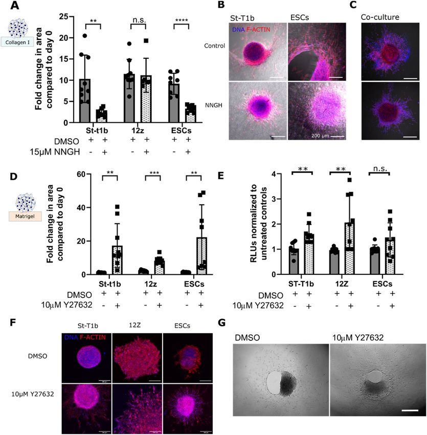
www.nature.com/scientificreports/
Figure 1. Endometriosis modelling in vitro. Left. Sampson’s theory of retrograde menstruation. Menstrual
tissue contains stromal condensates (dark blue) and collapsed epithelial glands (pink). Ectopic lesions are
frequently described to have a ‘bullet-like appearance’ Right. Spheroids generated using the hanging drop
method as a model of collapsed endometrium architecture are placed on either Matrigel or collagen I on day 4
and their phenotype on the hydrogels and the effect of pharmacological intervention is evaluated.
and low throughput23. The promising and integrative tissue engineering approach has so far recreated models of
decidual eutopic endometrium rather than lesions or menstrual stage e ndometrium19.
Menstrual stage endometrium is characterized by stromal reorganization into tightly packed cellular con-
densates sometimes referred to as ‘blue balls’, collapsed glands and blood debris24. We hypothesized that such
collapsed architecture could be modelled using the spheroid culture in vitro. Spheroid culture is a well-established
technique that commonly used to study malignancies25. Indeed, endometrial epithelial spheroids generated from
12Z, an endometriotic lesion derived epithelial cell line26 and endometriotic stromal cells27 have already been
shown to share histological similarities to lesions better than 2D culture. However, it has not been investigated
how endometrial spheroids interact with the ECM.
In this study, we show that endometrial spheroids create structures resembling lesions on collagen I and
Matrigel in vitro within 5–7 days. We demonstrate that this assay can dissect the effect of the cell and ECM type
as well as of small molecule- and RNA- drugs.
Results
An endometrial stromal cell line (St‑T1b), primary endometriotic stromal cells and the endo‑
metriotic epithelial cell line (12Z) self‑organize into spheroids in hanging drop culture. To
capture the heterogeneity of endometrial cells found in lesions, the cells we employed in this study were an
immortalized eutopic stromal cell line St-T1b28, primary endometriotic stromal cells (ESCs) and the ectopic
light red peritoneal lesion derived epithelial 12Z cell line that was previously shown to be invasive in a Matrigel
invasion assay29.
First, we validated that the cells retained their stromal and epithelial morphology in culture. Figure 2A shows
that while the St-T1b and ESCs cells have an elongated, fibroblast-like stromal morphology, 12Z cells have a
mostly polygonal shape and grow in clusters. Furthermore, on tissue culture (TC) plastic, the stromal cells exhibit
more defined actin fibers compared to the 12Z cells. Quantitative analysis (Fig. 2B) confirmed that 12Z cells are
significantly smaller (p < 0.0001) than St-T1b and ESCs, where the average area for St-T1b, 12Z and ESCs cells
on TC plastic were 2086 ± 904.1 µm2 (n = 29), 787.7 ± 380.9 µm2 (n = 32) and 1989 ± 889.5 µm2 (n = 30).
Recent studies suggested that spheroid culture offers several advantages over 2D culture and confirmed
that 12Z c ells26 and endometriotic stromal c ells27 can assemble into spheroids using the U-bottom 96 well
plates27. However, it has not been investigated whether also the hanging drop method can be used to fabricate
endometrial spheroids and whether there are any differences between spheroids fabricated from epithelial and
stromal endometrial cells alone or their co-culture. We, therefore, evaluated the hanging-drop method, each
drop containing 20,000 of stromal or epithelial cells or their co-culture in 20 µL of standard media and selected
day 4 as the harvesting day.
Bright-field images (Fig. 2C) show that all the studied cell types self-organized into spheroids. Interestingly,
the morphology of the spheroids varied across cell types. St-T1b and ESCs cells assembled into compact, round-
spheroids, while the 12Z spheroids were larger and sometimes exhibited slightly branching morphology. We
also generated co-culture spheroids from the epithelial 12Z and stromal St-T1b cell lines combined at 1:1 ratio
(Fig. 2D). Cell Tracker staining and confocal imaging suggest the two cell populations were homogeneously
distributed throughout the spheroid on day 4. Interestingly, while the size of individual 12Z cells in 2D is sig-
nificantly smaller compared to the ESCs and St-T1b cells, 12Z spheroids were significantly larger compared to
St-T1b and ESCs (n = 14, p < 0.0001 and p < 0.001) (Fig. 2E). To exclude that this is due to a cell-counting error,
the spheroid size was measured on spheroids prepared three independent times. The co-culture spheroids were
also significantly larger compared to St-T1b spheroids (n = 11) and had a higher metabolic activity that is indica-
tive of higher cell count and proliferation over the spheroid formation period (Fig. 2F,G).
Next, we evaluated whether the condensation into spheroids induces changes in gene expression. We analysed
a subset of genes related to ectopic tissue invasion. Gene expression analysis revealed that while organisation into
Scientific Reports | (2021) 11:4115 | https://doi.org/10.1038/s41598-021-83645-8 2
Vol:.(1234567890)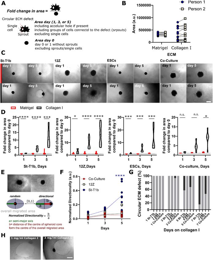
www.nature.com/scientificreports/

                                  Figure 2.  Spheroid formation by endometrial cells. (A) F-Actin (red) and nuclei (blue) stained St-T1b, 12Z and
                                  ESCs. (B) The projected area in 2D of St-T1b and ESCs is significantly larger than of 12Z cells (n = 29, 32 and
                                  30 cells, Kruskal–Wallis with Dunn’s multiple comparisons post hoc test. Data show mean ± s.d.). (C) Bright-
                                  field images of fixed spheroids that formed after 4-days using the hanging drop method. Scale bars 250 µm.
                                  (D) Co-Culture 1:1 St-T1b:12Z spheroids on day 4 stained by Cell Tracker. Red are St-T1b and green 12Z cells.
                                  Scale bar 200 µm. (E) The 12Z spheroids were significantly larger compared to St-T1b and ESCs spheroids
                                  (n = 14 prepared across three different preparations, Kruskal–Wallis with Dunn’s multiple comparisons post hoc
                                  test), the area was measured manually on bright-field images, 10 × magnification. (F) Metabolic-based assay
                                  suggests 12Z and Co-Culture spheroids on day 4 consist of a higher number of cells than St-T1b spheroids (n = 3
                                  independent wells and one preparation, one-way ANOVA with Tukey’s multiple comparisons test). (G) Spheroid
                                  projected area is also significantly larger in 12Z cells and co-culture groups than in the St-T1b group (n = 10–15
                                  independent wells from two different spheroid preparations, Kruskal–Wallis with Dunn’s multiple comparisons
                                  post hoc test). (H) qPCR analysis comparing gene expression in 2D and 3D spheroids on day 4 of the hanging
                                  drop culture in St-T1b cells and (I) qPCR analysis comparing gene expression in 2D and 3D spheroids on day 4
                                  of the hanging drop culture in 12Z cells (n = 3 independent preparations on the same cell lines, multiple t tests).
                                  For all figures in the panel *p < 0.05; **p < 0.01; ***p < 0.001, ****p < 0.0001 and not significant (n.s.) p > 0.05;
                                  Data shown as mean ± standard deviation (s.d.) or as mean + s.d.

                                  spheroids alters the gene expression of several markers in St-T1b cells, none of these markers was significantly
                                  altered in 12Z spheroids compared to monoculture across three independent preparations (Fig. 2H,I).

Scientific Reports |   (2021) 11:4115 |                https://doi.org/10.1038/s41598-021-83645-8                                                    3

                                                                                                                                                Vol.:(0123456789)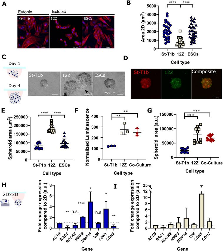
www.nature.com/scientificreports/
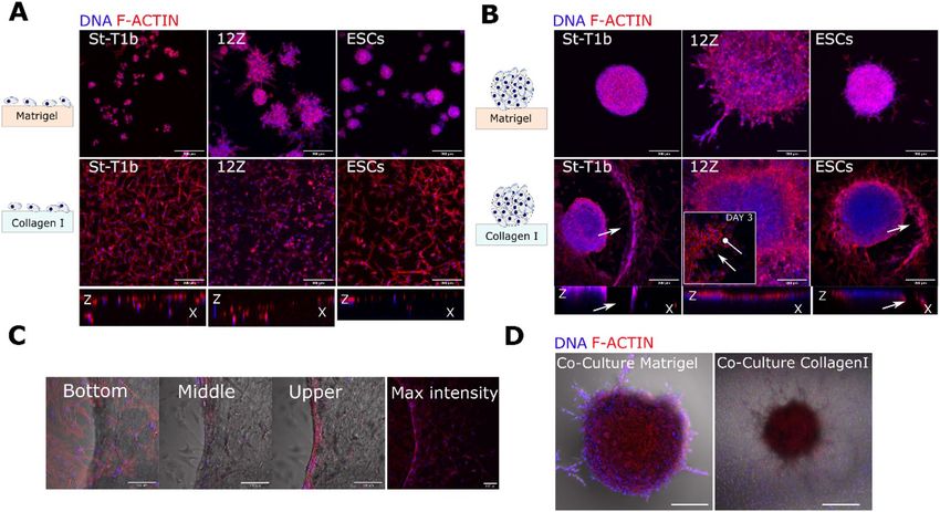
Figure 3. Lesion-like structures on collagen I and Matrigel. (A) Confocal images of a suspension of
endometrial cells after 3 days on Matrigel (top row). Stromal St-T1b and ESCs cellular aggregates consisted of
only a few cells and were highly circular and 12Z aggregates were larger and showed protrusions. All cell types
invaded collagen I (bottom row) as single cells (maximal intensity projection, scale bar 200 µm, f-actin red,
nuclei blue). (B) Confocal images of spheroids after 7 days on Matrigel and collagen I. 12Z exhibited the highest
number of protrusions on Matrigel. St-T1b and ESCs created circular defects in collagen I surrounded by cells,
whereas epithelial 12Z cells migrated as a sheet and confocal imaging revealed no invasion (maximal intensity
projection, scale bar, 200 µm, actin cytoskeleton red, nuclei blue). (C) Detail of three different imaging planes of
the edge of the circular defect in St-T1b spheroid in collagen I group. F-actin in red and DNA in blue. Scale bar
100 µm. (D) S-T1b: 12Z co-culture after 7 days on Matrigel (left) and collagen I (right). Scale bar 200 µm.
First, we examined the expression of Ras-related C3 botulinum toxin substrate 1(RAC1/Rac1), a small sig-
nalling G protein that directs actin-driven cellular protrusion, microtubule prolongation and the formation of
lamellipodia30 both in single cells and at the leading edge during collective migration31. The expression of RAC1
was significantly downregulated in 3D compared to 2D St-T1b (p < 0.01, n = 3) (Fig. 2H).
Spheroid St-T1b culture exhibited higher proteolytic gene expression compared to 2D (Fig. 2H). qPCR
analysis revealed that the spheroids exhibit higher expression of the secreted MMP2 (p < 0.0001, n = 3) and the
membrane-type metalloproteinase MMP14 (p < 0.05, n = 3) than cells grown in 2D.
As the epithelial to mesenchymal transition (EMT) and mesenchymal to epithelial transition (MET) have
been implicated in the progression of the disease, we further investigated the expression of mesenchymal mark-
ers vimentin (VIM) and cadherin-2 (CDH2) and the epithelial marker cadherin-1 (CDH1) (Fig. 2H). Vimentin
expression remained unchanged in both cell lines (p > 0.05, n = 3). The expression of CDH2, a cadherin known to
promote invasion in many cell t ypes32, was downregulated in St-T1b spheroids (p < 0.01, n = 3) while the expres-
sion of CDH1 was upregulated in St-T1b spheroids (< 0.05 = n = 3) compared to the 2D control.
Matrigel and collagen I trigger distinct phenotypes in single cells and spheroids where stromal
condensates create defects on collagen I. Having confirmed that endometrial stromal and epithelial
endometriotic cell line as well as their co-culture were able to form spheroids, we evaluated their invasive behav-
iour on two different ECM-derived hydrogels: Matrigel and collagen I using confocal imaging.
Single cells of all studied cells on Matrigel formed cellular aggregates by day 3(Fig. 3A). While these aggre-
gates remained mostly rounded in St-T1b and ESCs groups, the 12Z cell line aggregates consistently developed
multiple multicellular protrusions across several preparations. Cells seeded on collagen I were invading collagen
I as single cells (Fig. 3A).
We next evaluated the spheroid behaviour on Matrigel and collagen I. On the basement membrane (BM)
mimic Matrigel, the stromal St-T1b spheroids remained rounded with ESCs exhibiting few protrusions and only
the 12Z spheroids consistently developed multiple multicellular protrusions across several preparations. Confo-
cal imaging on day 7(Fig. 3B) revealed that the 12Z protrusive edges consisted of tightly packed cells (DNA in
blue) with scant cytoplasm (actin staining in red).
The response of all studied cell types to collagen I as spheroids was markedly different compared to single cells
(Fig. 3B). St-T1b and ESC spheroids on collagen I developed into invasive lesion-like structures (Fig. 3B). More
specifically, the St-T1b and ESC spheroids gradually invaded collagen I, leaving behind a circularly remodeled
matrix with a ring of tightly adhering cells at its margins (Fig. 3B,C). These rings appeared to stabilize the defect
and to limit further random cellular spreading outside of the defect in many but not all spheroids.
Scientific Reports | (2021) 11:4115 | https://doi.org/10.1038/s41598-021-83645-8 4
Vol:.(1234567890)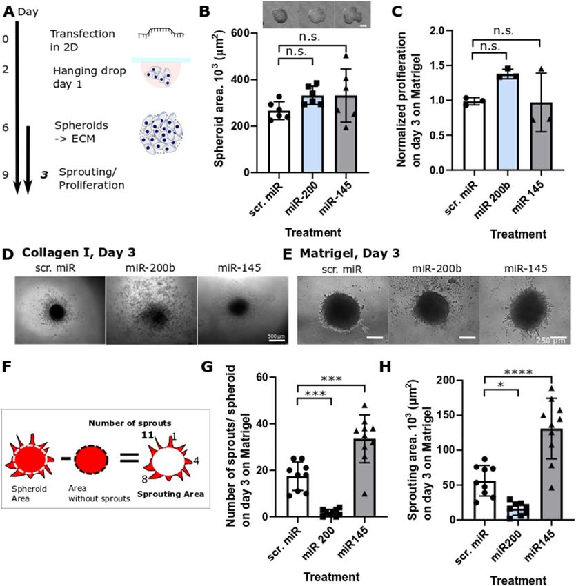
www.nature.com/scientificreports/
Interestingly, no matrix defect or directional spreading was detected in co-culture St-T1b:12Z spheroids
on collagen I (Fig. 3D). Co-culture spheroids on Matrigel developed protrusive edges similar to the 12Z-only
spheroids.
Directional invasion followed by the formation of a circular defect occurs in St‑T1b and ESCs
spheroids but not in St‑T1b:12Z co‑culture. Next, we quantified the invasive and migratory patterns
on Matrigel and collagen I using bright-field imaging and a parameter that we termed ‘Fold change in the area’
that we defined as the overall projected area, including matrix defects on the day of interest divided by the area
on the day 0 or 1 without any sprouts (Fig. 4A). All analysis was done manually in FIJI using the freehand selec-
tion tool. While manual measurement has its limitation, especially when the ‘Area’ increases and its margins
become irregular, no significant difference in measured areas was observed between different assessors (Fig. 4B).
Our data show that the ‘Fold change in area’ is significantly higher on collagen I compared to Matrigel across
all studied cell types by day 5 (Fig. 4C,D). Confocal imaging combined with brightfield microscopy suggested the
stromal spheroids invade (Fig. 3B,C) and migrate on the collagen I matrix directionally (Fig. 4C). To quantify this,
we used the parameter ‘Directionality’ that is calculated as the ratio of the distance of the centre of the spheroid
core from the centre of the overall migrated area b to the semi-major axis of the overall migrated area a (Fig. 4E).
The normalized directionality increased for St-T1b but not for 12Z or co-culture spheroids with time on collagen
I, especially between days 3 and 5 (Fig. 4F). The directional invasion was typically followed by matrix remodeling
resulting in a circular defect at the area with the densest stromal cell population (Fig. 4G). In our system (3 mg/
mL, 40 µL/well) this typically occurred around day 5 or 7 with 84.6% and 53.3% of St-T1b and ESCs, respectively,
having a defect on day 7 (n = 13–15 per time point) (Fig. 4G). The defects formed both on 1 mg/mL and 3 mg/
mL collagen I hydrogels, suggesting this behavior occurs across a range of collagen I concentrations (Fig. 4H).
Spheroid 3D culture as an effective tool to screen small molecule drug and microRNA‑based
therapeutics. We then evaluated the potential of the here presented endometrial spheroid in vitro assay to
screen the potential therapeutic effect of mechanoregulatory small molecules and micro RNAs (Supplementary
Table ST1).
The broad‑spectrum MMP inhibitor NNGH limits the invasive behaviour of stromal spheroids
on collagen I. Previous studies implicated that MMP signalling plays a role in the formation of early endo-
metriotic lesions15. Our study shows that the broad-spectrum MMP inhibitor 15 µM N-isobutyl-N-(4-methoxy-
phenylsulfonyl) glycyl hydroxamic acid (NNGH) significantly reduced ‘Fold change in the area’ on collagen I
from 10.4 fold to 2.3 fold (n = 6–9) and 9.2 fold to 3.3 fold (n = 6–9) in St-T1b and ESCs, respectively, but did not
significantly affect the ‘Fold change in the area’ in 12Z cells (n = 6–9) (Fig. 5A, Supplementary Figure S1). Fur-
thermore, it can be seen from Fig. 5B, that while NNGH treatment prevents the formation of the circular defect
on collagen I even after 7 days in culture, the migration of St-T1b and ESCs is not completely eliminated. The
effect of NNGH inhibitor on the St-T1b:12Z co-culture was less pronounced and neither the control nor NNGH
group formed matrix defects by day 5 (Fig. 5C).
ROCK inhibition significantly enhances spreading and invasion of all studied endometrial cell
types on Matrigel. The ROCK inhibitor Y27632 significantly (p < 0.01) increased the ‘Fold change in the
area’ of all studied cell types on Matrigel compared to DMSO (Fig. 5D). The area occupied by St-T1b, 12Z
and ESCs was 17.3, 6.6 and 22.3 fold larger compared to day 0 (Fig. 5E). Y27632 also affected the numbers of
metabolically active cells, which were significantly higher compared to controls for St-T1b and 12Z cells on day
5 on Matrigel. Moreover, Y27632 affected spheroid morphology (Fig. 5F). Y27632 on Collagen I resulted in a
disaggregation of the spheroid core in St-T1b and ESCs as shown in the Supplementary Figure S2. Treatment
with Y27632, in contrast to the MMP inhibitor NNGH, did not prevent Collagen I matrix remodeling in ESCs
(Fig. 5G) suggesting the directional remodeling is rather due to proteolytic action than acto-myosin contraction.
The spheroid model reveals context‑dependent roles of the mechanoregulatory microRNAs
miR‑200b and miR‑145 on the invasive behaviour of the endometriotic epithelial cell line 12Z
on Matrigel. We next investigated whether our in vitro model can be used as a tool to screen the functional
effect of various microRNAs on endometrial phenotype. In particular, we selected two microRNAs, miR-200b33
and miR-14534, that have been previously shown to be dysregulated in e ndometriosis35 and to modulate the
invasion and migration of 12Z cells in 2D and Transwell assays. miR-200b acts as a transcriptional repressor of
ZEB1/2 and thus downregulates EMT transition36. The miR-145 is upregulated in endometrial lesions and has
been described to modulate cytoskeletal dynamics in several cell types, including endometrial, and has many
validated targets, including beta and gamma actin, cofilin, fascin, myosin light chain 9 and Rho kinase Rock134,37.
The transfection was performed in monolayer culture before the fabrication of spheroids and the effect of micro-
RNAs on spheroid spreading was assessed after 3 days on Matrigel to minimize the effect of miR dilution and
degradation38 (Fig. 6A). It can be seen from Fig. 6B that microRNA transfection did not significantly alter the
ability of cells to form spheroids and the area of individual spheroids was not significantly different (p > 0.05)
across the treatment groups nor was the proliferation (Fig. 6C). We observed spheroid fragmentation of miR-
200b transfected cells on Collagen I which resulted in a discontinuous nature of the projected area the size of
which could not be reliably quantified (Fig. 6D). MiR-145 significantly reduced the spheroid area compared
to scr. miR controls on day 3 on collagen I (Supplementary Figure S3). On Matrigel, the microRNAs, affected
sprouting characteristics behaviour of 12Z cells as seen in the bright-field images in Fig. 6E. The miR-200b treat-
ment significantly decreased the number of sprouts per spheroid from ~ 17 to ~ 1, while miR-145 significantly
Scientific Reports | (2021) 11:4115 | https://doi.org/10.1038/s41598-021-83645-8 5
Vol.:(0123456789)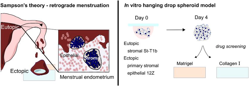
www.nature.com/scientificreports/
Figure 4. Quantification of spheroid behaviour on Matrigel and collagen I. (A) Schematic illustrating how the
‘Fold increase in the area’ was measured and calculated. (B) Validation of the manual measurement method
showed no significant difference between different assessors following the defined criteria (n matrigel = 13, n
collagen = 16, t test for each condition). (C) Brightfield images on day 1 and 5 of the spheroids on Matrigel (top
row) and collagen I (bottom row). Scale bar 500 µm. (D) Quantification of the fold change in area for individual
cell types and St-T1b:12Z co-culture (n = 12–15 independent wells per time point and condition collated from
three independent spheroid preparations, ncocultures = 4–5, one preparation. Two-way Repeated Measures (RM)
ANOVA, Šidák’s multiple comparisons tests). (E) Schematic illustrating how normalized directionality was
calculated. (F) Normalized directionally for St-T1b (circles), 12Z (squares) and co-cultures (diamonds) on day
1, 3 and 5 (n = 5–10 wells per experiment collated from two independent preparations, n co-culture = 5 from one
preparation). (G) Circular ECM defect quantification-grey colour represents the absence of macroscopic ECM
defect and the black colour a presence of a circular defect (n = 13–15 independent wells collated from three
separate spheroid preparations, nco-culture = 5 from one preparation. Two-way Repeated Measures (RM) ANOVA,
Šidák’s multiple comparisons test) (H) Directional matrix remodeling resulting in a circular defect occured on
both 1 mg/mL and 3 mg/mL collagen I hydrogels. For all figures in this panel *p < 0.05; **p < 0.01; ***p < 0.001,
****p < 0.0001, and n.s. p > 0.05.
Scientific Reports | (2021) 11:4115 | https://doi.org/10.1038/s41598-021-83645-8 6
Vol:.(1234567890)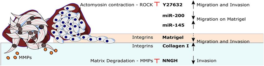
www.nature.com/scientificreports/
Figure 5. The effects of small molecule inhibitors on lesion formation. (A) The broad spectrum MMP inhibitor
NNGH significantly reduced the in vitro lesion size in St-T1b and ESCs but not in 12Z cells that migrated on
collagen I surface. The spheroid size was measured manually on days 0 and 5 (n = 6–9 independent wells across
two preparations, multiple t tests). (B) NNGH effectively prevented stromal cells from degrading collagen I
(bright field channel) but did not completely prevent the cells from migrating. Confocal images were obtained
on fixed samples after 7 days in culture. Scale bar, 200 µm. (C) Co-cultures on collagen I without (top) and with
(bottom) NNGH inhibitor on day 5 Scale bar, 200 µm. (D) The ROCK inhibitor Y27632 significantly increased
the spreading of endometrial cells on Matrigel after 5 days. Data were compared to the spheroid size on day
0 using bright-field images (n = 8–10 independent wells across two different spheroid preparations, multiple t
tests). (E) Y27632 significantly increases metabolic activity in all studied cell types on day 5 (n = 8–9, multiple
t tests, three independent preparations). (F) Confocal images demonstrating the increase in the projected area
of spheroids of all cell types on Matrigel upon Y27632 treatment Scale bar, 200 µm. (G) ESCs on Collagen I
on day 7 with and without Y27632. Y27632 did not prevent collagen I remodeling. Scale bar 500 µm *p < 0.05;
**p < 0.01; ***p < 0.001 and n.s. p > 0.05; Data shown as mean ± s.d.
increased the number of sprouts per spheroid to ~ 34 (Fig. 6F,G) and increased the overall sprouting area from
56.12 × 10³ ± 21.87 × 103 µm2 per scrambled control miR spheroid to 130.86 × 10³ ± 43.47 × 103 µm2 per miR-145
treated spheroids (p < 0.0001) (Fig. 6F,H). In line with previous findings on EMT-marker analysis in 2D-cultured
12Z cells31,33, qPCR analysis of miR-200b-treated 12Z spheroids indicated strong upregulation of CDH1 expres-
sion levels, however, the data were not significant due to high variability, since only minute amounts of RNA
could be isolated from the spheroids (Supplementary Figure S4).
Discussion
Endometriosis is a complex multifactorial disease1. The overall goal of this study was, therefore, to develop a
modular 3D in vitro model that makes it possible to study the interplay of different factors that have been pro-
posed to contribute to the pathogenesis of endometriosis and screen potential therapeutics in vitro.
Scientific Reports | (2021) 11:4115 | https://doi.org/10.1038/s41598-021-83645-8 7
Vol.:(0123456789)www.nature.com/scientificreports/
Figure 6. The effect of microRNA on 12Z sprouting on Matrigel. (A) Schematic of the workflow (B) none
of the microRNAs affected the ability of 12Z cells to self-organize into spheroids and all groups resulted in
spheroids with similar area (scale bar = 250 µm, n = 6 independent spheroids prepared across two preparations,
ANOVA). (C) None of the microRNA affected overall metabolic activity measured as luminescence compared
to scr.miR treated controls. Data are normalized to controls without any microRNA (n = 3 independent wells,
ANOVA, one repeat). (D) Representative images of microRNA treated 12Z spheroids after 3 days on collagen
I. Scale bar, 500 µm. (E) Representative images of microRNA treated 12Z spheroids after 3 days on Matrigel.
Scale bar, 250 µm. (F) A diagram showing how the number of sprouts and the sprouting area parameters were
calculated. (G) miR-200 significantly decreased while miR-145 significantly increased the number of sprouts per
spheroid after 3 days on Matrigel. (n = 9–10 independent wells across two independent preparations, ANOVA,
Tukey’s multiple comparisons). (H) The overall area occupied by sprouts was significantly larger and smaller
when treated with miR-145 and miR-200b, respectively, compared to scr.miR after 3 days on Matrigel (n = 8–10
independent wells across two different preparations, ANOVA, Tukey’s multiple comparisons, two independent
experiments), *p < 0.05; **p < 0.01; ***p < 0.001 and n.s. p > 0.05; data expressed as mean ± s.d.
First, we demonstrate that the hanging drop method makes it possible to generate endometrial spheroids
of reproducible size and thus provides a good alternative to the low-adhesion plate method26,39. Our data show
that the spheroid size is consistently cell-type specific, with stromal cells generating smaller spheroids than the
epithelial 12Z cells or their co-culture. This is likely due to proliferation of 12Z cell in spheroids as suggested
by the cell proliferation assay on spheroids on day 4. qPCR analysis revealed that the spheroid culture affects
gene expression. The stromal St-T1b had enhanced expression of the MMP2, MMP14 compared to 2D culture.
RAC1, on the other hand, was downregulated in St-T1b spheroids. Spheroids in which Rac1 production was
either inhibited or the gene was constitutively expressed had suppressed or enhanced migration in 3D matrices,
respectively40. We speculate that it is possible that RAC1 is temporarily downregulated in stromal cells cultured
as a suspension spheroid culture. While basal CDH1 expression was very low with a Ct value of 27, as expected
for a mesenchymal cell line, we observed a significantly increased expression in 3D culture. We could previously
Scientific Reports | (2021) 11:4115 | https://doi.org/10.1038/s41598-021-83645-8 8
Vol:.(1234567890)www.nature.com/scientificreports/
Figure 7. The invasiveness of endometrial spheroids depends both on the cell type (stromal St-T1b and ESCs
in brown, epithelial 12Z in blue) and ECM. Invasion and spreading is strongly enhanced by exposed collagen
I. Migration on Matrigel is modulated by microRNAs and ROCK inhibitor increases invasion and migration
on this substrate. Invasion on collagen is MMP-dependent. Red marks signify experimental intervention. Blunt
arrow signifies that an inhibitor was added. Black arrows show the effect of the intervention. Dashed arrow
suggest weak effect.
show that CDH1 mRNA expression can be induced in St-T1b cells by external stimuli such as seminal plasma41,
however, the upregulation in a 3D environment warrants further investigation. While 12Z cells have been initially
characterized as being CDH1 n egative28, low expression levels have been subsequently detected by our group in
cells authenticated by STR analysis32,33. Our experiments suggest there is no significant difference in any of the
analysed markers CDH133, RAC1, ROCK, MMP2 and MMP14 in the 12Z cell line. While another study showed
MMP2 expression is upregulated in spheroid compared 2D culture in 12Z cells26, this difference could be due
to different spheroid size and culture time.
Endometriosis is marked by the growth of endometrium at ectopic locations1. We, therefore, investigated
how epithelial 12Z, ESCs and St-T1b and co-culture spheroids; and single cells interact with two ectopic ECM
mimics Matrigel resembling the basement membrane and collagen I mimicking the exposed stroma. The ‘fold
increase in the area’ of the spheroids was markedly higher on collagen I than on Matrigel on day 5. Similarly,
single cells seeded on top of these hydrogels preferentially invaded collagen I hydrogels. Our results are in agree-
ment with previous studies conducted on cancer cells suggesting that collagen I alone can increase the invasive
cellular phenotype and show that this effect is significant across cell t ypes42–44. These data also tie well with the
previously reported clinical observations that tissue scarring either due to surgery or persistent microtrauma
could contribute to the pathogenesis of e ndometriosis7–9.
While the 12Z cell line was created from lesion-derived cells29 based on their ability to penetrate through
Matrigel coated invasion chambers, the 12Z single cells in our study only assembled into cellular aggregates with
processes and 12Z spheroids developed invasive edges. Previously, Pollock and colleagues also observed only low
levels of basal invasion in 12Z cells on Matrigel h ydrogels45. We speculate that the limited invasive capacity of 12Z
cell observed in this study could be due to the chemotactic gradient that is a key part of the invasion chamber
setup. Our group has indeed previously demonstrated that 12Z are invasive under a fetal calf serum g radient33.
Unexpectedly, there was a marked difference between the behaviour of stromal single-cell suspension and
spheroids on collagen I. The St-T1b and ESC spheroids but not single cells consistently migrated on, invaded
and remodelled collagen I in a directional manner leaving behind a circular defect in the material encircled by
the cells that visually resembled peritoneal endometriotic lesions. Given that this was the case for both the St-
T1b cell line derived from healthy cells and ectopic ESCs suggests such invasive behavior might be an inherent
property of stromal endometrial menstrual condensates and could be critical not only for the pathophysiology
of endometriosis but also for normal regeneration of endometrium.
Directional migration followed by matrix remodeling was not observed in the 12Z-spheroid or the 12Z: St-
T1b co-culture groups, suggesting the stromal-epithelial interactions modulate stromal invasiveness. While we
did not investigate the MMP levels of the co-culture spheroids, previous research determined that the co-culture
between endometrial Ishikawa epithelial and telomerase-immortalized stromal cells reduces the MMP2 levels in
stromal cells both in the absence of hormonal stimulation and in the presence of 10 nM estradiol concentration46.
In this paper, we further demonstrate that the endometrial spheroid-ECM platform can be used for drug
screening of small molecule drugs and micro-RNAs (Fig. 7). We show that the collagen I circular defect caused
by stromal cells arises due to matrix degradation via MMPs rather than due to cellular contraction. Both eutopic
and ectopic stromal cells had significantly upregulated MMP expression and the MMP inhibitor, NNGH, signifi-
cantly reduced the size of in vitro stromal lesions on collagen I. These results are in good agreement with Nap and
colleagues that demonstrated that inhibiting MMP activity prevents the development of endometriotic lesions
in a model combining chicken chorioallantoic membrane model and biopsies of menstrual stage endometrium
obtained from healthy d onors15. Our results refine this model and show that while, in agreement with the previ-
47
ous studies , the MMP inhibitor significantly slows down the invasion of spreading of stromal cells on collagen it
has little effect on the collective migration of 12Z cells. Another signaling molecule we targeted is ROCK, which
is a key regulator of the c ytoskeleton30,48. On Collagen I, ROCK inhibitor Y27632 treatment led to a rapid loss
of the spheroid core structure compared to controls and Y27632 did not prevent Collagen I remodeling by ESCs
suggesting the matrix remodeling is not primarily driven by matrix contraction but rather by MMP proteolytic
action. Y27632 further significantly increased the ‘fold change in area’ and cell numbers in vitro on Matrigel in
all studied cell types. Similar increase in cellular spreading following the treatment with ROCK inhibitors have
been described in microvascular endothelial c ells49, retinal pigment epithelial cells50 and osteoblastic cells51. It
Scientific Reports | (2021) 11:4115 | https://doi.org/10.1038/s41598-021-83645-8 9
Vol.:(0123456789)www.nature.com/scientificreports/
needs to be noted that Y27632 has a complex effect on p henotype52,53. For example, prior studies demonstrated
that Y27632 reduces endometriosis associated fibrosis in vitro54.
Another promising class of therapeutics targets microRNA s ignaling35,55. Given that a typical micro-RNA
has tens of targets, sequencing studies need to be accompanied by reliable functional assays to be biologically
meaningful56. In this study, we demonstrate that the spheroid assay can be used to reproducibly evaluate the
effect of individual microRNAs on the complex, multicellular spreading of endometriosis-mimicking constructs
over several days. We show that miR-200b treatment of 12Z cells resulted in a reduction of sprout formation,
which may be indicative of a less invasive phenotype. Our previous 2D data suggest that miR-200b may have
reverted the 12Z phenotype to an epithelial-state33, however, the paucity of RNA in the spheroids did not allow
us to unequivocally confirm this hypothesis, as we saw only a non-significant increase in expression of the epi-
thelial marker E-cadherin (Supplementary Figure S4). Our spheroid model further revealed that while miR-145
reduces the migrated area on Collagen I compared to controls (Supplementary Figure S3), results which are in
agreement with previous in vitro 2D assays34, the microRNA miR-145 up-regulated in ectopic lesions in vivo
increases 12Z sprouting on Matrigel in vitro. These findings were unexpected and investigating this into more
detail is beyond the main focus of this study. Nevertheless, there is an increasing appreciation that cells adopt
a host of invasive and migratory strategies that are highly context-dependent and enabled by distinct signal-
ing pathways. Liu and colleagues observed that miR-145 upregulation enhances angiogenesis, including the
sprouting from aortic rings and linked this to the suppression of tropomodulin 3 (TMOD3)57 while we observed
that miR-145 inhibits proliferation and migration in breast cancer and endometriotic cells using the Transwell
migration and scratch assays32,58. Therefore, miR-145 might influence cellular invasive behaviour not only in
cell-type but also invasive/migratory-mode manner and ECM-substrate-dependent manner. While the major-
ity of oncological studies on miR-145 function suggest that it reduces invasive growth by targeting a variety of
mRNAs, two studies in trophoblast cells have described invasion-promoting functions of miR-145, which were
attributed to a targeting of mucin 1 (MUC1) and leukemia inhibitory factor receptor (LIFR), r espectively59,60.
We can only speculate that the 3D spheroid culture compared to 2D culture of 12Z cells may have altered the
expression patterns of miR-145 target mRNAs in a way that alters the response to this epigenetic regulator. For
example, miR-145 may target new mRNAs that are not expressed in the 2D setting (or vice versa), resulting in
a different net response. Overall, we demonstrate that the spheroid assay can be used as an additional assay to
screen for both small molecule and RNA-based therapeutics.
A major limitation of our study is that it relies on cell lines that have been transformed and represent only
a limited subset of disease phenotypes and a more extensive primary cell pool will be required to confirm and
fully elucidate the here reported findings. We also did not investigate the influence of decidualization. Nota-
bly, our study did not incorporate primary endometrial epithelial cells with purely epithelial characteristics.
Additionally, the wider implementation of this assay for the study of endometriosis will rely on future advances
in the molecular characterization of spheroids and high-throughput image analysis. Furthermore, automated
image analysis would significantly increase the throughput of this assay. In recent years, the quality of image
processing algorithms has approached that of trained humans while significantly decreasing the time needed
to evaluate individual s amples61. It needs to be noted that for such algorithms either large training datasets or
pre-defined criteria are needed. Given the wide array of spheroid phenotypical responses, we have only started
to identify such criteria.
Overall, our screening platform provides evidence that the physiological condensation of endometrial stromal
cells into spheroids might play an important role in the development of a subset of endometriotic lesions. As
such a directional invasive phenotype in vitro is unlikely to arise by chance, endometrial stromal condensation
might also have currently unknown but likely important biological role in the cyclical regeneration of normal
endometrium. At the same time, our results show that the epithelial lesion-derived 12Z spheroids also rapidly
migrate on collagen I and stromal-epithelial interactions modulate the invasiveness of stromal cells. Previous
studies indeed revealed significant heterogeneity and variability among different endometriosis subtypes with
several sub-types staining predominantly for stromal m arkers62.
In conclusion, this study documents that endometrial stromal cell line St-T1b and primary endometriotic
stromal cells engage in directional migration with significant collagen I remodeling when cultured in spheroid
culture and that this behaviour is inhibited by the broad-spectrum MMP inhibitor NNGH. We anticipate that
this assay will be used to gain further insights into invasive processes involved in endometriosis and for the
screening of both small molecule and RNA-based drug candidates and their off-target effects.
Methods
Cell culture. The 12Z ectopic epithelial cell line17,29 was maintained in DMEM media (Sigma-Aldrich, cat.
No. D0819, Deisenhofen, Germany,) supplemented with 10% FBS (Biochrom GmbH, cat. no. S0615, Berlin,
Germany) and 1% Pen/Strep (Sigma-Aldrich, cat. No. P4333). The St-T1b cell line28 and primary ectopic lesion-
derived stromal cells (ESCs) were maintained in 70% DMEM/18% MCDB 105 media (Sigma-Aldrich, cat. No.
117-500) supplemented with 10% FBS, 1% Pen/Strep, 1% Glutamine and 5 µg/mL insulin (Sigma-Aldrich, cat.
No. 10516). Cells were routinely split twice a week. ESCs were prepared from ectopic lesions and characterized
as previously described63. Primary endometriotic stromal cells were prepared from a biopsy of a woman with
endometriosis who underwent surgical treatment at the Department of Gynecology and Obstetrics of Münster
University Hospital in 2013, and stored as aliquoted stocks in liquid nitrogen, which were freshly thawed and
passaged in routine culture two times prior to usage in the experiments described. The modified American Soci-
ety for Reproductive Medicine classification was used to assess endometriosis64. For all ESC experiments, stroma
cells derived from a lesion located at the pelvic wall (rASRM score II) of a 19-year-old patient were employed.
The study was carried out following the Declaration of Helsinki and approved by the local ethics commission
Scientific Reports | (2021) 11:4115 | https://doi.org/10.1038/s41598-021-83645-8 10
Vol:.(1234567890)www.nature.com/scientificreports/
MiR Specifications Cat. number Manufacturer
Scr. miR Pre-miR Negative Control 2 AM17111 Ambion, Darmstadt, Germany
miR-200b hsa-miR-200b-3p: MC 10492, mirVana, miRNA mimic 4464066 Ambion
miR-145 hsa-miR-145, Pre-miR miRNA Precursor AM17100 Ambion
Table 1. MicroRNAs used in the study.
(Ethikkommission der Ärztekammer Westfalen‐Lippe und der Medizinischen Fakultät der WWU; approval no.
1 IX Greb 1 from 19 September 2001, updated 2012). The participant gave written informed consent.
Spheroid formation. Spheroids were generated using the hanging drop method65, where 20 µL drops each
containing 20,000 cells were deposited on the top lid of a plastic Petri dish and the bottom chamber was filled
with sterile water or PBS (Sigma-Aldrich, cat. No. D1408). The spheroids were harvested after 4 days at 37 °C and
7.5% or 5% CO2. The co-culture spheroids were formulated at 1:1 12Z:St-T1b ratio.
Preparation of collagen I and Matrigel. A 3 mg/mL collagen I hydrogel was formed by neutralizing and
diluting the stock solution of Collagen Type I Rat Tail matrix (Corning, Bedford, MA, USA, cat. No. 354236,
4 mg/mL or 3.4. mg/mL batch) with 1 N NaOH (Applichem, cat. No. A1432, Darmstadt, Germany), 10 × PBS
(Sigma-Aldrich, cat. No. D1408) and chilled deionized water. The amount of 1 N NaOH was calculated as 1 N
NaOH volume = (volume of the stock collagen) × 0.023 mL. The amount of 10 × PBS was calculated as volume
10 × PBS = (final volume)/10. Phenol red-free Basement Membrane Matrix Growth Factor Reduced Matrigel
(Corning, cat. No. 356231) was thawed on ice prior to use. The gels were deposited into pre-chilled 96-wells at
35–40 µL per well in 9.2–9.4 mg/mL Matrigel. Each 96-well plate was subsequently sealed with parafilm and
the gels were left to solidify for 30–60 min at 37 °C. For higher magnification confocal imaging, collagen and
Matrigel were deposited on glass coverslips.
Spheroid response to collagen I/Matrigel. Following gel formation, the wells in a 96-well plate were
filled with 50 µL of phenol-red free DMEM (Gibco, cat. No. 21063-029, Darmstadt, Germany) supplemented
with 5% charcoal-treated FBS (Biochrom GmbH, cat. no. S0615) and 5 µg/mL insulin solution (Sigma-Aldrich,
cat. No. 10516). Subsequently, one to three spheroids per well were manually added to individual wells. The
media were changed every 3–5 days and the samples were kept in an incubator at 37 °C and 7.5% CO2. The
spheroids were imaged on day 1, 3, 5 and 7.
Metabolic activity measurement. Viability was assessed using the CellTiter-Glo 3D Viability assay (Pro-
mega, cat. No. G9681, Walldorf, Germany). Spheroids and surrounding medium were collected after four days
and transferred to an opaque-walled 96-well plate. A volume of CellTiter-Glo Reagent equal to the volume of cell
culture medium was added. The mix was incubated according to manufacturer instructions and luminescence
in the form of relative light units (RLUs) was recorded using a CLARIOstar Plus (BMG Labtech, Ortenberg,
Germany).
Inhibitors. The effects of three inhibitors on spheroid spreading were evaluated. The MMP inhibitor NNGH
(Merck, cat. No. SML0584, Darmstadt, Germany) was stored at 15 mM in DMSO and dissolved to the final con-
centration of 15 µM in media and the ROCK inhibitor Y27632 (Sigma-Aldrich, cat. No. Y0503, 10 mM stock)
at 10 µM. In all experiments, spheroids were added directly to inhibitor-containing media. Inhibitor-containing
5% charcoal-treated FBS/insulin media were exchanged every 3 days.
microRNA transfection. The transfection with negative control microRNA (Scr. miR), miR-200b and
miR-145 (Table 1) was performed in a 6-well plate on 60–70% confluent cells. Before transfection, the growth
media were exchanged for Opti-MEM I Reduced Serum Media (Gibco, cat. no. 31985-070, Thermo-Scientific,
Germany). The transfection with 20 nM microRNA of interest (Table 1) was conducted using the Dharmafect
reagent (Dharmacon, cat. no. T-2001-03, Lafayette, CO, USA). The cells were incubated with the transfection
mixture for 24 h when the media were exchanged for full growth media. MiR spheroids were fabricated 48 h after
the addition of transfection media.
Live cell staining and immunostaining. The F-actin cytoskeleton was visualized using Phalloidin Cru-
zFluor 594 Conjugate (Santa Cruz Biotechnology, cat. No. sc-363795, Santa Cruz, CA, USA) at 1:1000 dilution.
The nuclei were visualized using DAPI (Sigma-Aldrich, cat. No. D9564) diluted at 1:50,000. The cells were fixed
using 3.7% formaldehyde (Merck, cat. No. 1.04003.1000, Darmstadt, Germany) at 37 °C for 15 min. Following
washing with PBS (Sigma-Aldrich, cat. No. D1408), the cells were permeabilized with 0.1% Triton-X (Riedel-de-
Haen, cat. No. AG 56029, Seelze, Germany) for 5 min. Hydrogels in a 96-well plate were stained by adding 25 µL
of the 1:1000 phalloidin dye and incubated for 1 h at 37 °C. Live cells were stained either with CellTracker Green
CMFDA (Thermo Fischer, cat. No. C2925) or CellTracker Red CMTPX (Thermo Fischer, cat. No. C34552) at
a concentration of 5 µM according to manufacturer’s instructions prior to mixing two cell types to form a co-
culture.
Scientific Reports | (2021) 11:4115 | https://doi.org/10.1038/s41598-021-83645-8 11
Vol.:(0123456789)www.nature.com/scientificreports/
Forward Reverse
ACTB TCAAGATCATTGCTCCTCCTGAG ACATCTGCTGGAAGGTGGACA
RAC1 CGCCTCCTGTAGTCGCTTTG CACGCTGTATTCTCGCCAGTG
MMP14 CCATTGGGCATCCAGAAGAGAGC GGATACCCAATGCCCATTGGCCA
MMP2 GCCGTGTTTGCCATCTGTTT CTGCAGGGAGCAGAGATTCG
VIM TCAGCATCACGATGACCTTGAA CTGCAGAAAGGCACTTGAAAGC
CDH2 TTCTGACAACAGCTTTGCCTCTG TTTATTCAGAACGCTGGGGTCA
CDH1 CAAAGCCCAGAATCCCCAAG CACACCTGGAATTGGGCAAA
Table 2. Sybr Green PCR primers.
Actin hs99999903 m1
ROCK2 hs00153074 m1
Table 3. PCR primers Taqman.
Imaging. Cells were analysed for morphological and cytoskeletal markers. The bright-field images were
obtained using either an Axiovert100 (Carl Zeiss, Jena, Germany) or an inverted microscope (Leica, Wetzlar,
Germany) using 5 ×, 10 × and 20 × objectives. Confocal imaging was performed on fixed stained samples in a
96 well plate. Samples were imaged with the Zeiss LSM 880 inverted confocal microscope (10 ×, 0.45 NA) (Carl
Zeiss, Jena, Germany) equipped with ZEN 2 software and using 11.04 µm z-stack intervals and sequential scan-
ning (514 nm argon laser, 405 nm diode laser, Bright field). The number of sections was adjusted based on the
sample thickness.
Image analysis. All images were analysed in F IJI66. Confocal images are depicted as maximal intensity
projections. The spheroid area was measured manually by tracing the spheroids using the freehand tool and
measure function on Bright-field images of spheroids on Petri Dishes, glass slides or in a 96-well plate. Fold
increase in area was calculated as the spheroid area on a given day divided by spheroid size on day 0 or day 1. If
on day 1 any protrusions were present and the spheroid was used as a reference size for the given experiment,
the protrusions on day 1 were excluded from the analysis to better reflect the size of the original spheroid core.
The parameter directionality was calculated as the ratio between the distance in pixels between the centre of
the overall migrated area and the centre of the spheroid, divided by the semi-major axis of the overall migrated
area of the spheroid (Fig. 4E). The number of sprouts per image was counted manually and the sprouting area
was calculated as the total area occupied by an expanding spheroid with sprouts minus the area occupied by the
spheroid without any protrusions (Fig. 6F).
RNA extraction and cDNA synthesis. mRNA isolation was performed with InnuPREP RNA mini kit
(Analytikjena, cat. no. 845-KS-2040250, Jena, Germany) according to the supplier’s protocols. The quantity of
RNA was measured on an Eppendorf BioPhotometer (Eppendorf, Hamburg, Germany) and considered pure if
the absorbance at 260 nm/280 nm was more than 1.8. The concentration of 0.4 µg RNA/10 µL of dH2O was used.
cDNA synthesis was performed using High Capacity kit (Applied Biosystems, cat. No. 4368814, Foster City,
CA, USA) according to the manufacturer’s instructions on a TGradient thermocycler (Biometra, Göttingen,
Germany).
PCR. Quantitative RT-PCR analysis was performed using 20 ng cDNA per reaction using the Taqman Uni-
versal PCR Master Mix (Thermo Fisher, cat. No. 4304437) and SYBR Green PCR Master Mix (Thermo Fisher,
cat. No. 4344463). Gene expression values were calculated using the mean Ct values of the samples. The expres-
sion of target genes was normalized to the housekeeping gene ACT, and then to St-T1b cells line (2−ΔΔCt). The
primers were synthesized by Biolegio (Nijmegen, The Netherlands) and are listed in Tables 2 and 3.
Statistical analysis. Data were analysed using GraphPad Prism8 (GraphPad Software, San Diego, USA).
Normal distribution was tested using the Shapiro–Wilk test. A two-tailed unpaired Student’s t tests were used
to analyse statistical significance between two conditions in an experiment. For experiments with three or more
comparisons, an ordinary one-way ANOVA with a Tukey’s multiple comparisons test was used. For data that
were not normally distributed, the Kruskal–Wallis test followed by Dunn’s multiple comparisons test was used. A
two-way repeated-measures (RM) ANOVA with Šidák’s multiple comparisons test was used to evaluate the effect
of Matrigel and collagen I on spheroid size over time. Significance values were chosen as *p < 0.05; **p < 0.01;
***p < 0.001, ****p < 0.0001. Error bars represent the mean ± s.d or mean + s.d. All figure panels were assembled
in Inkscape 0.92.
Scientific Reports | (2021) 11:4115 | https://doi.org/10.1038/s41598-021-83645-8 12
Vol:.(1234567890)www.nature.com/scientificreports/
Received: 18 October 2019; Accepted: 29 January 2021
References
1. Zondervan, K. T. et al. Endometriosis. Nat. Rev. Dis. Prim. 4, 9 (2018).
2. Young, V. J., Brown, J. K., Saunders, P. T. K. & Horne, A. W. The role of the peritoneum in the pathogenesis of endometriosis. Hum.
Reprod. Update 19, 558–569 (2013).
3. Boretto, M. et al. Patient-derived organoids from endometrial disease capture clinical heterogeneity and are amenable to drug
screening. Nat. Cell Biol. 21, 1041–1051 (2019).
4. Abu-Asab, M., Zhang, M., Amini, D., Abu-Asab, N. & Amri, H. Endometriosis gene expression heterogeneity and biosignature:
A phylogenetic analysis. Obstet. Gynecol. Int. 2011, 1–12 (2011).
5. Sampson, J. A. Peritoneal endometriosis due to the menstrual dissemination of endometrial tissue into the peritoneal cavity. Am.
J. Obstet. Gynecol. 14, 422–469 (1927).
6. Leyendecker, G., Wildt, L. & Mall, G. The pathophysiology of endometriosis and adenomyosis: Tissue injury and repair. Arch.
Gynecol. Obstet. 280, 529–538 (2009).
7. Zhang, P. et al. Cesarean scar endometriosis: Presentation of 198 cases and literature review. BMC Womens Health 19, 14 (2019).
8. Gidwaney, R. et al. Endometriosis of abdominal and pelvic wall scars: Multimodality imaging findings, pathologic correlation,
and radiologic mimics. RadioGraphics 32, 2031–2043 (2012).
9. Khachani, I., Filali Adib, A. & Bezad, R. Cesarean scar endometriosis: An uncommon surgical complication on the rise? Case
report and literature review. Case Rep. Obstet. Gynecol. 2017, 1–4 (2017).
10. Griffith, J. S., Rodgers, A. K. & Schenken, R. S. Reviews: In vitro models to study the pathogenesis of endometriosis. Reprod. Sci.
17, 5–12 (2010).
11. Kasvandik, S. et al. Deep quantitative proteomics reveals extensive metabolic reprogramming and cancer-like changes of ectopic
endometriotic stromal cells. J. Proteome Res. 15, 572–584 (2016).
12. Krakhmal, N. V., Zavyalova, M. V., Denisov, E. V., Vtorushin, S. V. & Perelmuter, V. M. Cancer invasion: Patterns and mechanisms.
Acta Naturae 7, 17–28 (2015).
13. Grümmer, R. Models of endometriosis: In vitro and in vivo models. In Endometriosis 263–269 (Wiley-Blackwell, 2012). https://
doi.org/10.1002/9781444398519.ch25.
14. Witz, C., Monotoyarodriguez, I. & Schenken, R. Whole explants of peritoneum and endometrium: A novel model of the early
endometriosis lesion. Fertil. Steril. 71, 56–60 (1999).
15. Nap, A. W., Dunselman, G. A. J., de Goeij, A. F. P. M., Evers, J. L. H. & Groothuis, P. G. Inhibiting MMP activity prevents the
development of endometriosis in the chicken chorioallantoic membrane model. Hum. Reprod. 19, 2180–2187 (2004).
16. van der Linden, P. J. Q., de Goeij, A. F. P. M., Dunselman, G. A. J., Erkens, H. W. H. & Evers, J. L. H. Amniotic membrane as an
in vitro model for endometrium–extracellular matrix interactions. Gynecol. Obstet. Investig. 45, 7–11 (1998).
17. Nair, A. S. et al. Modeling the early endometriotic lesion: Mesothelium-endometrial cell co-culture increases endometrial invasion
and alters mesothelial and endometrial gene transcription. Fertil. Steril. https://doi.org/10.1016/j.fertnstert.2007.09.047 (2008).
18. Witzenbichler, B. et al. Vascular endothelial growth factor-C (VEGF-C/VEGF-2) promotes angiogenesis in the setting of tissue
ischemia. Am. J. Pathol. 153, 381–394 (1998).
19. Cook, C. D. et al. Local remodeling of synthetic extracellular matrix microenvironments by co-cultured endometrial epithelial
and stromal cells enables long-term dynamic physiological function. Integr. Biol. (Camb) 9, 271–289 (2017).
20. Orellana, R. et al. Important role of collective cell migration and nerve fiber density in the development of deep nodular endome-
triosis. Fertil. Steril. 107, 987-995.e5 (2017).
21. García-Solares, J., Dolmans, M.-M., Squifflet, J.-L., Donnez, J. & Donnez, O. Invasion of human deep nodular endometriotic lesions
is associated with collective cell migration and nerve development. Fertil. Steril. 110, 1318–1327 (2018).
22. Alzamil, L., Nikolakopoulou, K. & Turco, M. Y. Organoid systems to study the human female reproductive tract and pregnancy.
Cell Death Differ. https://doi.org/10.1038/s41418-020-0565-5 (2020).
23. Schäfer, W. R. et al. Critical evaluation of human endometrial explants as an ex vivo model system: A molecular approach. Mol.
Hum. Reprod. 17, 255–265 (2011).
24. McCluggage, W. G. Benign Diseases of the Endometrium. in Blaustein’s Pathology of the Female Genital Tract 305–358 (Springer
US, 2011). https://doi.org/10.1007/978-1-4419-0489-8_7.
25. Nath, S. & Devi, G. R. Three-dimensional culture systems in cancer research: Focus on tumor spheroid model. Pharmacol. Ther.
163, 94–108 (2016).
26. Brueggmann, D. et al. Novel three-dimensional in vitro models of ovarian endometriosis. J. Ovarian Res. 7, 17 (2014).
27. Yamanaka, K. et al. Dienogest inhibits aromatase and cyclooxygenase-2 expression and prostaglandin E2 production in human
endometriotic stromal cells in spheroid culture. Fertil. Steril. 97, 477–482 (2012).
28. Samalecos, A. et al. Characterization of a novel telomerase-immortalized human endometrial stromal cell line, St-T1b. Reprod.
Biol. Endocrinol. 7, 76 (2009).
29. Zeitvogel, A., Baumann, R. & Starzinski-Powitz, A. Identification of an invasive, N-cadherin-expressing epithelial cell type in
endometriosis using a new cell culture model. Am. J. Pathol. 159, 1839–1852 (2001).
30. Amano, M., Nakayama, M. & Kaibuchi, K. Rho-kinase/ROCK: A key regulator of the cytoskeleton and cell polarity. Cytoskeleton
(Hoboken). 67, 545–554 (2010).
31. Mayor, R. & Etienne-Manneville, S. The front and rear of collective cell migration. Nat. Publ. Gr. 17 (2016).
32. Derycke, L. D. M. & Bracke, M. E. N-cadherin in the spotlight of cell–cell adhesion, differentiation, embryogenesis, invasion and
signalling. Int. J. Dev. Biol. 48, 463–476 (2004).
33. Eggers, J. C. et al. microRNA miR-200b affects proliferation, invasiveness and stemness of endometriotic cells by targeting ZEB1,
ZEB2 and KLF4. Reprod. Biomed. Online 32, 434–445 (2016).
34. Adammek, M. et al. MicroRNA miR-145 inhibits proliferation, invasiveness, and stem cell phenotype of an in vitro endometriosis
model by targeting multiple cytoskeletal elements and pluripotency factors. Fertil. Steril. 99, 1346-1355.e5 (2013).
35. Ohlsson Teague, E. M. C. et al. MicroRNA-regulated pathways associated with endometriosis. Mol. Endocrinol. 23, 265–275 (2009).
36. Yu, J. et al. miR-200b suppresses cell proliferation, migration and enhances chemosensitivity in prostate cancer by regulating Bmi-1.
Oncol. Rep. 31, 910–918 (2014).
37. Xin, M. et al. MicroRNAs miR-143 and miR-145 modulate cytoskeletal dynamics and responsiveness of smooth muscle cells to
injury. Genes Dev. 23, 2166–2178 (2009).
38. Bartlett, D. W. & Davis, M. E. Insights into the kinetics of siRNA-mediated gene silencing from live-cell and live-animal biolumi-
nescent imaging. Nucleic Acids Res. 34, 322–333 (2006).
39. Shimizu, Y. et al. Dienogest, a synthetic progestin, inhibits prostaglandin E2 production and aromatase expression by human
endometrial epithelial cells in a spheroid culture system. Steroids 76, 60–67 (2011).
40. Mackay, J. L., Sood, A. & Kumar, S. Three-dimensional patterning of multiple cell populations through orthogonal genetic control
of cell motility. Soft Matter 10, 2372–2380 (2014).
Scientific Reports | (2021) 11:4115 | https://doi.org/10.1038/s41598-021-83645-8 13
Vol.:(0123456789)You can also read


