Heterotopic ossification in mice overexpressing Bmp2 in Tie2+ lineages
←
→
Page content transcription
If your browser does not render page correctly, please read the page content below
www.nature.com/cddis

ARTICLE                  OPEN

Heterotopic ossification in mice overexpressing Bmp2 in Tie2+
lineages
Belén Prados 1,2, Raquel del Toro2,3,4, Donal MacGrogan 1,2,9, Paula Gómez-Apiñániz1,2,9, Tania Papoutsi1,2,
                                                                                  ✉
Pura Muñoz-Cánoves 5,6, Simón Méndez-Ferrer 7,8 and José Luis de la Pompa 1,2

© The Author(s) 2021

    Bone morphogenetic protein (Bmp) signaling is critical for organismal development and homeostasis. To elucidate Bmp2 function in the
    vascular/hematopoietic lineages we generated a new transgenic mouse line in which ectopic Bmp2 expression is controlled by the Tie2
    promoter. Tie2CRE/+;Bmp2tg/tg mice develop aortic valve dysfunction postnatally, accompanied by pre-calcific lesion formation in valve
    leaflets. Remarkably, Tie2CRE/+;Bmp2tg/tg mice develop extensive soft tissue bone formation typical of acquired forms of heterotopic
    ossification (HO) and genetic bone disorders, such as Fibrodysplasia Ossificans Progressiva (FOP). Ectopic ossification in Tie2CRE/+;Bmp2tg/tg
    transgenic animals is accompanied by increased bone marrow hematopoietic, fibroblast and osteoblast precursors and circulating pro-
    inflammatory cells. Transplanting wild-type bone marrow hematopoietic stem cells into lethally irradiated Tie2CRE/+;Bmp2tg/tg mice
    significantly delays HO onset but does not prevent it. Moreover, transplanting Bmp2-transgenic bone marrow into wild-type recipients
    does not result in HO, but hematopoietic progenitors contribute to inflammation and ectopic bone marrow colonization rather than to
    endochondral ossification. Conversely, aberrant Bmp2 signaling activity is associated with fibroblast accumulation, skeletal muscle fiber
    damage, and expansion of a Tie2+ fibro-adipogenic precursor cell population, suggesting that ectopic bone derives from a skeletal
    muscle resident osteoprogenitor cell origin. Thus, Tie2CRE/+;Bmp2tg/tg mice recapitulate HO pathophysiology, and might represent a useful
    model to investigate therapies seeking to mitigate disorders associated with aberrant extra-skeletal bone formation.

    Cell Death and Disease (2021)12:729 ; https://doi.org/10.1038/s41419-021-04003-0

INTRODUCTION                                                                               immune infiltration in damaged connective tissue, which is
Bone morphogenetic proteins (BMPs) regulate fundamental                                    eventually replaced by endochondral bone through fibroblast
processes in development and organismal homeostasis [1]. During                            proliferation, mesenchymal condensation, and chondro-
canonical Bmp signaling, BMPs ligands bind to BMP type I                                   osteogenic differentiation [6, 7]. Subsequently, the woven bone
receptors (BMPRIs), or activin-like kinase (ALK) 2,3, or 6. This                           gives way to the lamellar bone and marrow stroma, with
complex binds to BMP type II receptor (BMPRII), which phosphor-                            hematopoietic progenitors, adipocytes, osteoblasts and osteo-
ylates BMPRI, which in turn phosphorylates regulatory-Smads                                clasts, while capillary-like vessels give rise to bone marrow
(Smad1/5/8). Phosphorylated Smad1/5/8 binds to nuclear Smad4,                              (BM) sinusoid-type vessels.
forming a nuclear complex that accumulates in the nucleus, where                              Acquired HO is relatively common but its etiology is poorly
it is recruited to transcriptional complexes to mediate BMP-driven                         understood [6]. In contrast, genetic forms, like Fibrodysplasia
gene expression [2]. BMPs were discovered owing to their                                   ossificans progressiva (FOP; OMIM #135100, ORPHA337) are rare,
fundamental role in bone formation and homeostasis [1], and                                but provide mechanistic insight [8–10]. FOP patients have
BMP2 is critical for chondrocyte proliferation and endochondral                            progressive spontaneous and injury-induced HO resulting in
bone maturation, and necessary for bone fracture healing [3, 4].                           complete mobility loss. FOP is caused by a mutation in the gene
    Heterotopic ossification (HO) is bone formation at extra-skeletal                       encoding the type I ACVR1/ALK2 BMP receptor [11]. The ACVR1-
sites, including muscle, tendon, ligament, and other connective                            R206H mutant receptor acquires the ability to respond to the
tissues, and a complication of injury and surgery [5, 6]. HO occurs                        TGFß family ligand Activin A [12, 13], and becomes sensitive to
through intramembranous and endochondral bone formation,                                   other BMPs [14–16]. ACVR1 mutations alone cannot explain the
resembling fracture repair processes. Lesions are characterized by                         recurrent “flare-ups” resulting in extra skeletal ossification

1
 Intercellular Signaling in Cardiovascular Development & Disease Laboratory, Centro Nacional de Investigaciones Cardiovasculares (CNIC), Melchor Fernández Almagro 3, 28029
Madrid, Spain. 2CIBER de Enfermedades Cardiovasculares, Madrid, Spain. 3Centro Nacional de Investigaciones Cardiovasculares (CNIC), Madrid, Spain. 4Cardiovascular
Physiophatology group, Instituto de Biomedicina de Sevilla-IBIS, (Hospital Universitario Virgen del Rocío/CSIC/Universidad de Sevilla). Manuel Siurot, s/n, 41013 Sevilla, Spain.
5
 Tissue Regeneration Laboratory, Centro Nacional de Investigaciones Cardiovasculares (CNIC), Madrid, Spain. 6Department of Experimental & Health Sciences, Universidad
Pompeu Fabra (UPF), ICREA and CIBERNED, Dr. Aiguader 88, Barcelona, Spain. 7Wellcome Trust-Medical Research Council Cambridge Stem Cell Institute and Department of
Haematology, University of Cambridge, and National Health Service Blood and Transplant, Cambridge Biomedical Campus, Cambridge CB2 0AW, UK. 8National Health Service
Blood and Transplant, Cambridge Biomedical Campus, Cambridge CB2 0PT, UK. 9These authors contributed equally: Donal MacGrogan, Paula Gómez-Apiñániz.
✉email: jlpompa@cnic.es
Edited by M. Agostini

Received: 5 March 2021 Revised: 6 July 2021 Accepted: 7 July 2021

Official journal of CDDpress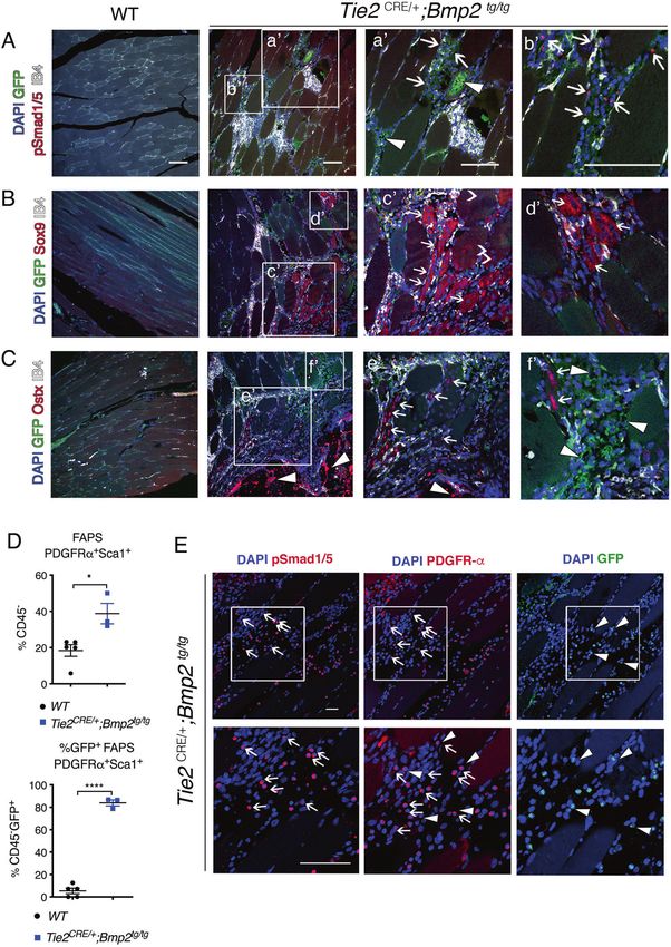
B. Prados et al.
                  2
                      following trauma, muscular fatigue, or other inflammatory insults,           HO lesions were observed in 95% (19/20) Tie2CRE/+;Bmp2tg/tg
                      which also trigger acquired forms of HO. The innate immune                  animals by nano PET-CT (Supplementary Table 1, Supplementary
                      system [17, 18] and local “niche” soft tissue microenvironment              Videos 1–5). Only one of 20 mice remained HO-free (Supplemen-
                      [19, 20] need to be further characterized to help clarify this issue.       tary Table 1). HO was manifested as fused cervical spine and
                      Moreover, Activin A seems to play a significant role in the initial          scapula extensions (47.2%) resulting in severe scoliosis (Fig. 2A;
                      steps of FOP during immune infiltration subsequent to injury.                Supplementary Videos 1,2), and as bony plaques adjacent to the
                      However, once ectopic bone is fused to the normal bone skeleton,            chest wall (55%; Supplementary Videos 3). Several animals (65%)
                      additional canonical BMP ligands may be required to sustain                 displayed bone outgrowths in hindlimbs (Supplementary Table 1;
                      ectopic bone development.                                                   Supplementary Movies 4,5). At 16 weeks, 70% of animals
                         The identification of bone osteoprogenitors has generated                 presented HO in multiple areas, while by 20 weeks, 100%
                      considerable interest [21, 22]. Skeletal muscle-resident cells including    presented severe HO lesions extending to most of the areas
                      myoblasts [23, 24], satellite cells [25] or fibroadipogenic progenitors      described (Supplementary Fig. 1C, Supplementary Table 1 and
                      (FAPs) have osteogenic differentiation ability [26, 27]. Hematopoietic      Supplementary Video 6). Of 7 heterozygous Tie2CRE/+;Bmp2tg/+
                      progenitors participate in bone formation at sites of tissue                animals analyzed by nano PET-CT at 16 and 32 weeks, only 2
                      inflammation, but are insufficient to initiate this process [28, 29].         developed mild lesions by 32 weeks (Supplementary Table 1 and
                      Endothelial cells in mice constitutively expressing ACVR1-R206H,            Supplementary Fig. 1D). Circulating Bmp2 levels for heterozygous
                      transform into mesenchymal cells with progenitor properties, that           transgenic mice at 24 weeks was 1.5-fold compared with WT
                      give rise to ectopic bone [30]. However, lineage tracing using a            (Supplementary Fig. 1E), while Bmp2 levels of homozygous
                      Tie2CRE driver line and local transplantation of Tie2CRE-derived            transgenic Tie2CRE/+;Bmp2tg/tg animals were 5–6 fold, suggesting
                      endothelial cells into skeletal muscle have excluded the endothelium        that HO is highly sensitive to Bmp2 levels.
                      as a source of ectopic bone formation [31]. Rather, non-endothelial            HO was associated with typical histopathological changes,
                      Tie2+ resident skeletal muscle stem cells including FAP osteopro-           including mononuclear cell infiltration (Fig. 2B, a and a’),
                      genitors appear to be the principal FOP cells-of-origin [31, 32].           fibroblast accumulation (Fig. 2B, b and b’), and chondro-
                         In view of ACVR1 receptor activation complexity, the uncer-              osteogenic overgrowths (Fig. 2B, c, c’, d and d’) and development
                      tainty of target osteo-progenitors, and modulating “niche” factors,         of a BM-like stroma (Fig. 2B, d and d’). Chondrogenic tissue
1234567890();,:

                      disease modeling is necessary to better understand both acquired            organization in the epiphysis and head of the femur and tibia
                      and genetic HO. Bmp2 plays fundamental roles in cardiac valves              was similar in Tie2CRE/+;Bmp2tg/tg and WT animals (Fig. 2C, a, a’, b).
                      formation and heart chamber patterning [33, 34], but its cardiac            However, additional cartilage was present in joints of Tie2CRE/+;
                      overexpression causes lethality [35, 36]. To study Bmp2 vascular            Bmp2tg/tg animals, including osteochondral patches surrounding
                      gain-of-function postnatally, we generated Tie2CRE/+;Bmp2tg/tg              the tibial head and neck (Fig. 2C, b and b’). Ectopic bone was
                      mice, which overexpress Bmp2 in hematopoietic/endothelial                   present adjacent to the normal bone in transgenic animals
                      lineages. These mice survive birth, develop pre-calcific valve               (Fig. 2C), and bone mass was increased in Tie2CRE/+;Bmp2tg/tg
                      disease and a systemic bone disorder in skeletal muscle and other           compared to WT animals (Fig. 2C, c, c’, d and d’). We measured
                      connective tissues, resulting in severe skeletal deformities whose          bone mass density (BMD) in tibia and femur bones in WT and
                      nature we have investigated.                                                Tie2CRE/+;Bmp2tg/tg animals [39]. BMD was increased about 20% in
                                                                                                  Tie2CRE/+;Bmp2tg/tg mice compared to WT (9226 vs. 7720
                                                                                                  Hounsfield Units; Supplementary Fig. 2A, B). Ossification of the
                      RESULTS                                                                     meniscus (Fig. 2C, d and d’) would likely result in limb
                      Endothelial Bmp2 overexpression results in valve dysfunction                immobilization in transgenic animals. Thus, Tie2CRE/+;Bmp2tg/tg
                      We previously generated a transgenic mouse line in which CAG-               mice show similar histopathological lesions found in human HO.
                      driven Bmp2 expression is activated upon Cre-mediated removal of
                      a β-Geo-stop cassette [35] (Supplementary Fig. 1A). We crossed the          Hematopoietic progenitors contribution to HO in Tie2CRE/+;
                      CAG-Bmp2 allele with Tie2CRE line, which is active in hematopoietic/        Bmp2tg/tg mice
                      endothelial lineages from E7.5 onwards [37]. Vascular GFP reporter          Tie2+ cells are potential osteogenic progenitors in HO [21, 30, 31]. To
                      expression was observed at E9.5, confirming Cre-mediated recom-              characterize the hematopoietic contribution to HO in Tie2CRE/+;
                      bination (Supplementary Fig. 1B).                                           Bmp2tg/tg mice, bone marrow (BM) was isolated from normal, and
                         Ectopic Bmp2 signaling leads to osteogenic differentiation of            ectopic bone of fore- and hindlimbs, scapulae, hips, and sternum of
                      valve interstitial cells [38]. To determine the effect of increased         Tie2CRE/+;Bmp2tg/tg mice, and processed separately. The combined
                      endothelial Bmp2 expression on valve function, we generated                 total BM (normal and ectopic bone) cell number was increased in
                      Tie2CRE/+;Bmp2tg/tg mice. At 16 weeks, circulating Bmp2 levels were         Tie2CRE/+;Bmp2tg/tg mice (Fig. 3A, left panel), although there were
                      almost six-fold higher than in WT animals (Fig. 1A). Tie2CRE/+;             important variations in the total hematopoietic cell number in ectopic
                      Bmp2tg/tg mice showed shortened pulmonary acceleration time                 bone (Fig. 3A, right panel) due to uneven HO in individual animals.
                      and acceleration to ejection time ratio by ultrasound (Fig. 1B),               Granulocyte-monocyte (GM) precursors colony forming-units
                      indicating pulmonary hypertension potentially leading to respira-           (CFU) were increased in Tie2CRE/+;Bmp2tg/tg mice (Fig. 3B), suggest-
                      tory insufficiency. Tie2CRE/+;Bmp2tg/tg mice displayed significantly          ing that enforced Bmp2 expression in Tie2+ cells drives hemato-
                      increased aortic valve mean, peak velocity and pressure gradient            poietic progenitor expansion. Interestingly, ectopic bone marrow
                      (Fig. 1C, D). Three of seven animals displayed chondrogenic and             formed CFU-GM, to a lesser extent than normal WT or tg BM
                      lipid droplet islands at the leaflet base (Fig. 1E), indicative of pre-      (Fig. 3B, right panel). CFU-GM isolated from transgenic spleen and
                      calcific disease. These results indicate that ectopic endothelial            peripheral blood (PB) were unchanged (Supplementary Fig. 1D).
                      and/or hematopoietic Bmp2 expression leads to aortic valve                     To support the evidence of increased hematopoietic progenitors
                      dysfunction compatible with a pre-calcific valve stage.                      in Tie2CRE/+;Bmp2tg/tg BM, we analyzed Lin-Sca1+c-Kit+ (LSK) cells.
                                                                                                  This population was increased in total BM (normal and ectopic
                      Hematopoietic/endothelial Bmp2 overexpression causes a HO                   bones) of Tie2CRE/+;Bmp2tg/tg mice (Fig. 3C, left panel), and present in
                      During these studies, we found that Tie2CRE/+;Bmp2tg/tg mice                ectopic BM (Fig. 3C, middle panel). Consistent with widespread Tie2
                      develop severe scoliosis and ankylosis with depressed locomotor             expression in hematopoietic progenitors [37], more than 93% of the
                      behavior and respiratory insufficiency. Extensive HO was diag-               LSK population in tg normal and ectopic BM was GFP+ (Fig. 3C, right
                      nosed by PET-CT at 16 weeks of age and confirmed at autopsy                  panel), reflecting an important contribution of hematopoietic cells to
                      (Fig. 2A; Supplementary Fig. 1C; Supplementary Table 1). Localized          ectopic Bmp2 production and BM formation.

                                                                                                                       Cell Death and Disease (2021)12:729
B. Prados et al.
                                                                                                                                                   3

Fig. 1 Constitutive endothelial Bmp2 overexpression results in aortic valve dysfunction and pre-calcification. A Circulating Bmp2 levels
detected by ELISA in WT and Tie2 CRE/+;Bmp2 tg/tg adult mice serum. B Quantification of pulmonary acceleration time (PAT, left panel), and PAT-
ejection time ratio (PAT/PET, right panel) measured by ultrasound on 16-week-old WT and Tie2 CRE/+;Bmp2 tg/tg mice. C Quantification of the
aortic valve velocity (AoV Mean and Peak Vel), and pressure gradient (AoV Mean and Peak Grad) measured by ultrasound. D Representative
images of acquired data of the aortic velocity peaks detected by ultrasound in WT (≈1000 mm/s) and tg animals (≈1200 mm/s). E Top panels:
Masson trichromic staining on consecutive sections of aortic valve from 18-week-old WT and Tie2 CRE/+;Bmp2 tg/tg mice. Chondrocyte island
(arrow) in aortic annulus at the base of the leaflet. Bottom panels: Localization of lipid droplets (arrowheads) identified by Oil Red O staining.
Unpaired t test, two tails, mean ± SD *P < 0.05; ***P < 0.001; ****P < 0.0001; ns, non-significant. Scale bar 200 µm.

Cell Death and Disease (2021)12:729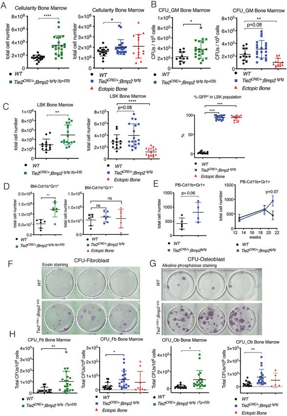
B. Prados et al.
4

       We queried whether inflammation was required for HO in                  Tie2CRE/+;Bmp2tg/tg BM. We monitored inflammation onset in 4 WT
    Tie2CRE/+;Bmp2tg/tg mice. FACS analysis revealed that the pro-            and 4 Tie2CRE/+;Bmp2tg/tg mice for 7 weeks (at 13, 19, and 21 weeks;
    inflammatory CD11b+Gr1+ cell population was increased in                   Fig. 3E, right panel), and by the time all the Tie2CRE/+;Bmp2tg/tg mice
    combined (normal and ectopic) Tie2CRE/+;Bmp2tg/tg BM (Fig. 3D,            had developed HO, their inflammatory cell numbers had increased
    left panel), whereas there was no difference in tg or WT BM               in PB (Supplementary Fig. 3E, right panel).
    (Fig. 3D, right panel). Circulating CD11b+Gr1+ cells were margin-            We tested fibroblast (Fb) and osteoblast (Ob) colony–forming
    ally increased (p = 0.06; Fig. 3E, left panel), although their numbers    potential in Tie2CRE/+;Bmp2tg/tg normal and ectopic BM. Fb (Fig. 3F)
    increased in PB (Supplementary Fig. 1E, left panel), indicating that      and Ob (Fig. 3G) CFUs readily formed from WT and transgenic
    a greater proportion of inflammatory cells are mobilized from              mice BM, from ectopic BM of transgenic animals (Supplementary

                                                                                                   Cell Death and Disease (2021)12:729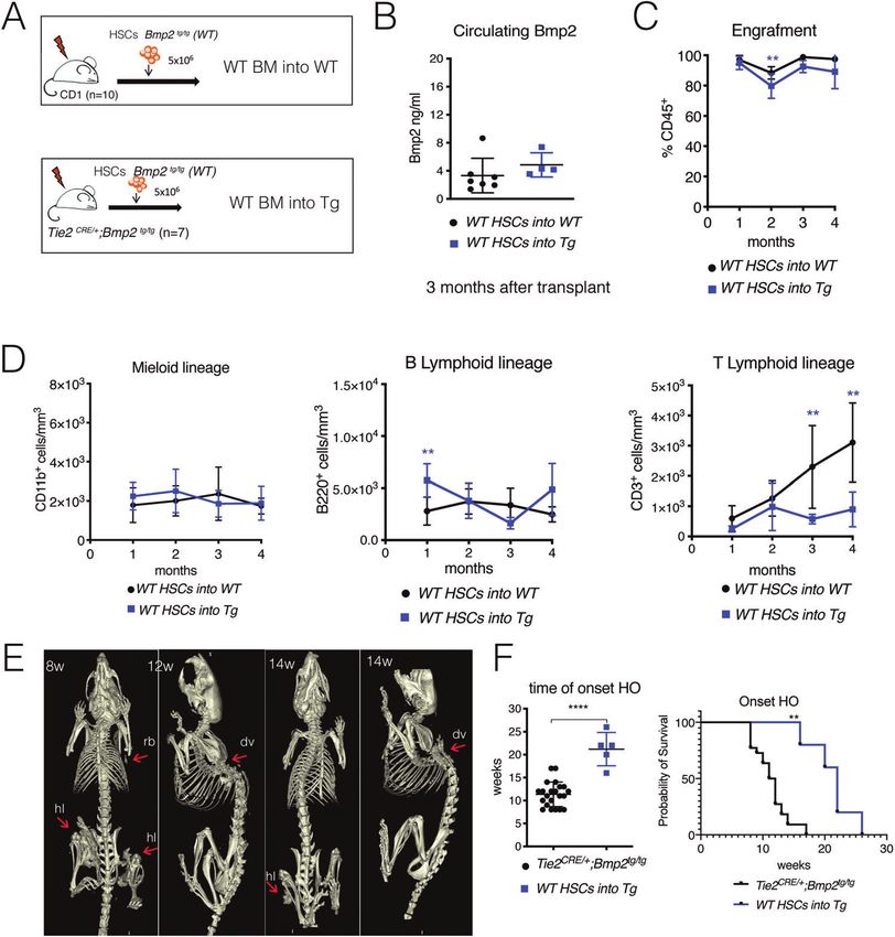
B. Prados et al.
                                                                                                                                                      5
Fig. 2 Constitutive Tie2-driven Bmp2 expression causes HO lesions in mice. A Nano PET-computed tomography (CT) images of 16-week-old
WT and Tie2 CRE/+;Bmp2 tg/tg mice showing ectopic bone lesions close to ribs, scapulae, and neck (red arrows, ribs, rb; nk, neck; dorsal vertebrae,
dv). B H&E staining on sections of skeletal muscle of the hindlimbs of Tie2 CRE/+;Bmp2 tg/tg mice showing histological features typical of HO
lesions. a. Evidence of inflammation in HO lesion. a’ Damaged skeletal muscle fibers with central nuclei (arrows), mononuclear infiltration
(black arrowheads), and fat cells (white arrowheads). b, b’ Area of massive fibroblast accumulation. c Ectopic bone in skeletal muscle (arrows)
next to tibia. c’ Chondro-osteogenic areas with chondrocytes (white arrowhead), and osteoblasts (black arrowheads). d, d’ Mature ectopic
bone with colonizing bone marrow cells (arrows), chondrocytes (white arrowhead), and osteoblasts (black arrowhead). C Top panels: Alcian
blue staining of sections of WT and Tie2 CRE/+;Bmp2 tg/tg knee joint. a, a’ In WT, chondrocyte tissue (in blue) is located in the epiphysis region,
tip of the bone (black arrowheads), and head of the fibula (arrow). b, b’ In Tie2 CRE/+;Bmp2 tg/tg, chondrocyte tissue is located in the epiphysis
region at the tip of the bone (black arrowheads), accumulated in connective tissue (arrow) of the meniscus and head of the fibula
(white arrow) and chondrogenic areas (white arrowhead) inside the skeletal muscle. Bottom panels: Alizarin red staining of WT and Tie2 CRE/+;
Bmp2 tg/tg knee joint. c, c’ In WT, osteogenic tissue (in red) is located in the epiphysis region and tip of the bone complementary to
chondrogenic areas (white arrowheads). d, d’ Intense staining in the head of Tie2 CRE/+;Bmp2 tg/tg joints (white arrowheads in (d’)). Extra
ossification inside the skeletal muscle and head of the fibula (arrows in (d)), and the meniscus (black arrowhead). Scale bar 200 µm.

Fig. 1F). Both CFU-Fbs and CFU-Obs were increased in combined               (Supplementary Fig. 4C, left panel) and excluded. Three surviving
tg BM (normal and ectopic) and tg BM (Fig. 3F, G), suggesting that          mice expressed GFP (Supplementary Fig. 3C, right panel). Circulating
forced Bmp2 expression in Tie2+ cells expands the fibro-                     Bmp2 levels in two of these mice were comparable to those found
osteoblastic BM progenitor population.                                      in Tie2CRE/+;Bmp2tg/tg mice (5.9-fold increased) (Supplementary Fig.
   BMP2 is known to promote erythropoiesis [40]. By FACS                    4B). Transgenic mice transplanted with WT HSCs had comparable
analysis, ProE, EryA, EryC, and erythrocytes (high-Ter119+) were            Bmp2 levels to controls (Fig. 4B), confirming the hematopoietic
all significantly increased in tg mice BM (Supplementary Fig. 1E),           origin of circulating Bmp2.
suggesting that forced BMP2 expression in Tie2+ cells promotes                 To determine lineage contributions to BM reconstitution, blood
erythropoiesis in vivo.                                                     samples were analyzed by FACS monthly over five months
                                                                            following transplantation. CD45+ cell engraftment in control and
Hematopoietic cells overexpressing BMP2 do not trigger HO                   WT groups transplanted with Tie2CRE/+;Bmp2tg/tg HSCs (n = 3) was
in wild-type mice                                                           comparable, reaching 90-98% (Supplementary Fig. 4C). Tie2CRE/+;
We queried if Tie2+ Bmp2-expressing BM hematopoietic cells can              Bmp2tg/tg animals transplanted with WT HSCs reached 80–95%
give rise to HO in wild-type (WT) mice. Eight-week-old lethally             engraftment (Fig. 4C). Myeloid CD11b+ cell engraftment was
irradiated C57/BL6 WT mice were transplanted with BM hemato-                similar between groups (Supplementary Fig. 4D and Fig. 4D, left
poietic stem cells (HSCs) from either WT (Bmp2tg/+) or Tie2CRE/+;           panel). Lymphoid B220+ cells increased during the first month
Bmp2tg/+ mice (Supplementary Fig. 3A). Hematopoietic cell                   compared to controls (Supplementary Fig. 4D, and Fig. 4D, middle
(CD45+) engraftment, was examined by FACS five months after                  panel). In contrast, CD3+ T lymphoid cells were not reconstituted
BM transplantation and thereafter bimonthly for over a year. WT             to WT levels when transplanted with Tie2CRE/+;Bmp2tg/tg HSCs
mice transplanted with WT HSCs engrafted between 95-100 % of                (Supplementary Fig. 4D, right panel), as observed previously
CD45+ cells. WT mice transplanted with Tie2CRE/+;Bmp2tg/+ HSC               (Supplementary Fig. 3B). Tie2CRE/+;Bmp2tg/tg animals transplanted
engrafted 80-90 % of CD45+ cells (Supplementary Fig. 3B). Bmp2              with WT HSCs did not recover normal CD3+ levels compared to
increased almost four-fold after 10 months in WT animals                    controls (Fig. 4D, right panel). Thus, B Lymphoid cells were initially
transplanted with Bmp2-overexpressing hematopoietic cells                   depleted in Tie2 CRE/+;Bmp2 tg/tg mice transplanted with WT HSC
(Supplementary Fig. 3C). Hematopoietic-derived Bmp2 accounted               but recovered, while T cells remained depleted for the entire
for ~50% Bmp2 levels found in Tie2CRE/+;Bmp2tg/tg mice (compare             duration of the experiment.
Fig. 1A and Supplementary Fig. 3B).                                            Tie2CRE/+;Bmp2tg/tg mice transplanted with WT hematopoietic
   We analyzed multi-lineage reconstitution in PB (CD11b+, B220+            cells eventually develop HO, likely because HO was already taking
and CD3+) by FACS every month for 12 months. Whilst myeloid                 place at the time of transplantation (8 weeks). Five animals
reconstitution was unaffected, B and T lymphoid cells were                  presented typical HO with variable severity, including complete
decreased at all time points (Supplementary Fig. 3D). BMP2/4 has            hindlimb immobilization by 16 weeks (n = 1), dorsal vertebrae at
been shown to antagonize T-cell lineage differentiation [41, 42].           20 weeks (n = 1), and hindlimb and dorsal vertebrae at 22 weeks
None of the transplanted animals developed HO by Nano-PET-CT                (n = 2) (Fig. 4E). One mouse presented a fat cyst dorsally but no
(data not shown), even 12 months after the BM transplant assay.             HO, and another developed HO in dorsal vertebrae at 26 weeks
Thus, Bmp2 secreted by hematopoietic Tie2CRE/+;Bmp2tg/+ cells in            (not shown). One mouse remained asymptomatic 37 weeks after
WT mice is not sufficient to drive HO. Alternatively, local                  transplant. Thus, 5 out of 7 Tie2CRE/+;Bmp2tg/tg mice reconstituted
expression of other Tie2-Cre-targeted cells is crucial to initiate          with WT hematopoietic cells developed HO, with delayed onset
flare-ups and HO.                                                            compared to non-transplanted animals. Disease onset in Tie2CRE/+;
                                                                            Bmp2tg/tg mice occurred at 16–28 weeks in transplanted mice,
Transplanting WT BM into Tie2CRE/+;Bmp2tg/tg mice delays HO                 versus 8–18 weeks in non-transplanted ones (Fig. 4F, left panel).
onset                                                                       Tie2CRE/+;Bmp2tg/tg mice transplanted with WT HSCs survive
We asked whether Bmp2 dosage affected HO formation in Tie2CRE/+;            10 weeks longer than non-transplanted transgenics (Fig. 4F, right
Bmp2tg/tg mice. Eight weeks-old lethally irradiated CD1 mice were           panel). Therefore, Bmp2 expression in non-hematopoietic cells is
used for transplant assays, because Tie2CRE/+;Bmp2tg/tg mice were in        essential for HO development, and concomitant Bmp2 expression
a CD1-enriched mixed background. WT mice were transplanted with             by hematopoietic cells accelerates this process.
5 million BM nucleated cells from WT (Bmp2tg/tg; n = 10) or Tie2CRE/+;
Bmp2tg/tg (n = 10) mice (Fig. 4A and Supplementary Fig. 4A). A              Chondro-osteogenic differentiation is associated with BMP
second group of 8-week-old lethally irradiated Tie2CRE/+;Bmp2tg/tg          signaling activation in FAP cells
mice (n = 7) was transplanted with WT (Bmp2tg/tg) HSCs in CD1-              Tie2 marks a subset of resident skeletal muscle cells [31], which
enriched mixed background (Fig. 4A). Three transplanted WT mice             potentially contribute to HO. Tie2CRE/+;Bmp2tg/tg cartilage and
died after 19 days, and could not be analyzed further. In four, GFP         bone lesions showed GFP+ co-staining with IB4+ (Fig. 5A), co-
was either not expressed or expressed in 15–20% of total HSCs               localizing with Tie2+ fibroblastic/adipocytic skeletal-muscle cells

Cell Death and Disease (2021)12:729
B. Prados et al.
6
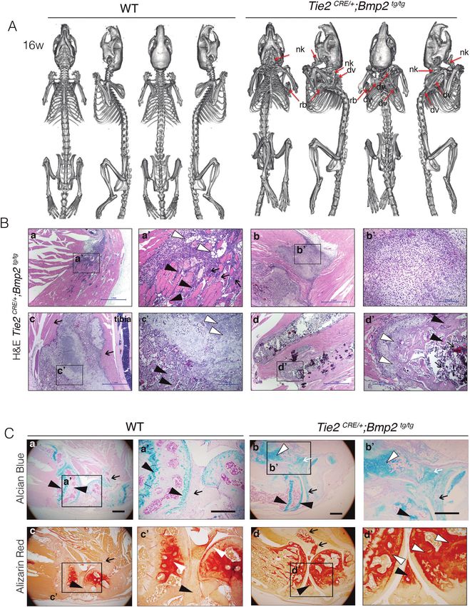
(Supplementary Fig. 5A, white arrowheads). GFP+ infiltrating The chondrogenic marker Sox9 was expressed in ectopic bone
inflammatory cells expressing IB4 were detected in fibroprolifera- chondrocyte nuclei (Supplementary Fig. 5C, left panel). Scattered
tive areas (Supplementary Fig. 5A), and have been previously Sox9+ nuclei were also found in GFP-expressing fibroblasts
described [43]. GFP-expressing fat cells were identified based on (Supplementary Fig. 5C, d’) and damaged skeletal muscle fiber
their peripheral nuclei (Supplementary Fig. 5B, b’). Phospho- cytoplasm identified by central nuclei (Fig. 5B, c’ and d’, and
Smad1/5-stained nuclei were detected in scattered cells in Supplementary Fig. 5C, c’). This expression pattern was also
fibroproliferative areas (Fig. 5A, a’ and b’ and Supplementary observed in areas next to HO, although involving very few
Fig. 5B, a’), as well as among fat cells (Supplementary Fig. 5B, b’). nuclei (Supplementary Fig. 5C, c’, open arrowheads). The
Bmp2 activation may be non-cell autonomous because p-Smad- osteogenic marker Osterix, was also detected amongst Sox9-
positive cells did not express GFP (Fig. 5A, a’ and b’ and expressing cells in consecutive sections (Fig. 5C, e’, and f’) and in
Supplementary Fig. 5B, a’). ectopic bone osteoblasts (Supplementary Fig. 5D, arrowheads).
Cell Death and Disease (2021)12:729B. Prados et al.
7
Fig. 3 Hematopoietic stem cells contribute to HO in Tie2 CRE/+;Bmp2 tg/tg mice. A Quantification of bone marrow (BM) cellularity. Left, total
BM (combined BM of transgenic (tg) and ectopic bone (EB)) cellularity is increased in Tie2 CRE/+;Bmp2 tg/tg mice. Right, cellularity of WT BM, tg
BM, and ectopic BM. Ectopic BM cellularity is highly variable. B Quantification of granulocyte, monocyte colony forming units (CFU_GM). Left,
total BM (tg + EB) CFU-GM was increased in Tie2 CRE/+;Bmp2 tg/tg mice. Right, ectopic BM can give rise to CFU-GMs. C Quantification of the
FACS-isolated Lin-Sca1+cKit+ (LSK) population. Left, the LSK population is increased in Tie2 CRE/+;Bmp2 tg/tg BM. Middle, LSK progenitors in
ectopic BM are detectable and below normal. Right, the bulk (93.6% in tg and 93.2% in ectopic BM) of the LSK population in Tie2Cre/+;Bmp2tg/tg
BM is GFP+. D Quantification of the FACS-isolated pro-inflammatory Cd11b+Gr1+ cells from BM. Left, the Cd11b+Gr1+ population is increased
in total Tie2 CRE/+;Bmp2 tg/tg BM (tg + EB). Right, varying presence of Cd11b+Gr1+ cells in ectopic BM. E. FACS-isolated pro-inflammatory
Cd11b+Gr1+ cells from PB. Left, Cd11b+Gr1+ cells are marginally increased in Tie2 CRE/+;Bmp2 tg/tg PB with HO. Right, Cd11b+Gr1+ population
at different time points, 13, 19, and 21 weeks. At 21 weeks, when mice present HO, pro-inflammatory cells increase in Tie2 CRE/+;Bmp2 tg/tg.
F Representative eosin staining of CFU-fibroblast (Fb). G Representative alkaline phosphatase staining of CFU-osteoblast (Ob). H Quantification
showing increased CFU-Fb and CFU-Ob in Tie2 CRE/+;Bmp2 tg/tg BM. A, B, C, D, H, for WT and Tie2 CRE/+;Bmp2 tg/tg tg or tg + EB BM groups,
unpaired t test, two tails, mean ± SD *P < 0.05; **P < 0.01; ***P < 0.001; ****P < 0.0001, ns non-significant. E Right panel, two-way ANOVA and
Sidak’s correction.
Fig. 4 Transplanting WT BM into transgenic Tie2Cre/+;Bmp2tg/tg mice delays HO onset. A Schematic representation of transplant assays. WT
(CD1) and Tie2Cre/+;Bmp2tg/tg mice were transplanted with WT BM cells. B Circulating Bmp2 levels are maintained in each group 3 months after
transplant. C Hematopoietic cell engraftment. FACS-quantification of CD45+ cells from PB. Tie2CRE/+;Bmp2tg/tg and WT mice show similar HSC
engraftment. D FACS-quantification of myeloid (CD11b+), B Lymphoid (B220+) and T Lymphoid (CD3+) cells in the two groups of transplanted
animals. E Nano-PET-CT imaging of transgenic Tie2CRE/+;Bmp2 tg/tg mice transplanted with WT HSC. These animals develop HO (red arrows) in
ribs, hind limbs, and dorsal vertebrae (ribs, rb; hind limbs, hl; and dorsal vertebrae, dv) at 8, 12, and 14 weeks after transplant, suggesting that
ectopic bones formation started before transplant. F Representation of the variability of HO onset in non transplanted Tie2CRE/+;Bmp2tg/tg mice
and Tie2CRE/+;Bmp2tg/tg transplanted with WT HSCs showing significant delay in the onset of HO (8–18 weeks versus 16-28w). Kaplan-Meier
curve showing that Tie2CRE/+;Bmp2tg/tg mice transplanted with WT HSCs survive 10 weeks longer on average non-transplanted transgenic mice.
C, D Two-way ANOVA and Sidak’s correction **P < 0.01; F unpaired t test, two tails, mean ± SD. t test ****P < 0.0001. E Survival curve and
survival curve analysis, **P < 0.01.
Cell Death and Disease (2021)12:729B. Prados et al.
8
Fig. 5 Tie2Cre/+;Bmp2tg/tg resident skeletal muscle cells are characterized by pSmad1/5 and chondro-osteogenic marker expression, and
increased fibro-adipogenic progenitors. Immunodetection of indicated osteo-chondrogenic marker proteins (red), with GFP (green), IB4
(white) and DAPI (blue), on consecutive WT or Tie2 CRE/+;Bmp2 tg/tg hindlimb skeletal muscle sections, A Nuclear immunostaining of GFP+
pSmad 1/5 (arrows) interspersed in muscle interstitium (a’ and b’). White arrowheads indicate GFP+ adipocytes. B Cytoplasmic Sox9
immunostaining in cells (arrows in c’ and d’) surrounded by damaged skeletal muscle fibers identified as central nuclei (open arrowheads in
c’). C. Nuclear immunostaining of Osterix (Osx) in cells surrounded by fibers (arrows in e’ and f’). Arrowheads indicate strong Osx
immunostaining in ectopic bone emerging areas. Arrowheads in f’ indicate GFP+ adipocytes. D Top, FACS quantification of the
CD45-PDGFRα+Sca1+ fibro-adipogenic (FAP) population in Tie2 CRE/+;Bmp2 tg/tg skeletal muscle. Bottom, FACS quantification showing that 84%
of the CD45-GFP+ cells are PDGFRα+Sca1+ cells. E Immunostainings of pSmad1/5/8 and PDGFRα (red), GFP (green), and DAPI (blue), on
consecutive Tie2 CRE/+;Bmp2 tg/tg hindlimb skeletal muscle sections showing expression in connective tissue surrounding muscle fibers. Bottom
panels are magnifications of areas indicated in top panels. Arrows indicate co-localization of pSmad1/5 and PDGFRα cells. Arrowheads
indicate co-localization of PDGFRα and GFP+ cells. Cells expressing the three markers are indicated by arrows and arrowheads (middle bottom
panel). Scale bars 200 µm. Unpaired t test, two tails, mean ± SD *P < 0.05; ****P < 0.0001.
Cell Death and Disease (2021)12:729B. Prados et al.
9
Moreover, Osterix staining was observed in larger central nuclei “niche” in damaged skeletal muscle of Tie2CRE/+;Bmp2tg/tg mice is
of damaged fibers next to ectopic bone (Supplementary Fig. 5D, suggested by the presence of central nuclei, co-immunostaining
e’, and f’). of isolectin B4 (also a leukocyte marker), pSmad1/5, Sox9, and
The expression of chondro-osteogenic markers in damaged osterix, in chondro-osteogenic areas of differentiation. Tie2+ FAP
skeletal muscle fibers adjacent to ectopic bone, suggests an active osteoprogenitors are identifiable by PDGFRα and Sca1 cell surface
repair process. The number of satellite cells labeled by expression [32], bipotent fibro/adipogenic potential [26, 27], and
CD45+Sca1-CD34+α7int+ was not significantly different between contribution to ectopic cartilage and bone [31, 32]. HO lesions in
Tie2CRE/+;Bmp2tg/tg and WT hindlimb skeletal muscle (Fig. 5D, left Tie2CRE/+;Bmp2tg/tg mice might be attributed to local Bmp2 release
panel). There were no GFP+ satellite cells among Tie2CRE/+;Bmp2tg/tg through autocrine/paracrine mechanisms. Conversely, resident
skeletal muscle cells (Fig. 5D, right panel), suggesting that satellite skeletal muscle satellite cells (CD45−CD34+α7int+) that give rise to
cells are not overexpressing Bmp2 and probably not directly differentiated myocytes [51] were unchanged, suggesting that
implicated in HO in Tie2CRE/+;Bmp2tg/tg mice. muscle regenerative potential is not altered. Further studies are
Resident FAPs are CD45-Tie2+PDGFRα+Sca1+ have been required to characterize other progenitor populations residing in
proposed as HO cells-of-origin [26]. FAPs were readily increased skeletal muscle, ligaments and tendons [31, 52] in the context of
in Tie2CRE/+;Bmp2tg/tg skeletal muscle (Fig. 5D, top panel), Bmp2-driven HO.
accounting for 84% of the resident Tie2+ cells (Fig. 5D, bottom Previous BMP overexpression studies using a variety of
panel). In fact, among fibroproliferative areas, the majority of promoters failed to cause HO (reviewed in [53]), because the
pSmad1/5-, PDGFRα- (Fig. 5A, a’, b’; 5E, left panels; Supplementary relevant progenitor cell type had not been targeted. One
Fig. 5B, a’, b’ and 5E, middle panels), and Sox9-expressing cells exception is the transgenic mouse overexpressing BMP4 under
were GFP+ (Supplementary Fig. 5C, c’, d’ and 5E right panels). In control of neuron-specific enolase (Nse) promoter [53, 54]. Our
contrast, Sox9-or Ostx- expressing damaged fibers did not express Tie2CRE/+;Bmp2tg/tg mice resembles this model, matching a
GFP (Fig. 5B c’, d’; 5C e’, f’ and Supplementary Fig. 5C c’; 5D, e’, f’), stereotyped spreading pattern of HO formation. Neither the
suggesting that most FAPs are Tie2+ cells, and/or that Tie2 NSE-BMP4 model, nor our Tie2CRE/+;Bmp2tg/tg model recapitulate
expression is silenced during FAP differentiation. any of the congenital phenotypes associated with FOP. Moreover,
direct versus indirect Bmp effects on target stem/progenitor cell
populations cannot be assessed using these models. Nevertheless,
DISCUSSION Tie2CRE/+;Bmp2tg/+ mice are viable, and develop HO within weeks.
Tie2CRE/+;Bmp2tg/tg mice develop HO, apparently spontaneously This HO model does not require surgical procedures involving the
within 4 months. Normal skeletogenesis is not perturbed, with no implantation of a BMP-loaded matrix and Bmp2 dosage can be
obvious HO in juvenile mice, while adult mice present a variety of modulated via copy number gene expression. Tie2CRE/+;Bmp2tg/tg
skeletal deformities including scoliosis, and spinal defects mice are maintained on a heterozygote background, so that a
resembling lesions found in HO patients. Tie2CRE/+;Bmp2tg/tg mice single cross allows for the generation of experimental animals.
may allow for further studies of therapies designed to mitigate the
effects of HO.
MATERIALS AND METHODS
Bmp2 is a key pathway activated and causally linked to calcific
Mouse strains and genotyping
aortic valve disease in animal models [38, 44–46]. Tie2CRE/+;Bmp2tg/tg The following mouse strains were used: male and female mixed
mice present pulmonary valve hypertension and pre-calcific aortic background C56BL/6-CD1 R26CAGBmp2tg [35] and Tie2CRE [37]. For
valve dysfunction by 16–18 weeks, characterized by fibrosis, lipid simplicity, R26CAGBmp2tg/+ and R26CAGBmp2tg/tg are abbreviated in the
deposition and chondrogenesis. Bmp2 overexpression promotes text and figures as Bmp2tg/+ and Bmp2tg/tg, respectively. Details of
ectopic EMT in developing Nkx2.5CRE/+;Bmp2tg/+ embryos [35], genotyping will be provided upon request. Recipient transplanted animals
suggesting that EMT might contribute to pro-calcific disease in were WT C56BL/6, CD1 or Tie2Cre/+;Bmp2tg/+ animals. Animal studies were
adults. However, there is lack of evidence for EMT contributing to approved by the CNIC Animal Experimentation Ethics Committee and by
adult valve disease pathology [47], and further studies are the Community of Madrid (Ref. PROEX 83.8/20). Animal procedures
required. Tie2CRE/+;Bmp2tg/tg mice develop systemic HO between conformed to EU Directive 2010/63EU and Recommendation 2007/526/
EC regarding the protection of animals used for experimental and scientific
8–18 weeks precluding a meaningful study of valve changes, purposes, enforced in Spanish law under Real Decreto 53/2013.
but the effect of ageing over a longer-period might be assessed in
Tie2CRE/+;Bmp2tg/+ heterozygotes, which are healthy up to
32 weeks. ELISA
Our transplant studies indicate that while Bmp2 transgenic Blood samples were taken by submandibular vein puncture from different
groups: control WT and Tie2Cre/+;Bmp2tg/tg mice at 16 weeks of age (n = 10/
progenitors contribute BM stem cell components, the HO each group); control WT and heterozygous Tie2Cre/+;Bmp2tg/+ at 24 weeks
chondro-osteogenic stem/progenitor cell is not BM-derived, of age (n = 6/each group). 1 year after transplant, WT animals transplanted
consistent with previous studies [28, 29]. Otherwise, hematopoie- with WT BMCs, n = 6, and WT animals transplanted with Tie2Cre/+;Bmp2tg/+
tic/endothelial Bmp2 overexpression profoundly affects hemato- BMCs, n = 8; 3 months after transplant WT animals transplanted with WT
poiesis. LSK stem cells, myeloid CFU-GM and Cd11b/Gr11 cells, BMCs (n = 7) and Tie2Cre/+;Bmp2tg/+ animals transplanted with WT BMCs
and fibroblast and osteoblast progenitors are all expanded in (n = 4) were analyzed. Serum was obtained by centrifugation at 4000 rpms
transgenic mice. Tie2CRE/+;Bmp2tg/tg mice display increased Cd11b/ for 10 min at RT. Circulating Bmp2 was measured by using the human
Gr11 cells in BM and PB, and increased erythroid lineage BMP2 ELISA construction kit (Antigenix America Inc. RHF913CKC) under the
manufacturer´s instructions.
differentiation, consistent with in vitro findings [40]. Overall
increased BM cellularity is consistent with HSC niche enlargement,
enhancement of HSC self-renewal and pool size. Within the BM Ultrasound
niche, non-canonical BMP signaling regulates intrinsic HSC Mice were anaesthetized by inhalation of isoflurane and oxygen (1.25%
maintenance in vivo [48–50], and is implicated in determining and 98.75% respectively) and examined by a 30 MHz transthoracic
the HSC fate, by promoting a pro-lymphoid transcriptional echocardiography probe. Images were obtained with VEVO 2100
(VisualSonics, Toronto, Canada) from Tie2Cre/+;Bmp2tg/tg (n = 10) and WT
program and sustaining lymphoid-biased HSC commitment (n = 9) littermates. Short axis and long axis, B Mode, and 2D M-Mode views
[48, 49]. were obtained from the M mode by an expert in ultrasound in a blind
The CD45-PDGFRα+Sca1+ population expansion in skeletal fashion as described previously [55]. From these images, left ventricle (LV)
muscle identifies FAPs as potential cells of origin in Tie2CRE/+; function was estimated by fractional shortening (FS) and ejection fraction
Bmp2tg/tg mice. Evidence for a soft tissue chondro-osteoprogenitor (EF). For FS measurements a long or short-axis view of the heart was
Cell Death and Disease (2021)12:729B. Prados et al.
10
selected to obtain an M mode registration in a line perpendicular to the LV and bones were dissected from posterior and anterior limbs, hips, and
septum and posterior wall at the level of the mitral chordae tendinea. sternum. Extra bone formations from transgenic animals were dissected
Pulmonary acceleration time (PAT) and ejection time (PET) were measured and processed separately. BM cells were obtained by crushing bones with
in the parasternal short-axis view by pulsed-wave Doppler of pulmonary a mortar in PBS. The solution containing BM cells was separated, and the
artery flow [56]. B-mode and color-Doppler guided pulsed-wave Doppler remaining bone was treated with Collagenase I for 45 min at 37 °C in a
was used to record the maximal transvalvular jet velocity. Specifically, to shaking bath to obtain stromal cells. All samples were 70 μm–filtered. Red
avoid Doppler misalignment, coaxial interrogation of the aortic flow was blood cells were removed from BM and spleen samples using lysis buffer
ensured by the operator, and all the measurements were obtained using (0.15 M NH4Cl for 10 min at 4 °C) and cell number was determined.
an angle of interrogationB. Prados et al.
11
560501), Sca1-PE-Cy7 (1:200, BD Biosciences 558162), α7-Int-PE (1:100, 20. Huang Y, Wang X, Lin H. The hypoxic microenvironment: a driving force for
AbLab AB10STMW215), CD34-Alexa647 (1:40, BD Pharmigen 560230), heterotopic ossification progression. Cell Commun Signal. 2020;18:20.
CD140a (PDGFRA) Monoclonal Antibody (APA5), Biotin (1ː200, Thermo- 21. Lounev VY, Ramachandran R, Wosczyna MN, Yamamoto M, Maidment AD, Shore
Fisher 13-1401-82), streptavidin-APC-Cy7 (1:100, BD Biosciences 554063) EM, et al. Identification of progenitor cells that contribute to heterotopic skele-
and 7AAD (1:100, BD Pharmigen 559925) for viability. Satellite cells were togenesis. J Bone Jt Surg Am. 2009;91:652–63.
identified as CD45+Sca1-CD34+α7int+ and fibro-adipogenic cells as 22. Pulik L, Mierzejewski B, Ciemerych MA, Brzoska E, Legosz P. The survey of cells
CD45-PDGFRα+Sca1+. responsible for heterotopic ossification development in skeletal muscles-human
and mouse models. Cells 2020;9:1324.
23. Katagiri T, Yamaguchi A, Komaki M, Abe E, Takahashi N, Ikeda T, et al. Bone
Statistics morphogenetic protein-2 converts the differentiation pathway of C2C12 myo-
Due to the high variability in HO onset and severity, the analysis was made
blasts into the osteoblast lineage. J Cell Biol. 1994;127:1755–66.
in groups n > 10 in some cases. Statistical assessment is indicated in the
24. Akiyama S, Katagiri T, Namiki M, Yamaji N, Yamamoto N, Miyama K, et al. Con-
figure legends. For each experiment comparing two groups, a mean ± SD is stitutively active BMP type I receptors transduce BMP-2 signals without the
represented and a two-tailed t test was performed. For experiments ligand in C2C12 myoblasts. Exp Cell Res. 1997;235:362–9.
comparing two groups at different time points, a mean ± SD is represented 25. Wada MR, Inagawa-Ogashiwa M, Shimizu S, Yasumoto S, Hashimoto N. Genera-
at each timepoint and a two-way ANOVA followed by Sidak’s correction tion of different fates from multipotent muscle stem cells. Development.
was performed. *P < 0.05; **P < 0.01; ***P < 0.001; **** P < 0.0001.
2002;129:2987–95.
26. Joe AW, Yi L, Natarajan A, Le Grand F, So L, Wang J, et al. Muscle injury activates
resident fibro/adipogenic progenitors that facilitate myogenesis. Nat Cell Biol.
DATA AVAILABILITY 2010;12:153–63.
Data sharing is not applicable to this article as no datasets were generated or 27. Uezumi A, Fukada S, Yamamoto N, Takeda S, Tsuchida K. Mesenchymal pro-
analyzed during the current study. genitors distinct from satellite cells contribute to ectopic fat cell formation in
skeletal muscle. Nat Cell Biol. 2010;12:143–52.
28. Kaplan FS, Glaser DL, Shore EM, Pignolo RJ, Xu M, Zhang Y, et al. Hematopoietic
stem-cell contribution to ectopic skeletogenesis. J Bone Jt Surg Am.
REFERENCES 2007;89:347–57.
1. Wang RN, Green J, Wang Z, Deng Y, Qiao M, Peabody M, et al. Bone Morpho- 29. Suda RK, Billings PC, Egan KP, Kim JH, McCarrick-Walmsley R, Glaser DL, et al.
genetic Protein (BMP) signaling in development and human diseases. Genes Dis. Circulating osteogenic precursor cells in heterotopic bone formation. Stem Cells.
2014;1:87–105. 2009;27:2209–19.
2. Brazil DP, Church RH, Surae S, Godson C, Martin F. BMP signalling: agony and 30. Medici D, Shore EM, Lounev VY, Kaplan FS, Kalluri R, Olsen BR. Conversion of
antagony in the family. Trends Cell Biol. 2015;25:249–64. vascular endothelial cells into multipotent stem-like cells. Nat Med. 2010;16:1400–6.
3. Shu B, Zhang M, Xie R, Wang M, Jin H, Hou W, et al. BMP2, but not BMP4, is crucial 31. Wosczyna MN, Biswas AA, Cogswell CA, Goldhamer DJ. Multipotent progenitors
for chondrocyte proliferation and maturation during endochondral bone devel- resident in the skeletal muscle interstitium exhibit robust BMP-dependent
opment. J Cell Sci. 2011;124:3428–40. osteogenic activity and mediate heterotopic ossification. J Bone Min Res.
4. Halloran D, Durbano HW, Nohe A. Bone morphogenetic protein-2 in development 2012;27:1004–17.
and bone homeostasis. J Dev Biol. 2020;8:19. https://doi.org/10.3390/jdb8030019. 32. Lees-Shepard JB, Yamamoto M, Biswas AA, Stoessel SJ, Nicholas SE, Cogswell CA,
5. Edwards DS, Kuhn KM, Potter BK, Forsberg JA. Heterotopic ossification: a review of et al. Activin-dependent signaling in fibro/adipogenic progenitors causes fibro-
current understanding, treatment, and future. J Orthop Trauma. 2016;30:S27–30. dysplasia ossificans progressiva. Nat Commun. 2018;9:471.
6. Meyers C, Lisiecki J, Miller S, Levin A, Fayad L, Ding C, et al. Heterotopic ossifi- 33. Rivera-Feliciano J, Tabin CJ. Bmp2 instructs cardiac progenitors to form the heart-
cation: a comprehensive review. JBMR. 2019;3:e10172. valve-inducing field. Dev Biol. 2006;295:580–8.
7. Kraft CT, Agarwal S, Ranganathan K, Wong VW, Loder S, Li J, et al. Trauma- 34. Luna-Zurita L, Prados B, Grego-Bessa J, Luxan G, del Monte G, Benguria A, et al.
induced heterotopic bone formation and the role of the immune system: a Integration of a Notch-dependent mesenchymal gene program and Bmp2-driven
review. J Trauma Acute Care Surg. 2016;80:156–65. cell invasiveness regulates murine cardiac valve formation. J Clin Invest.
8. Kaplan FS, Le Merrer M, Glaser DL, Pignolo RJ, Goldsby RE, Kitterman JA, et al. 2010;120:3493–507.
Fibrodysplasia ossificans progressiva. Best Pr Res Clin Rheumatol. 2008;22:191–205. 35. Prados B, Gomez-Apinaniz P, Papoutsi T, Luxan G, Zaffran S, Perez-Pomares JM,
9. Pignolo RJ, Shore EM, Kaplan FS. Fibrodysplasia ossificans progressiva: clinical et al. Myocardial Bmp2 gain causes ectopic EMT and promotes cardiomyocyte
and genetic aspects. Orphanet J Rare Dis. 2011;6:80. proliferation and immaturity. Cell Death Dis. 2018;9:399.
10. Kaliya-Perumal AK, Carney TJ, Ingham PW. Fibrodysplasia ossificans progressiva: 36. Papoutsi T, Luna-Zurita L, Prados B, Zaffran S.de la Pompa JL. Bmp2 and Notch
current concepts from bench to bedside. Dis Model Mech. 2020;13:dmm046441. cooperate to pattern the embryonic endocardium. Development. 2018;145:
https://doi.org/10.1242/dmm.046441. dev163378.
11. Shore EM, Xu M, Feldman GJ, Fenstermacher DA, Cho TJ, Choi IH, et al. A 37. Kisanuki YY, Hammer RE, Miyazaki J, Williams SC, Richardson JA, Yanagisawa M.
recurrent mutation in the BMP type I receptor ACVR1 causes inherited and Tie2-Cre transgenic mice: a new model for endothelial cell-lineage analysis
sporadic fibrodysplasia ossificans progressiva. Nat Genet. 2006;38:525–7. in vivo. Dev Biol. 2001;230:230–42.
12. Hatsell SJ, Idone V, Wolken DM, Huang L, Kim HJ, Wang L, et al. ACVR1R206H 38. Nigam V, Srivastava D. Notch1 represses osteogenic pathways in aortic valve
receptor mutation causes fibrodysplasia ossificans progressiva by imparting cells. J Mol Cell Cardiol. 2009;47:828–34.
responsiveness to activin A. Sci Transl Med. 2015;7:303ra137. 39. Pascau J, Vaquero, JJ, Abella M, Cacho R, Desco M, Lage E. Multimodality work-
13. Hino K, Ikeya M, Horigome K, Matsumoto Y, Ebise H, Nishio M, et al. Neofunction station for small animal image visualization and analysis. Mol Imaging Biol.
of ACVR1 in fibrodysplasia ossificans progressiva. Proc Natl Acad Sci USA. 2006;8:97–8.
2015;112:15438–43. 40. Maguer-Satta V, Bartholin L, Jeanpierre S, Ffrench M, Martel S, Magaud JP, et al.
14. Kaplan FS, Pignolo RJ, Shore EM. Granting immunity to FOP and catching het- Regulation of human erythropoiesis by activin A, BMP2, and BMP4, members of
erotopic ossification in the Act. Semin Cell Dev Biol. 2016;49:30–6. the TGFbeta family. Exp Cell Res. 2003;282:110–20.
15. Hildebrand L, Stange K, Deichsel A, Gossen M, Seemann P. The Fibrodysplasia 41. Cejalvo T, Sacedon R, Hernandez-Lopez C, Diez B, Gutierrez-Frias C, Valencia J,
Ossificans Progressiva (FOP) mutation p.R206H in ACVR1 confers an altered et al. Bone morphogenetic protein-2/4 signalling pathway components are
ligand response. Cell Signal. 2017;29:23–30. expressed in the human thymus and inhibit early T-cell development. Immu-
16. Allen RS, Tajer B, Shore EM, Mullins MC. Fibrodysplasia ossificans progressiva nology. 2007;121:94–104.
mutant ACVR1 signals by multiple modalities in the developing zebrafish. Elife. 42. Hager-Theodorides AL, Ross SE, Sahni H, Mishina Y, Furmanski AL, Crompton T.
2020;9:e53761. Direct BMP2/4 signaling through BMP receptor IA regulates fetal thymocyte
17. Alessi Wolken DM, Idone V, Hatsell SJ, Yu PB, Economides AN. The obligatory role progenitor homeostasis and differentiation to CD4+CD8+ double-positive cell.
of Activin A in the formation of heterotopic bone in Fibrodysplasia Ossificans Cell Cycle. 2014;13:324–33.
Progressiva. Bone. 2018;109:210–7. 43. Lyons SA, Pastor A, Ohlemeyer C, Kann O, Wiegand F, Prass K, et al. Distinct
18. Kan C, Yang J, Na D, Xu Y, Yang B, Zhao H, et al. Inhibition of immune checkpoints physiologic properties of microglia and blood-borne cells in rat brain slices after
prevents injury-induced heterotopic ossification. Bone Res. 2019;7:33. permanent middle cerebral artery occlusion. J Cereb Blood Flow Metab.
19. Haupt J, Stanley A, McLeod CM, Cosgrove BD, Culbert AL, Wang L, et al. ACVR1 2000;20:1537–49.
(R206H) FOP mutation alters mechanosensing and tissue stiffness during het- 44. Mohler ER 3rd, Gannon F, Reynolds C, Zimmerman R, Keane MG, Kaplan FS. Bone
erotopic ossification. Mol Biol Cell. 2019;30:17–29. formation and inflammation in cardiac valves. Circulation. 2001;103:1522–8.
Cell Death and Disease (2021)12:729B. Prados et al.
12
45. Nus M, Macgrogan D, Martinez-Poveda B, Benito Y, Casanova JC, Fernandez-Aviles FUNDING
F, et al. Diet-induced aortic valve disease in mice haploinsufficient for the notch Grants PID2019-104776RB-I00, SAF2016-78370-R, CB16/11/00399 (CIBER CV), and
pathway effector RBPJK/CSL. Arterioscler Thromb Vasc Biol. 2011;31:1580–8. RD16/0011/0021 (TERCEL) from the Spanish Ministry of Science, Innovation, and
46. Gomez-Stallons MV, Wirrig-Schwendeman EE, Hassel KR, Conway SJ, Yutzey KE. Universities (MCIU) to JLDLP. The cost of this publication was supported in part with
Bone morphogenetic protein signaling is required for aortic valve calcification. FEDER funds. The CNIC is supported by the MCIU and the Pro-CNIC Foundation, and
Arterioscler Thromb Vasc Biol. 2016;36:1398–405. is a Severo Ochoa Center of Excellence (SEV-2015-0505). The authors declare no
47. Kim AJ, Alfieri CM, Yutzey KE. Endothelial cell lineage analysis does not provide competing interests.
evidence for EMT in adult valve homeostasis and disease. Anat Rec. 2019;302:125–35.
48. Crisan M, Kartalaei PS, Vink CS, Yamada-Inagawa T, Bollerot K, van IW, et al. BMP
signalling differentially regulates distinct haematopoietic stem cell types. Nat ETHICS
Commun. 2015;6:8040. Animal studies were approved by the CNIC Animal Experimentation Ethics
49. Naka K, Hirao A. Regulation of hematopoiesis and hematological disease by TGF- Committee and by the Community of Madrid (Ref. PROEX 83.8/20). Animal
beta family signaling molecules. Cold Spring Harb Perspect Biol. 2017;9:a027987. procedures conformed to EU Directive 2010/63EU and Recommendation 2007/526/
https://doi.org/10.1101/cshperspect.a027987. EC regarding the protection of animals used for experimental and scientific purposes,
50. Warsi S, Blank U, Dahl M, Grahn THM, Schmiderer L, Andradottir S, et al. BMP enforced in Spanish law under Real Decreto 53/2013.
signaling is required for postnatal murine hematopoietic stem cell self-renewal.
Haematologica. 2020. https://doi.org/10.3324/haematol.2019.236125.
51. Biferali B, Proietti D, Mozzetta C, Madaro L. Fibro-adipogenic progenitors cross- COMPETING INTERESTS
talk in skeletal muscle: the social network. Front Physiol. 2019;10:1074. The authors declare no competing interests.
52. Dey D, Bagarova J, Hatsell SJ, Armstrong KA, Huang L, Ermann J, et al. Two tissue-
resident progenitor lineages drive distinct phenotypes of heterotopic ossification.
Sci Transl Med. 2016;8:366ra163. ADDITIONAL INFORMATION
53. Kan L, Hu M, Gomes WA, Kessler JA. Transgenic mice overexpressing BMP4 Supplementary information The online version contains supplementary material
develop a fibrodysplasia ossificans progressiva (FOP)-like phenotype. Am J available at https://doi.org/10.1038/s41419-021-04003-0.
Pathol. 2004;165:1107–15.
54. Kan L, Kessler JA. Animal models of typical heterotopic ossification. J Biomed Correspondence and requests for materials should be addressed to J.L.d.l.P.
Biotechnol. 2011;2011:309287.
55. Cruz-Adalia A, Jimenez-Borreguero LJ, Ramirez-Huesca M, Chico-Calero I, Barreiro Reprints and permission information is available at http://www.nature.com/
O, Lopez-Conesa E, et al. CD69 limits the severity of cardiomyopathy after reprints
autoimmune myocarditis. Circulation. 2010;122:1396–404.
56. Thibault HB, Kurtz B, Raher MJ, Shaik RS, Waxman A, Derumeaux G, et al. Noninvasive Publisher’s note Springer Nature remains neutral with regard to jurisdictional claims
assessment of murine pulmonary arterial pressure: validation and application to in published maps and institutional affiliations.
models of pulmonary hypertension. Circ Cardiovasc Imaging. 2010;3:157–63.
57. Al-Ghamdi AM, Melendez LJ, Massel D. Ejection fraction velocity ratio as an
indicator of aortic stenosis severity. Echocardiography 2005;22:195–202.
Open Access This article is licensed under a Creative Commons
Attribution 4.0 International License, which permits use, sharing,
ACKNOWLEDGEMENTS
adaptation, distribution and reproduction in any medium or format, as long as you give
We thank A. Galicia for taking care of our mice and S. Alonso-Martín for helping with
appropriate credit to the original author(s) and the source, provide a link to the Creative
skeletal muscle isolation and sorting.
Commons license, and indicate if changes were made. The images or other third party
material in this article are included in the article’s Creative Commons license, unless
indicated otherwise in a credit line to the material. If material is not included in the
AUTHOR CONTRIBUTIONS article’s Creative Commons license and your intended use is not permitted by statutory
BP and JLdlP performed study concept and design; BP and RdT performed development regulation or exceeds the permitted use, you will need to obtain permission directly
of methodology. BP and RdT provided acquisition, analysis, and interpretation of data, from the copyright holder. To view a copy of this license, visit http://creativecommons.
and statistical analysis; PG-A and TP provided technical assistance and data acquisition; org/licenses/by/4.0/.
BP and DM drafted the manuscript; BP, RdT, DM, SM-F, PM-C, and JLdlP revised the
manuscript; PM-C, SM-F, and JLdlP provided funding and material support. All authors
reviewed the manuscript during its preparation and approved the final version. © The Author(s) 2021
Cell Death and Disease (2021)12:729You can also read