Intestinal Flora is a Key Factor in Insulin Resistance and Contributes to the Development of Polycystic Ovary Syndrome
←
→
Page content transcription
If your browser does not render page correctly, please read the page content below
Endocrinology, 2021, Vol. 162, No. 10, 1–16
                                                                                            doi:10.1210/endocr/bqab118
                                                                                                        Research Article

Research Article

Intestinal Flora is a Key Factor in Insulin
Resistance and Contributes to the Development

                                                                                                                                                                 Downloaded from https://academic.oup.com/endo/article/162/10/bqab118/6305268 by guest on 22 September 2021
of Polycystic Ovary Syndrome
Yue-Lian Yang,1,2,* Wei-Wei Zhou,3,* Shan Wu,4 Wen-Li Tang,5
Zong-Wei Wang,6 Zu-Yi Zhou,1 Ze-Wen Li,1 Qing-Fa Huang,1 Yan He,1,* and
Hong-Wei Zhou1,*
1
 Microbiome Medicine Center, Division of Laboratory Medicine, Zhujiang Hospital, Southern Medical
University, Guangzhou 510282, China; 2Department of Gerontology, Zhujiang Hospital, Southern Medical
University, Guangzhou 510282, China; 3Department of Gastroenterology, The First Affiliated Hospital of
South China University, Hengyang 421000, China; 4Guangdong Pharmaceutical University, Guangzhou
510310, China; 5Shenzhen Fun-Poo Biotech Co., Ltd., Shenzhen 518000, China; and 6Affiliated Shenzhen
Maternity & Child Healthcare Hospital, Southern Medical University, Shenzhen 518000, China
ORCiD numbers: 0000-0002-3618-3326 (Y.-L. Yang); 0000-0003-2472-8541 (H.-W. Zhou), 0000-0002-6611-2959 (Y. He).

*Yue-Lian Yang and Wei-Wei Zhou contributed equally to this work.

Accession numbers: Gut microbiome 16S rRNA gene sequences have been deposited in the European Nucleotide
Archive under the accession numbers PRJEB38647 and PRJEB38648.

Abbreviations: AUC, area under the curve; CDCA, chenodeoxycholic acid; CV, coefficient of variation; FGF15, fibroblast
growth factor 15; FINS, fasting plasma insulin; FMT, fecal microbiota transplantation; FPG, fasting plasma glucose; FXR,
farnesoid X receptor; GTT, glucose tolerance test; GUDCA, glycoursodeoxycholic acid; HOMA-beta, homeostasis model
assessment for beta cell function; HOMA-IR, homeostasis model assessment for insulin resistance index; LET, letrozole;
OTU, operational taxonomic unit; PCoA, principal coordinate analysis; PCOS, polycystic ovary syndrome; qPCR, quantitative
PCR
Received: 15 December 2020; Editorial Decision: 9 June 2021; First Published Online: 19 June 2021; Corrected and Typeset:
19 August 2021.

Abstract
Context: The key gut microbial biomarkers for polycystic ovarian syndrome (PCOS) and
how dysbiosis causes insulin resistance and PCOS remain unclear.
Objective: To assess the characteristics of intestinal flora in PCOS and explore whether
abnormal intestinal flora can affect insulin resistance and promote PCOS and whether
chenodeoxycholic acid (CDCA) can activate intestinal farnesoid X receptor (FXR),
improving glucose metabolism in PCOS.
Setting and design: The intestinal flora of treatment-naïve PCOS patients and hormo-
nally healthy controls was analyzed. Phenotype analysis, intestinal flora analysis, and
global metabolomic profiling of caecal contents were performed on a letrozole-induced
PCOS mouse model; similar analyses were conducted after 35 days of antibiotic treat-
ment on the PCOS mouse model, and glucose tolerance testing was performed on the

ISSN Online 1945-7170
© The Author(s) 2021. Published by Oxford University Press on behalf of the Endocrine Society.
This is an Open Access article distributed under the terms of the Creative Commons Attribution-NonCommercial-
NoDerivs licence (http://creativecommons.org/licenses/by-nc-nd/4.0/), which permits non-commercial reproduction              https://academic.oup.com/endo   1
and distribution of the work, in any medium, provided the original work is not altered or transformed in any way, and
that the work is properly cited. For commercial re-use, please contact journals.permissions@oup.com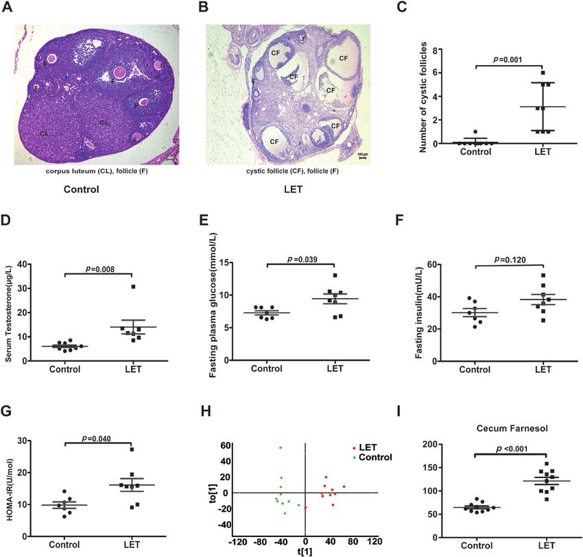
2                                                                                        Endocrinology, 2021, Vol. 162, No. 10

PCOS mouse model after a 35-day CDCA treatment. Mice receiving fecal microbiota
transplants from PCOS patients or healthy controls were evaluated after 10 weeks.
Results: Bacteroides was significantly enriched in treatment-naïve PCOS patients. The
enrichment in Bacteroides was reproduced in the PCOS mouse model. Gut microbiota
removal ameliorated the PCOS phenotype and insulin resistance and increased relative
FXR mRNA levels in the ileum and serum fibroblast growth factor 15 levels. PCOS stool-
transplanted mice exhibited insulin resistance at 10 weeks but not PCOS. Treating the
PCOS mouse model with CDCA improved glucose metabolism.
Conclusions: Bacteroides is a key microbial biomarker in PCOS and shows diagnostic
value. Gut dysbiosis can cause insulin resistance. FXR activation might play a beneficial
rather than detrimental role in glucose metabolism in PCOS.

                                                                                                                                  Downloaded from https://academic.oup.com/endo/article/162/10/bqab118/6305268 by guest on 22 September 2021
Key Words: PCOS, FXR, insulin resistance, intestinal flora

Polycystic ovary syndrome (PCOS) is a common gyneco-               More research is needed to determine the characteristics of
logical endocrine disease and a major cause of anovulatory         the gut microbiota in PCOS.
infertility in reproductive-aged women (1). However, in               The gut microbiota plays a key role in regulating en-
addition to being a reproductive disease, PCOS is also as-         ergy balance and is involved in the development and pro-
sociated with a wide range of metabolic disorders. Women           gression of obesity, diabetes, and metabolic syndrome (20,
with PCOS have a high risk of obesity (2), diabetes (2),           21). In addition, Lactobacillus has been found to be rele-
metabolic syndrome (3), nonalcoholic fatty liver disease           vant to fasting blood glucose and central adiposity (22).
(4), cardiovascular disease (5), and endometrial cancer (6).       Moreover, fecal microbiota transplantation (FMT) from
Although increased ovarian and/or adrenal androgen secre-          obese into germ-free mice can result in an obese phenotype
tion (7, 8), partial folliculogenesis arrest (8, 9), insulin re-   (23). However, the community structure and function of
sistance (10-12), neuroendocrine axis dysfunction (8), and         the intestinal flora in PCOS and the role and mechanism
genetic factors (13) have been suggested to be involved in         of the intestinal flora in the pathogenesis of PCOS remain
the development of PCOS, the precise underlying triggers           obscure. The role of gut microbiota in PCOS needs to be
for these key biochemical and metabolic disturbances re-           further determined. Recent reports have revealed that the
main largely unclear.                                              gut microbiota influences the farnesoid X receptor (FXR)-
    In recent years, with the progress of next-generation          fibroblast growth factor 15 (FGF15) axis in mice (24).
sequencing technology and the verification of aseptic ani-         Some studies have found that intestinal FXR is involved
mals, intestinal flora has been shown to be directly related       in glucose and lipid metabolism and insulin resistance and
to many diseases and even plays a role in various diseases,        that the activation of the FXR signalling pathway can im-
such as obesity, diabetes, atherosclerosis, and inflamma-          prove glucose metabolism and lipid metabolism disorders
tory enteritis (14). Recent studies have shown that the gut        (25, 26). After newly diagnosed type 2 diabetes was treated
microbiota of individuals with PCOS differs from those of          with metformin for 3 days, intestinal FXR was inhibited
healthy controls. However, analyses of the composition of          by the intestinal FXR antagonist glycoursodeoxycholic
the gut microbiota in women with PCOS have yielded in-             acid (GUDCA), and metabolic dysfunction, including
consistent results (15-18). Beza et al. found higher relative      hyperglycemia, was improved (27). The role of intestinal
abundances of Streptococcaceae and lower Bacteroidaceae            FXR in glucose metabolism in PCOS needs to be further
and Porphyromonadaceae in PCOS (15). By contrast,                  determined. Bile acids are physiological ligands for FXR,
Xinyu et al. found that Bacteroides vulgatus was markedly          and the strongest activator of FXR is the primary bile
elevated in the gut microbiota of individuals with PCOS            acid chenodeoxycholic acid (CDCA) (28). In the gut, pri-
(17). Interestingly, other studies found that the relative         mary bile acids are transformed into secondary bile acids
abundances of the genera Catenabacterium and Kandleria             by the metabolic activities of gut anaerobic bacteria (29).
(18) or Parabacteroides and Clostridium were enriched in           Letrozole (LET) treatment of female mice during puberty
PCOS (16). The reasons for the inconsistent results may            resulted in reproductive hallmarks of PCOS, including
be complex and diverse. Our group found that, among                hyperandrogenemia, anovulation, and polycystic ovaries
phenotypes, host location showed strong associations with          (30), and did not alter food intake (31). Therefore, the
microbiota variations (19). Host location may partially            LET-induced PCOS mouse model provides an opportunity
explain the inconsistencies in the intestinal flora in PCOS.       to study the relation of the gut microbiome and insulin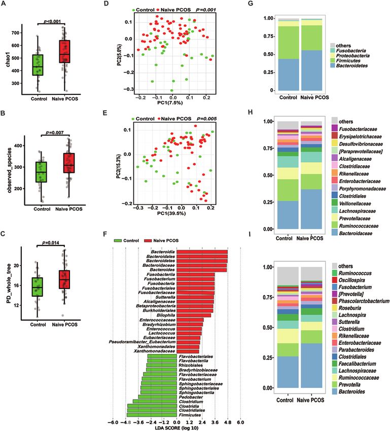
Endocrinology, 2021, Vol. 162, No. 10                                                                                       3

resistance in a diet-independent setting. Our hypothesis is      [CV] 1.45%; LH sensitivity range from 0.3 IU/L to 200
that gut microbiota dysbiosis can result in insulin resist-      IU/L, intra-assay CV 1.54%; testosterone sensitivity range
ance and contributes to the development of PCOS and that         from 0.025 μg/L to 15.0 μg/L, intra-assay CV 3.37%) at the
dysbiosis promotes glucose metabolism disorder possibly          Department of Clinical Chemistry in Zhujiang Hospital of
through the bile acid-intestinal FXR signalling pathway.         Southern Medical University. Serum glucose was measured
The purpose of our study was to accumulate more data on          by an automatic biochemical analyser (BS2001, Mindray,
the characteristics of the intestinal flora in PCOS, explore     China; sensitivity range from 0.6 mmol/L to 33.0 mmol/L,
whether abnormal intestinal flora can affect insulin resist-     intra-assay CV 3.0%, interassay CV 5.0%). The homeostasis
ance and promote the development of PCOS, and deter-             model assessment for insulin resistance index (HOMA-IR)
mine whether activation of intestinal FXR by CDCA can            and homeostasis model assessment for beta cell function
improve glucose metabolism in PCOS. Our research will            (HOMA-beta) were calculated using the following formulas:

                                                                                                                                  Downloaded from https://academic.oup.com/endo/article/162/10/bqab118/6305268 by guest on 22 September 2021
establish experimental evidence for the relationship among
the intestinal flora, CDCA, intestinal FXR, and PCOS.            HOMA-IR = fasting plasma glucose (FPG) (mM) × fasting
                                                                   plasma insulin (FINS) (mIU/L)/22.5;
                                                                 HOMA-beta = 20 × FINS (mIU/L)/ [FPG (mM) - 3.5] ×
                                                                   100%.
Methods
Human subjects
The study and all experimental procedures were approved
by the Ethics Committee at the Zhujiang Hospital of              DNA extraction
Southern Medical University. All patients were recruited         Fecal samples were obtained for DNA extraction. A frozen
from the Gynecology and Obstetrics outpatient clinic at the      aliquot (200 mg) of each fecal sample was processed using
Zhujiang Hospital of Southern Medical University. We re-         the PowerSoil DNA Extraction Kit (Shenzhen Bioeasy
cruited 56 individuals with PCOS and 31 healthy controls.        Biotechnologies Co., Ltd., China). The DNA concentra-
Written informed consent was obtained from all participants.     tions were measured using a NanoDrop system (Thermo
Women with PCOS were diagnosed according to the 2003             Fisher Scientific Co.).
Rotterdam criteria, which require the presence of at least 2
of the following: (1) oligo-ovulation and/or anovulation; (2)
clinical and/or biochemical signs of hyperandrogenism; and       16S sequencing and bioinformatics analysis
(3) polycystic ovaries. Diagnoses of PCOS were made after        Real-time quantitative PCR (qPCR) analysis was performed
the exclusion of other etiologies for hyperandrogenemia or       using the SYBR Green PCR master mix (Vazyme) and the ABI
ovulatory dysfunction (Cushing syndrome, 21-hydroxylase          ViiA 7 real-time PCR system (Applied Biosystems). We used
deficiency, congenital adrenal hyperplasia, androgen-            the barcoded V4F 5′ GTGTGYCAGCMGCCGCGGTAA
secreting tumors, thyroid disease, and hyperprolactinemia).      3′ and V4R 5′ CCGGACTACNVGGGTWTCTAAT 3′
All individuals with PCOS were first-visit patients and had      primers to amplify bacterial 16S rRNA V4 fragments. All
not received PCOS-related treatment. The individuals had         qPCRs were carried out with 2 μL of template DNA in
not received any antibiotic treatment for at least 1 month       a final volume of 20 μL following the manufacturer’s in-
before the sample collection. The healthy controls were from     structions (Invitrogen Life Technology Co., Ltd). The amp-
the general community and had regular menstrual cycles,          lification thermal cycling conditions were as follows: initial
normal ovarian morphology, and normal levels of hormones.        temperature of 94°C for 5 minutes, followed by 30 cycles of
Women who were breastfeeding or pregnant within the past         94°C for 30 seconds, 52°C for 30 seconds, and 72°C for 45
year or who took medication within the past 3 months were        seconds, and a final extension step of 72°C for 5 minutes.
excluded from the study. Height, body weight, waist circum-          All samples were sequenced using the paired-end
ference, and hip circumference were measured, and the body       strategy on the Illumina platform. Processing of the raw
mass index and waist-to-hip ratio were calculated. Peripheral    Illumina paired-end sequences was mainly performed
blood samples were collected from all subjects during days 2     on the QIIME (v1.9.1) platform (32, 33). The resulting
through 5 of menstruation after an overnight fast. Stool sam-    sequences were demultiplexed, and barcodes and pri-
ples were collected using stool collection tubes and stored in   mers of each sequence were then removed. We performed
a -80°C freezer.                                                 reference-based operational taxonomic unit (OTU) clus-
    Levels of serum FSH, LH, testosterone, and insulin were      tering against the Greengenes database v13_8 with a simi-
tested by an automatic chemiluminescent analyzer (Unicel         larity of 97% (32, 34, 35). R programming software and
DxI 800, Beckman Coulter, USA; FSH sensitivity range from        SPSS 24 were used for the statistical analysis, and P < 0.05
0.3 IU/L to 200 IU/L, intra-assay coefficient of variation       was considered statistically significant.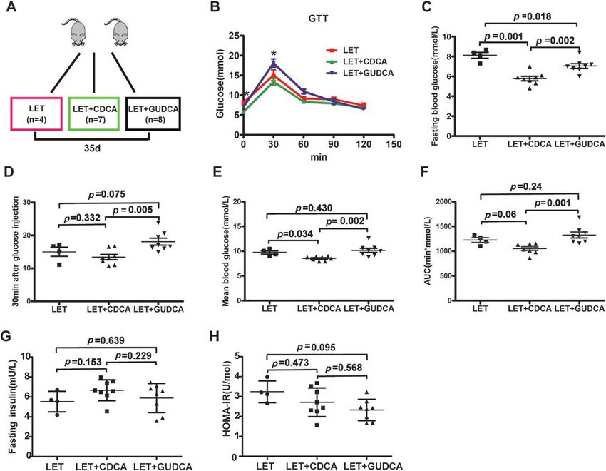
4                                                                                     Endocrinology, 2021, Vol. 162, No. 10

Animal models                                                  the end of the experiment. At the end of the experiments,
Four-week-old C57BL/6 female mice were randomly div-           the mice were anesthetized; blood was collected via retro-
ided into different groups, housed 4 to 5 per cage, and        orbital bleeding, centrifuged at 4000 rpm for 10 minutes
maintained under controlled temperature, lighting (12-         at 4°C, and stored at -80°C for subsequent serum analyses;
hour light: 12-hour dark cycle), and standard laboratory       the ovaries were dissected, fixed in 4% paraformaldehyde
conditions with free access to rodent feed and water. All      at 4°C overnight, and processed for histology; the intestines
animal experimental procedures were approved by the            and livers were quickly frozen and stored at -80°C.
Laboratory Animal Center and Ethics Committee at the
Zhujiang Hospital of Southern Medical University ac-
                                                               Glucose tolerance tests
cording to the national legislation for animal care. (1) The
                                                               Mice were fasted for 12 hours before the GTT. Glucose levels

                                                                                                                               Downloaded from https://academic.oup.com/endo/article/162/10/bqab118/6305268 by guest on 22 September 2021
mice were randomly divided into 2 groups and treated
for 5 weeks. The control group received vehicle only 1%        were measured by tail vein blood sampling using an Accu-
aqueous solution of carboxymethylcellulose, once daily         Chek Performa blood glucose analyser (Roche Diagnostics,
by oral gavage. The PCOS model mouse group was orally          sensitivity range from 0.3 mmol/L to 33.3 mmol/L). After
gavaged with LET (Sigma-Aldrich, catalog #PHR1540)             measurement of fasting glucose levels, the mice were IP in-
2.8 mg/kg-1 body weight, which was dissolved in 1%             jected with glucose (2 g/kg-1 body weight) for the GTT, and
carboxymethylcellulose once daily (36, 37). (2) The mice       tail blood samples were collected at 30, 60, 90 and 120
were randomly divided into a control FMT group, a PCOS         minutes after the IP injection for glucose level detection.
FMT group, LET+control FMT group, and LET+ PCOS
FMT group. The mice were treated with an antibiotic cock-
                                                               Serum analysis
tail (20 mg/mL vancomycin, 40 mg/mL neomycin sulfate,
40 mg/mL metronidazole, 40 mg/mL ampicillin) (38) for          Serum was collected to measure sex hormone and in-
5 days before human stool transplantation experiments.         sulin levels. The levels of testosterone were tested by an
The human stool samples were mixed with saline solution        automatic chemiluminescent analyser (Unicel DxI 800,
(20 mg/mL), vortexed, and centrifuged to collect the super-    Beckman Coulter; sensitivity range from 0.025 μg/L to
natant. The mice received another 10 weeks of treatment        15.0 μg/L, intra-assay CV 3.37%). Insulin was determined
by oral gavage with 100 µL fecal suspension from healthy       by ELISA kits (MM-0579M2, Meimian, China; sensitivity
controls for the control FMT group or gavage with 100 µL       range from 0.3 mIU/L to 8.0 mIU/L) for mice.
fecal suspension from PCOS patients for the PCOS FMT
group. The mice received another 5 weeks of treatment by
                                                               Morphology
oral gavage with LET and 10 weeks of treatment by oral
gavage with 100 µL fecal suspension from healthy controls      The ovarian tissues were processed for hematoxylin and
for the LET+control FMT group or gavage with 100 µL            eosin staining. Ovaries were quickly collected, fixed in 4%
fecal suspension from PCOS patients for the LET+PCOS           paraformaldehyde, subjected to gradient ethanol hydra-
FMT group. (3) The mice were randomly divided into a           tion, and embedded in paraffin. The sections were prepared
control group, a PCOS model mouse group, and a PCOS            and stained with hematoxylin and eosin. The ovaries were
with antibiotics group and treated for 5 weeks. The PCOS       longitudinally and serially sectioned into 5-μm sections
with antibiotics group was administered LET plus an anti-      (CM2016; Leica); all of the sections were mounted onto a
biotic cocktail (20 mg/mL vancomycin, 40 mg/mL neo-            glass slide and observed for histomorphological examination
mycin sulfate, 40 mg/mL metronidazole, and 40 mg/mL            under a light microscope (NIKON DS-U3, Nikon Eclipse
ampicillin) once daily by oral gavage. (4) The mice were       E100; Nikon). The numbers of cystic follicles were counted.
randomly divided into the PCOS model mouse group,
the LET+CDCA group, and the LET+GUDCA group and
treated for 5 weeks. Mice in the LET+CDCA group were           Real-time qPCR analysis
administered LET and CDCA (Targetmol, catalog #T0847)          Phenol-chloroform extraction was performed to isolate total
at a dosage of 50 mg/kg/d (27) once daily by oral gavage.      RNA from mouse liver and ileum tissue with TRIzol reagent
Mice in the LET+GUDCA group were administered LET              (Invitrogen, catalog #15596-026). cDNA was synthesized
and GUDCA (Sigma-Aldrich, catalog #06863) at a dosage          from 2 μg of total RNA with a Reverse Transcription Kit
of 50 mg/kg/d (27) once daily by oral gavage. The fecal pel-   (Takara, catalog #RR036A). Real-time qPCR analysis was
lets were collected from each mouse on day 35, flash frozen    performed using SYBR Green PCR master mix (Vazyme,
in liquid nitrogen, and stored at -80°C. Glucose tolerance     catalog #Q111-02) with the ABI ViiA 7 real-time PCR
tests (GTTs) were performed on the mice the day before         system (Applied Biosystems). The qPCR primers used in this
Endocrinology, 2021, Vol. 162, No. 10                                                                                                                       5

study were FXR-F 5′-TGGGCTCCGAATCCTCTTAGA-3′                                      nonnormally distributed data to evaluate the statistical signifi-
and FXR-R 5′-TGGTCCTCAAATAAGATCCTTGG-3′. All                                      cance of differences among 3 groups. The data among the 3
qPCRs were carried out in a final volume of 20 μL. The                            groups were analyzed by homogeneity tests of variances. The
amplification thermal cycling conditions were as follows:                         homogeneity of variance among the 3 groups was determined
95°C for 5 minutes; 40 cycles at 95°C for 10 seconds and                          with the least significant difference test, and missing vari-
60°C for 30 seconds; and a final extension step of 95°C for                       ance was determined with the Dunnett test. For more than 2
15 seconds and 60°C for 1 minute.                                                 groups, multiple comparisons were adjusted by the false dis-
                                                                                  covery rate. A 2-tailed Student t test was used to evaluate sig-
Metabolomics                                                                      nificant differences between 2 groups. For the nonparametric
                                                                                  tests, the 2-tailed Mann-Whitney U test was used to evaluate
Global metabolomics profiling was performed for cecum
                                                                                  significant differences between 2 groups. Data are shown

                                                                                                                                                                  Downloaded from https://academic.oup.com/endo/article/162/10/bqab118/6305268 by guest on 22 September 2021
contents (ultra-high-performance liquid chromatograph and
                                                                                  as the means ± SDs or as medians with interquartile ranges.
high-resolution mass spectrometer; Waters). Samples were
                                                                                  P < 0.05 was considered statistically significant.
separated on a chromatographic column with mobile phase
A, 0.1% formic acid in water, and mobile phase B, 0.1%
formic acid in acetonitrile, and analyzed in positive and
                                                                                  Results
negative heated electrospray ionization mode at m/z 50 to
1500 as separate injections. The injection volume was 5 μL.                       Bacteroides is increased significantly in PCOS
The flow rate was 400 μL/min with a 45°C column tempera-                          patients, and the microbial characteristics have
ture. MassLynx identified and aligned features. Data were                         potential diagnostic value
exported to Progenesis QI for analysis. Data analyses were                        Laboratory, anthropometric, and patient history data are
performed using DataFactory. Partial least squares discrim-                       summarized in Table 1. Treatment-naïve PCOS patients
inant analysis was used to determine differential metabolites.                    had significantly higher waist circumference, hip circum-
                                                                                  ference, and waist-to-hip ratio (P = 0.001, 0.024, and
                                                                                  0.015, respectively) levels than controls, whereas no dif-
Statistics                                                                        ference was found for age, weight, height, body mass
GraphPad Prism version 5.0 (GraphPad Software) and SPSS                           index, or HOMA-beta (P = 0.069, 0.090, 0.660, 0.072,
version 23.0 were used for statistical analysis. The sample                       and 0.749, respectively). HOMA-IR, FPG, and FINS were
distribution was determined by the Kolmogorov-Smirnov                             higher in the treatment-naïve PCOS group (P = 0.001,
normality test. One-way ANOVA was used for normally                               0.029, and 0.0001, respectively). Treatment-naïve PCOS
distributed data and the Kruskal-Wallis test was used for                         patients showed a characteristic dysregulation of LH and

Table 1. Characteristics of the study participants and hormone levels

Parameters                                            Control (n = 31)                                  PCOS (n = 56)                                P values

Age, y                                              26.00 (24.00, 27.00)                             24.00 (22.00, 27.00)                              0.069
Height, cm                                         160.23 ± 3.40                                    160.69 ± 5.36                                      0.660
Weight, kg                                          52.00 (48.50, 54.00)                             55.60 (47.63, 64.88)                              0.090
Waist circumference, cm                             69.06 ± 5.23                                     74.72 ± 10.23                                     0.001
Hip circumference, cm                               89.24 ± 3.62                                     92.62 ± 9.84                                      0.024
BMI, kg/m2                                          19.81 (18.76, 21.16)                             21.07 (18.75, 24.41)                              0.072
WHR                                                  0.77 ± 0.06                                      0.81 ± 0.06                                      0.015
HOMA-IR                                              1.03 (0.80, 1.38)                                1.61 (0.94, 2.33)                                0.001
HOMA-β                                              90.92 ± 122.88                                   96.88 ± 49.14                                     0.749
FPG, mmol/L                                          4.95 ± 0.46                                      5.18 ± 0.46                                      0.029
FINS, mIU/L                                          4.96 (3.53, 5.99)                                7.01 (4.27, 10.21)                               0.001
FSH, IU/L                                            6.82 ± 2.86                                      7.07 ± 1.45                                      0.580
LH, IU/L                                             4.50 (3.03, 5.96)                                8.67 (4.77, 12.97)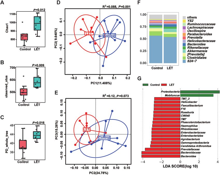
6                                                                                                          Endocrinology, 2021, Vol. 162, No. 10

testosterone secretion, with higher LH and testosterone                          There was a significant difference in bacterial alpha
levels than controls (P = 0.000 and 0.033). FSH was not                       diversity between treatment-naïve PCOS patients and
significantly different between the 2 groups (P = 0.580),                     healthy controls (Fig. 1A-C). Beta (β) diversity of the mi-
whereas LH/FSH was higher in treatment-naïve PCOS pa-                         crobial communities based on principal coordinate ana-
tients than in controls (P = 0.000).                                          lysis (PCoA, unweighted UniFrac) of the UniFrac metric

                                                                                                                                                           Downloaded from https://academic.oup.com/endo/article/162/10/bqab118/6305268 by guest on 22 September 2021

Figure 1. Characteristics of the gut microbiota in individuals with PCOS. Comparison of the stool microbiome of women with treatment-naïve PCOS
with that of healthy controls (treatment-naïve PCOS patients: 56; controls: 31). (A-C) α-diversity of the microbiota in the feces. Data are presented as
the mean ± SEM, P < 0.05, Wilcoxon rank-sum test. (D, E) Two-dimensional plot of PCoA for the microbiota. (F) Differentially abundant taxa identified
using LEfSe analysis, P < 0.05, Kruskal-Wallis rank sum test. (G-I) The gut microbiota average relative abundance of predominant bacterial taxa at the
phylum, family, and genus levels. PCoA, principal coordinate analysis; PCOS, polycystic ovarian syndrome.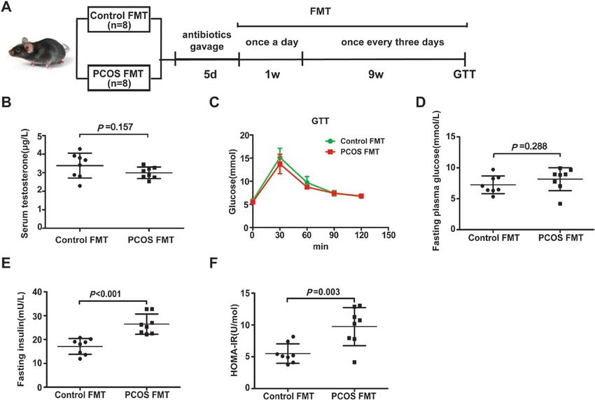
Endocrinology, 2021, Vol. 162, No. 10                                                                                                             7

(Fig. 1D, E, P = 0.001 and 0.005) revealed a separation be-                  exhibited follicles in various stages and fresh corpora lutea.
tween treatment-naïve PCOS patients and healthy controls.                    The granulosa within the follicles showed multiple layers
Furthermore, the abundance of Bacteroides was markedly                       (Fig. 4A). The PCOS mouse ovaries follicles showed a disor-
higher in treatment-naïve PCOS patients than in healthy                      ganized granulosa cell compartment with irregular granulosa
controls (Fig. 1F-I). To explore the potential ability of the                cell layer thickness, characteristic of atretic antral follicles.
intestinal flora to identify treatment-naïve PCOS, we con-                   The PCOS mouse ovaries appeared to lack corpora lutea and
structed a random forest model based on the intestinal                       have more antral follicles. Many cystic follicles showed no
flora. The performance of the model was assessed using re-                   granulosa layer or scant granulosa (Fig. 4B). The numbers of
ceiver operating characteristic analysis, achieving an area                  cystic follicles, testosterone levels, fasting blood glucose, and
under the curve (AUC) value of 0.78 (95% CI, 0.74-0.83).                     HOMA-IR in PCOS model mice were significantly higher
Subsequently, we further analyzed which bacteria have the                    than those in controls (Fig. 4C-G). To investigate the possible

                                                                                                                                                        Downloaded from https://academic.oup.com/endo/article/162/10/bqab118/6305268 by guest on 22 September 2021
ability to identify PCOS. According to the ranking of im-                    causes of insulin resistance resulting from intestinal flora dis-
portance scores in the random forest models, the results                     order in PCOS model mice, we collected cecum contents from
showed that the top 10 OTUs with the potential to identify                   PCOS model mice and controls for the metabolomics test.
PCOS had 4 OTUs belonging to Bacteroides (Fig. 2A, B).                       Metabolomics analysis further revealed that cecum farnesol
                                                                             increased significantly in PCOS model mice (Fig. 4H-I).

High abundance of Bacteroides, insulin
resistance, and cecum farnesol increase in PCOS                              Intestinal flora is a key factor in insulin resistance
model mice                                                                   and contributes to the development of PCOS
There was a significant difference in bacterial alpha diversity              To investigate the effect of the gut microbiota on insulin
between the PCOS model mice and controls (Fig. 3A-C). The                    sensitivity in PCOS, stools from healthy controls or indi-
β diversity of the microbial communities (based on PCoA,                     viduals with PCOS were transplanted into mice by oral
unweighted UniFrac) of the UniFrac metric (Fig. 3D, E) re-                   gavage for 10 weeks (Fig. 5A). The testosterone levels were
vealed a separation between PCOS model mice and controls.                    not different between the mice transplanted with stool
Furthermore, the abundance of Bacteroides was markedly                       from healthy controls and the mice transplanted with stool
higher in PCOS model mice than in controls, and this finding                 from individuals with PCOS (Fig. 5B). Compared with mice
was similar to the intestinal flora of PCOS patients (Fig.                   transplanted with stool from healthy controls, mice trans-
3F-G). Under light microscopy, the control mouse ovaries                     planted with stool from individuals with PCOS displayed

Figure 2. The gut microbiota signature can be used to discriminate between treatment-naïve PCOS patients and healthy controls. A random forest
model based on the intestinal flora of treatment-naïve PCOS patients and healthy controls was used to explore the potential ability of the intestinal
flora to identify treatment-naïve PCOS patients (treatment-naïve PCOS patients: 56; controls: 31). (A) A random forest model assessed using receiver
operating characteristic analysis (area under the curve [AUC] = 0.78). (B) The top 10 OTUs with the potential to identify treatment-naïve PCOS pa-
tients. OTU, operational taxonomic unit; PCOS, polycystic ovarian syndrome.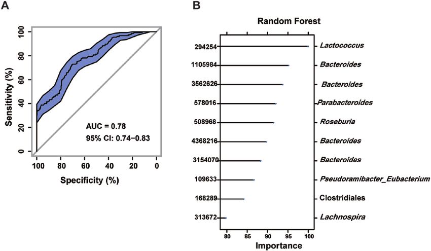
8 Endocrinology, 2021, Vol. 162, No. 10
Downloaded from https://academic.oup.com/endo/article/162/10/bqab118/6305268 by guest on 22 September 2021
Figure 3. Characteristics of the gut microbiota in PCOS model mice. Comparison of the stool microbiome between letrozole (LET)-induced PCOS
model mice and controls (PCOS model mice: 8; controls: 9). (A-C) α-diversity of the microbiota in the feces. Data are presented as the mean ± SEM,
P < 0.05, Wilcoxon rank-sum test. (D, E) Two-dimensional plot of PCoA for the microbiota. (F) The gut microbiota average relative abundance of the
predominant bacterial taxa at the genus level. (G) Differentially abundant taxa identified using LEfSe analysis, P < 0.05, Kruskal-Wallis rank sum test.
PCoA, principal coordinate analysis; PCOS, polycystic ovarian syndrome; SEM, standard error of the mean.
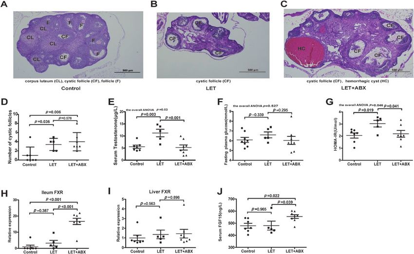
insulin resistance but not disordered glucose metabolism (Fig. 7A-D). The testosterone levels in the antibiotic-treated
(Fig. 5C-F). However, there was no difference between PCOS model mice were significantly lower than those in
PCOS model mice transplanted with stool from healthy the PCOS model mice (Fig. 7E). Administration of anti-
controls and PCOS model mice transplanted with stool biotic cocktail to the mice improved insulin resistance (Fig.
from individuals with PCOS (39). To further determine the 7F-G). FXR mRNA levels in the ileum were significantly
role of the intestinal flora in the PCOS phenotype in the increased in the presence of the antibiotic cocktail, but no
host, a continuous antibiotic cocktail was administered by differences were observed in the liver. Moreover, we found
gavage for 35 days (Fig. 6E). Microbial diversity and com- that administration of antibiotic cocktails increased serum
position were also apparently altered by antibiotic cocktail FGF15 levels (Fig. 7H-J). This finding indicated that the
treatment from the results of the chao1, observed_otus, removal of Bacteroidetes or Firmicutes affected intestinal
PD_whole_tree, and Shannon indexes; the PCoA of the FXR expression in PCOS.
Bray-Curtis distances; and species comparison (Fig. 6A-D,
F-G). Bacteroidetes and Firmicutes in PCOS mice were re-
moved completely after antibiotic cocktail treatment (Fig. Activation of FXR improves glucose metabolism
6H-I). Under light microscopy, the antibiotic-treated PCOS in PCOS model mice
model mouse ovaries still showed more antral follicles. Next, we measured the effects of intestinal FXR on glucose
Large cysts showed no granulosa layer or scant granulosa metabolism in the PCOS model mice (Fig. 8A). We foundEndocrinology, 2021, Vol. 162, No. 10 9
Downloaded from https://academic.oup.com/endo/article/162/10/bqab118/6305268 by guest on 22 September 2021
Figure 4. Insulin resistance and cecum farnesol are increased in PCOS model mice. Comparing the phenotype and cecal contents by global
metabolomics between letrozole (LET)-induced PCOS model mice and controls (PCOS model mice: 8; controls: 9). (A, B) Hematoxylin and eosin
staining of ovaries, corpus luteum (CL), cystic follicle (CF), and follicle (F). (C) Quantitative analysis of cystic follicles. (D) Serum testosterone levels.
(E) Fasting blood glucose. (F) Fasting insulin. (G) HOMA-IR. (H) Orthogonal partial least squares-discriminate analysis (OPLS-DA) of cecum content
global metabolomics. (I) Cecum farnesol. (C-I) P values were determined by 2-tailed Student t test, and P < 0.05 was considered statistically signifi-
cant. HOMA-IR, homeostasis model assessment for insulin resistance index; PCOS, polycystic ovarian syndrome.
that administration of the FXR agonist CDCA to LET- significantly higher fasting blood glucose, glucose 30 min-
treated mice affected glucose metabolism, as revealed by the utes after glucose injection, mean blood glucose, and AUC
GTT (Fig. 8B). The fasting blood glucose and mean blood of the GTT. These results confirmed that CDCA therapy
glucose from the GTT in the CDCA-treated PCOS model can improve glucose metabolism in PCOS model mice.
mice were significantly lower than those in the PCOS model
mice (Fig. 8C-F). There were no differences in fasting in-
sulin levels and HOMA-IR among the PCOS model group, Discussion
LET+CDCA group, and LET+GUDCA group (Fig. 8G-H). In this study, we identified dysbiosis of the intestinal flora
Compared with PCOS model mice treated with CDCA, in treatment-naïve PCOS patients. First, we observed an
mice treated with the FXR antagonist GUDCA displayed altered gut microbial pattern in treatment-naïve PCOS10 Endocrinology, 2021, Vol. 162, No. 10
Downloaded from https://academic.oup.com/endo/article/162/10/bqab118/6305268 by guest on 22 September 2021
Figure 5. Effects of PCOS fecal microbiota transplantation on the disruption of insulin sensitivity. Mice transplanted with stool suspensions from
healthy controls and women with PCOS were defined as the control-FMT and PCOS-FMT groups, respectively. After treatment with an antibiotic
cocktail (20 mg/mL vancomycin, 40 mg/mL neomycin sulfate, 40 mg/mL metronidazole, 40 mg/mL ampicillin intragastrically once daily) for 5 days,
the mice were orally gavaged with stool suspensions once a day for 1 week and once every 3 days for 9 weeks (control-FMT: 8; PCOS-FMT: 8). (A)
Schematic representation of the experimental design. (B) Serum testosterone levels. (C) GTT. (D) Fasting blood glucose. (E) Fasting insulin. (F)
HOMA-IR. (B-E) P values were determined by 2-tailed Student t test, and P < 0.05 was considered statistically significant. FMT, fecal microbiota trans-
plantation; GTT, glucose tolerance test; HOMA-IR, homeostasis model assessment for insulin resistance index; PCOS, polycystic ovarian syndrome.
patients when compared with the gut microbial pat- taxa in PCOS model mice and controls and then found
terns of controls. Second, the LEfSe tool revealed that the that the PCOS model mice had a high relative abundance
treatment-naïve PCOS patients had a high relative abun- of Bacteroidetes, which is in accordance with the find-
dance of Bacteroidetes; this finding was in accordance ings among PCOS patients. A high relative abundance of
with a recent study investigating the intestinal flora in in- Bacteroidetes may be characteristic of PCOS intestinal
dividuals with PCOS (40, 41). Third, 4 of the top 10 OTUs flora. This result may provide more data on the intestinal
with the highest importance in random forest model be- flora characteristics of PCOS.
longed to Bacteroides. The combination of these 10 OTUs In our work, compared with controls, women with
discriminated PCOS patients from healthy controls with PCOS and PCOS model mice had insulin resistance, in ac-
high accuracy. We noted that the gut microbiome-based cord with previous studies that considered that insulin re-
analysis could be used to predict the disease as a classifier sistance plays a significant etiological role in PCOS (42,
with an AUC of 0.78, implying that the microbial signa- 43). Moreover, the intestinal flora disorder of women with
ture identified may be a potentially powerful tool for the PCOS and that of PCOS model mice are similar. Evidence
diagnosis of PCOS and that integrating clinical markers is accumulating that the gut microbiota is involved in the
and microbial profiles may further improve the discrim- etiology of insulin resistance (44, 45). Mice transplanted
inative ability. with stool from individuals with PCOS displayed insulin
PCOS model mice had dysbiosis of the intestinal flora resistance only and did not show abnormal glucose metab-
compared to the intestinal flora of the control mice. We olism, contradicting a recent study that showed abnormal
used LEfSe to determine differentially abundant bacterial glucose metabolism in mice transplanted with stool fromEndocrinology, 2021, Vol. 162, No. 10 11
Downloaded from https://academic.oup.com/endo/article/162/10/bqab118/6305268 by guest on 22 September 2021
Figure 6. Characteristics of the gut microbiota in PCOS mice after treatment with antibiotic cocktails. Intestinal flora analysis of PCOS model mice
after antibiotic treatment (20 mg/mL vancomycin, 40 mg/mL neomycin sulfate, 40 mg/mL metronidazole, 40 mg/mL ampicillin intragastrically once
daily) removed the intestinal flora for 35 days (control: 7; LET: 5; LET+ABX: 7). (A) Schematic representation of the above experimental design. (B-E)
α-diversity of the microbiota in the feces. Data are presented as the mean ± SEM, P < 0.05, Wilcoxon rank-sum test. (F, G) Two-dimensional plot of
PCoA for the microbiota. (H) The gut microbiota average relative abundance of the predominant bacterial taxa at the genus level. (I) Differentially
abundant taxa identified using LEfSe analysis, P < 0.05, Kruskal-Wallis rank-sum test. ABX, antibiotic treatment; LET, letrozole; PCoA, principal coord-
inate analysis; PCOS, polycystic ovarian syndrome; SEM, standard error of the mean.12 Endocrinology, 2021, Vol. 162, No. 10
Downloaded from https://academic.oup.com/endo/article/162/10/bqab118/6305268 by guest on 22 September 2021
Figure 7. Removal of the intestinal flora can improve insulin resistance and suppress the development of PCOS. Phenotype analysis of PCOS model
mice after antibiotic treatment (20 mg/mL vancomycin, 40 mg/mL neomycin sulfate, 40 mg/mL metronidazole, 40 mg/mL ampicillin intragastrically
once daily) removed the intestinal flora for 35 days (control: 7; LET: 5; LET+ABX: 7). (A-C) Hematoxylin and eosin staining of the ovary, corpus luteum
(CL), cystic follicle (CF), and hemorrhagic cyst (HC). (D) Quantitative analysis of cystic follicles. (E) Serum testosterone levels. (F) Fasting blood glu-
cose. (G) HOMA-IR. (H) mRNA levels of ileum FXR. (I) mRNA levels of liver FXR. (J) Serum FGF15 levels. (D-J) P values were determined by 1-way
ANOVA for normally distributed data and the Kruskal-Wallis test for nonnormally distributed data. P < 0.05 was considered statistically significant.
ABX, antibiotic treatment; FGF15, fibroblast growth factor 15; FXR, farnesoid X receptor; HOMA-IR, homeostasis model assessment for insulin re-
sistance index; LET, letrozole; PCOS, polycystic ovarian syndrome.
individuals with PCOS for 3 weeks and suggesting the need by farnesol is very weak. FXR can be activated only by a
for further experimental verification (17). However, no dif- superphysiological dose of farnesol (47), which means that
ference was found between the PCOS mice transplanted farnesol is an antagonist of FXR. Intestinal L-cell secretion
with fecal microbiota from control women and the PCOS of GLP-1 decreased, and blood glucose and HOMA-IR
mice transplanted with fecal microbiota from women with increased after FXR was suppressed (48, 49). Cecum
PCOS. This result may be because the drugs used for PCOS farnesol-FXR may be related to insulin resistance in PCOS
modelling were too strong or the time for FMT was in- model mice.
sufficient. When we removed Bacteroidetes and Firmicutes FXR is widely distributed in organs such as the liver,
from the intestinal flora via a continuous 35-day anti- kidney, and intestines (28). FXR mRNA levels in the
biotic cocktail treatment, the PCOS model mice exhibited ileum, but not in the liver, were significantly increased in
improved phenotypes and insulin resistance, which was antibiotic-treated PCOS mice compared with those in the
similar to the finding that a prominent reduction in the PCOS model mice. The serum FGF15 level was significantly
abundance of Firmicutes and Bacteroidetes with vanco- increased synchronously. Intestinal FXR activation induces
mycin and bacitracin would ameliorate insulin resistance the expression of FGF15/19, and it has been demonstrated
in diet-induced obesity (46). The gut microbiota appeared that FGF15/19 affects glucose and energy homeostasis (48,
to be an interrelated key factor in insulin resistance and 49). FGF19 transgenic mice showed increased hepatic β
may promote the pathogenesis of PCOS. oxidation, reduced adipose tissue weight, and improved
Furthermore, we identified and analyzed cecum me- glucose tolerance and insulin sensitivity (50). Our results
tabolites and found that cecum farnesol was significantly suggest that intestinal flora may play an important role in
increased in PCOS model mice. Farnesol is a natural the pathogenesis and insulin resistance of PCOS via the
ligand of FXR; however, the activity of FXR activated FXR signalling pathway.Endocrinology, 2021, Vol. 162, No. 10 13
Downloaded from https://academic.oup.com/endo/article/162/10/bqab118/6305268 by guest on 22 September 2021
Figure 8. Intestinal FXR plays an important role in glucose metabolism in PCOS model mice. Mice were divided into 3 groups (LET, LET+CDCA,
and LET+GUDCA) and were treated for 35 days to explore the effects of intestinal FXR (LET: 4, LET+CDCA: 7, and LET+GUDCA: 8). (A) Schematic
representation of the experimental design. (B) GTT. (C) Fasting blood glucose. (D) Thirty minutes after glucose injection. (E) Mean blood glucose. (F)
Area under the curve (AUC) of the GTT. (G) Fasting insulin. (H) HOMA-IR. (B-H) P values were determined by 1-way ANOVA with the least significant
difference (LSD) multiple comparison post hoc test. P < 0.05 was considered statistically significant. CDCA, chenodeoxycholic acid; FXR, farnesoid
X receptor; GTT, glucose tolerance test; GUDCA, glycoursodeoxycholic acid; HOMA-IR, homeostasis model assessment for insulin resistance index;
LET, letrozole; PCOS, polycystic ovarian syndrome.
Bile acids are physiological ligands for FXR and can intestinal flora affects the properties and functions of bile
regulate multiple metabolic diseases by binding to FXR acids. The abundance of Bacteroidetes, which can hydro-
(28, 29). The primary bile acid CDCA is a strong agonist lyze CDCA by microbial bile salt hydrolases, is high in
of FXR (51), and the secondary bile acid GUDCA, trans- PCOS patients and PCOS model mice. We considered that
formed from primary bile acids by the metabolic activities the metabolism of CDCA mediated by Bacteroides was
of gut anaerobic bacteria, is an antagonist of FXR (27). In enhanced in PCOS patient and PCOS model mice so that
our study, we observed that oral administration of CDCA activated intestinal FXR was downregulated. Thus, the
improved glucose intolerance. Oral GUDCA supplemen- metabolic disorders of PCOS may in part be due to a lack
tation did not affect glucose metabolism in PCOS mice. In of intestinal FXR activation. The observations of similar
the gut, primary bile acid CDCA is successively converted metabolic effects of antibiotics and CDCA in PCOS model
by microbial bile salt hydrolase, an enzyme expressed pre- mice suggest that the intestinal flora–bile acid–intestinal
dominantly by Bacteroides, Lactobacillus, Bifidobacteria, FXR signalling pathway might be an important mech-
and Clostridium, and by bacterial 7α-dehydroxylase, an anism of glucose metabolic disorder in PCOS. However,
enzyme mainly expressed by Eubacterium and Clostridium, no differences in fasting insulin levels and HOMA-IR were
into the secondary bile acid lithocholic acid (52). The found among the PCOS model group (LET), LET+CDCA14 Endocrinology, 2021, Vol. 162, No. 10
group, and LET+GUDCA group. Thus, the mechanisms by none of the fecal samples were verified by collecting 2 sam-
which the gut microbiota affect insulin resistance are in- ples from the same woman, all fecal samples were collected
deed complex and might involve individual susceptibility, in strict accordance with the same fecal collection standards.
which should be investigated further.
The present study explored the characteristics of the in-
testinal flora in individuals with PCOS and the effect of the Acknowledgments
intestinal flora on PCOS. We concluded that a high rela- Financial Support: This study was supported by the National Key
tive abundance of Bacteroidetes may be characteristic of R&D Program of China (2019YFA0802300 and 2017YFC1310600),
the National Natural Science Foundation of China (NSFC31570497,
the PCOS intestinal flora. In addition, we found that re-
NSFC 31322003 and NSFC 81800746), the National Projects of Major
moving the gut microbiota decreased serum testosterone Infectious Disease Control and Prevention (2017ZX10103011), the Sci-
levels, ameliorated insulin resistance, and increased relative ence and Technology Program of Guangzhou, China (201904010091),
Downloaded from https://academic.oup.com/endo/article/162/10/bqab118/6305268 by guest on 22 September 2021
FXR mRNA levels in the ileum. PCOS stool-transplanted and the Natural Science Foundation of Hunan Province (2018JJ3467).
mice exhibited insulin resistance at 10 weeks. Treating the
PCOS model mice with CDCA improved fasting blood glu-
cose and mean blood glucose levels. The intestinal flora Additional Information
is a key factor in the development of insulin resistance in Correspondence: Hong-Wei Zhou, Division of Laboratory Medicine,
PCOS, and it promotes the glucose metabolism disorder Zhujiang Hospital, 253 Gongye Avenue, Haizhu District, Guangzhou
of PCOS possibly through the Bacteroidetes–bile acid–in- City, Guangdong Province510282, China. Email: biodegradation@
testinal FXR signalling pathway; moreover, FXR activa- gmail.com; or Yan He, Division of Laboratory Medicine, Zhujiang
Hospital, 253 Gongye Avenue, Haizhu District, Guangzhou City,
tion may have a beneficial, rather than detrimental, effect
Guangdong Province 510282, China. Email: 197053351@qq.com.
on PCOS glucose metabolism. The intestinal flora shows Disclosures: The authors declare that there are no conflicts of
promise as a potential target for PCOS treatment. interest.
Data Availability: The data used to support the findings of this
study are available from the corresponding author on reasonable
Limitations request.
Women with PCOS were diagnosed according to the 2003
Rotterdam criteria; PCOS subtypes were not identified.
Subtype analyses can determine if the microbiome is different References
between different PCOS subtypes. In this study, we recruited 1. Escobar-Morreale HF. Polycystic ovary syndrome: defin-
56 individuals with PCOS and 31 healthy controls. We did ition, aetiology, diagnosis and treatment. Nat Rev Endocrinol.
not perform a subtype analysis because the sample size for 2018;14(5):270-284.
2. Jacewicz-Święcka M, Kowalska I. Polycystic ovary syndrome
each subtype was too small to produce reliable results; future
and the risk of cardiometabolic complications in longitudinal
studies with larger sample sizes should be conducted to explore
studies. Diabetes Metab Res Rev. 2018;34(8):e3054.
the influence of PCOS subtypes. When studying insulin resist- 3. Spinedi E, Cardinali DP. The polycystic ovary syndrome and the
ance induced by intestinal flora, the addition of bile acid sup- metabolic syndrome: a possible chronobiotic-cytoprotective ad-
plementation and antibiotic treatments with the PCOS fecal juvant therapy. Int J Endocrinol. 2018;2018:1349868.
transplantation experiment would have further strengthened 4. Kumarendran B, O’Reilly MW, Manolopoulos KN, et al.
our findings. Antibiotic treatment of PCOS model mice can de- Polycystic ovary syndrome, androgen excess, and the risk of
crease serum testosterone levels, ameliorate insulin resistance, nonalcoholic fatty liver disease in women: a longitudinal study
based on a United Kingdom primary care database. Plos Med.
and increase relative FXR mRNA levels in the ileum; treating
2018;15(3):e1002542.
the PCOS model mice with CDCA improved fasting blood glu-
5. Bajuk Studen K, Jensterle Sever M, Pfeifer M. Cardiovascular
cose and mean blood glucose levels, but the addition of bile risk and subclinical cardiovascular disease in polycystic ovary
acid supplementation and antibiotic treatments to the control syndrome. Front Horm Res. 2013;40:64-82.
mice would have better demonstrated our experimental results. 6. Dumesic DA, Lobo RA. Cancer risk and PCOS. Steroids.
Our study would have been improved by strictly excluding 2013;78(8):782-785.
vegetarians, and those with probiotic, prebiotic, and antibiotic 7. Goodarzi MO, Carmina E, Azziz R. DHEA, DHEAS and PCOS.
J Steroid Biochem Mol Biol. 2015;145:213-225.
use before specimen collection. However, it is difficult to re-
8. Rosenfield RL, Ehrmann DA. The pathogenesis of polycystic
quire participants to standardize their diet before collecting
ovary syndrome (PCOS): the hypothesis of PCOS as functional
the stool samples. We reduced the impact of diet on the fecal ovarian hyperandrogenism. Endocr. Rev. 2016;37(5):467-520.
samples by strictly following the standard that the individuals 9. Garg D, Tal R. The role of AMH in the pathophysiology
could not have received treatment with any antibiotic for at of polycystic ovarian syndrome. Reprod Biomed Online.
least 1 month before collecting the fecal samples. Although 2016;33(1):15-28.Endocrinology, 2021, Vol. 162, No. 10 15
10. Miller WL, Tee MK. The post-translational regulation of 17,20 27. Sun L, Xie C, Wang G, et al. Gut microbiota and intestinal
lyase activity. Mol Cell Endocrinol. 2015;408:99-106. FXR mediate the clinical benefits of metformin. Nat Med.
11. Diamanti-Kandarakis E, Dunaif A. Insulin resistance and the 2018;24(12):1919-1929.
polycystic ovary syndrome revisited: an update on mechanisms 28. Gege C, Makishima M, Okamoto AY, et al. Identification of a nu-
and implications. Endocr Rev. 2012;33(6):981-1030. clear receptor for bile acids. Science. 1999;284(5418):1362-1365.
12. Wu S, Divall S, Nwaopara A, et al. Obesity-induced in- 29. Matsubara T, Li F, Gonzalez FJ. FXR signaling in the entero-
fertility and hyperandrogenism are corrected by deletion hepatic system. Mol Cell Endocrinol. 2013;368(1-2):17-29.
of the insulin receptor in the ovarian theca cell. Diabetes. 30. Torres PJ, Skarra DV, Ho BS, et al. Letrozole treatment of adult
2014;63(4):1270-1282. female mice results in a similar reproductive phenotype but dis-
13. Crespo RP, Bachega TASS, Mendonça BB, Gomes LG. An up- tinct changes in metabolism and the gut microbiome compared
date of genetic basis of PCOS pathogenesis. Arch Endocrinol to pubertal mice. BMC Microbiol. 2019;19(1):57.
Metab. 2018;62(3):352-361. 31. Skarra DV, Hernández-Carretero A, Rivera AJ, Anvar AR,
Downloaded from https://academic.oup.com/endo/article/162/10/bqab118/6305268 by guest on 22 September 2021
14. Nagpal R, Yadav H, Marotta F. Gut microbiota: the next-gen Thackray VG. Hyperandrogenemia induced by letrozole treat-
frontier in preventive and therapeutic medicine? Front Med ment of pubertal female mice results in hyperinsulinemia
(Lausanne). 2014;1:15. prior to weight gain and insulin resistance. Endocrinology.
15. Jobira B, Frank DN, Pyle L, et al. Obese adolescents with 2017;158(9):2988-3003.
PCOS have altered biodiversity and relative abundance in 32. McDonald D, Price MN, Goodrich J, et al. An improved
gastrointestinal microbiota. J. Clin. Endocrinol. Metab. Greengenes taxonomy with explicit ranks for ecological
2020;105(6):dgz263. and evolutionary analyses of bacteria and archaea. Isme J.
16. Zhang J, Sun Z, Jiang S, et al. Probiotic bifidobacterium lactis 2012;6(3):610-618.
V9 regulates the secretion of sex hormones in polycystic ovary 33. Caporaso JG, Kuczynski J, Stombaugh J, et al. QIIME allows
syndrome patients through the gut-brain axis. mSystems. analysis of high-throughput community sequencing data. Nat
2019;4(2): e00017-e00019. Methods. 2010;7(5):335-336.
17. Qi X, Yun C, Sun L, et al. Gut microbiota–bile acid–inter- 34. Edgar RC. Search and clustering orders of magnitude faster
leukin-22 axis orchestrates polycystic ovary syndrome. Nat. than BLAST. Bioinformatics. 2010;26(19):2460-2461.
Med. 2019;25(9):1225-1233. 35. He Y, Caporaso JG, Jiang XT, et al. Stability of operational
18. Insenser M, Murri M, Del Campo R, Martínez-García MÁ, taxonomic units: an important but neglected property for ana-
Fernández-Durán E, Escobar-Morreale HF. Gut microbiota and lyzing microbial diversity. Microbiome. 2015;3:20.
the polycystic ovary syndrome: influence of sex, sex hormones, 36. Kauffman AS, Thackray VG, Ryan GE, et al. A novel letrozole
and obesity. J Clin Endocrinol Metab. 2018;103(7):2552-2562. model recapitulates both the reproductive and metabolic pheno-
19. He Y, Wu W, Zheng HM, et al. Regional variation limits appli- types of polycystic ovary syndrome in female mice. Biol Reprod.
cations of healthy gut microbiome reference ranges and disease 2015;93(3):69.
models. Nat Med. 2018;24(10):1532-1535. 37. Kafali H, Iriadam M, Ozardali I, Demir N. Letrozole-induced
20. Parekh PJ, Arusi E, Vinik AI, Johnson DA. The role and influ- polycystic ovaries in the rat: a new model for cystic ovarian dis-
ence of gut microbiota in pathogenesis and management of ease. Arch Med Res. 2004;35(2):103-108.
obesity and metabolic syndrome. Front Endocrinol (Lausanne). 38. Rakoff-Nahoum S, Paglino J, Eslami-Varzaneh F, Edberg S,
2014;5:47. Medzhitov R. Recognition of commensal microflora by toll-
21. Sonnenburg JL, Bäckhed F. Diet-microbiota interactions as mod- like receptors is required for intestinal homeostasis. Cell.
erators of human metabolism. Nature. 2016;535(7610):56-64. 2004;118(2):229-241.
22. Lim MY, You HJ, Yoon HS, et al. The effect of heritability and 39. Yuelian Y. Data from: Effects of PCOS faecal microbiota trans-
host genetics on the gut microbiota and metabolic syndrome. plantation on the disruption of insulin sensitivity in PCOS
Gut. 2017;66(6):1031-1038. model mice. Harvard Dataverse. 2020. Deposited October 7,
23. Turnbaugh PJ, Ley RE, Mahowald MA, Magrini V, 2020. doi:10.7910/DVN/CAILIS.
Mardis ER, Gordon JI. An obesity-associated gut microbiome 40. Qi X, Yun C, Sun L, et al. Gut microbiota–bile acid–inter-
with increased capacity for energy harvest. Nature. leukin-22 axis orchestrates polycystic ovary syndrome. Nat.
2006;444(7122):1027-1031. Med. 2019;25(8):1225-1233.
24. Degirolamo C, Rainaldi S, Bovenga F, Murzilli S, Moschetta A. 41. Liu R, Zhang C, Shi Y, et al. Dysbiosis of gut microbiota asso-
Microbiota modification with probiotics induces hepatic bile ciated with clinical parameters in polycystic ovary syndrome.
acid synthesis via downregulation of the Fxr-Fgf15 axis in mice. Front Microbiol. 2017;8:324.
Cell Rep. 2014;7(1):12-18. 42. Diamanti-Kandarakis E, Papavassiliou AG. Molecular mechan-
25. Gadaleta RM, van Erpecum KJ, Oldenburg B, et al. Farnesoid isms of insulin resistance in polycystic ovary syndrome. Trends
X receptor activation inhibits inflammation and preserves Mol Med. 2006;12(7):324-332.
the intestinal barrier in inflammatory bowel disease. Gut. 43. Hutchison SK, Harrison C, Stepto N, Meyer C, Teede HJ.
2011;60(4):463-472. Retinol-binding protein 4 and insulin resistance in polycystic
26. Cipriani S, Mencarelli A, Palladino G, Fiorucci S. FXR activa- ovary syndrome. Diabetes Care. 2008;31(7):1427-1432.
tion reverses insulin resistance and lipid abnormalities and pro- 44. Pedersen HK, Gudmundsdottir V, Nielsen HB, et al.; MetaHIT
tects against liver steatosis in Zucker (fa/fa) obese rats. J Lipid Consortium. Human gut microbes impact host serum metabolome
Res. 2010;51(4):771-784. and insulin sensitivity. Nature. 2016;535(7612):376-381.16 Endocrinology, 2021, Vol. 162, No. 10
45. Khan MT, Nieuwdorp M, Bäckhed F. Microbial modulation of 49. Taoka H, Yokoyama Y, Morimoto K, et al. Role of bile acids
insulin sensitivity. Cell Metab. 2014;20(5):753-760. in the regulation of the metabolic pathways. World J Diabetes.
46. Hwang I, Park YJ, Kim YR, et al. Alteration of gut microbiota 2016;7(13):260-270.
by vancomycin and bacitracin improves insulin resistance 50. Tomlinson E, Fu L, John L, et al. Transgenic mice expressing human
via glucagon-like peptide 1 in diet-induced obesity. Faseb J. fibroblast growth factor-19 display increased metabolic rate and
2015;29(6):2397-2411. decreased adiposity. Endocrinology. 2002;143(5):1741-1747.
47. Forman BM, Goode E, Chen J, et al. Identification of a nu- 51. Jung D, Podvinec M, Meyer UA, et al. Human organic anion
clear receptor that is activated by farnesol metabolites. Cell. transporting polypeptide 8 promoter is transactivated by the
1995;81(5):687-693. farnesoid X receptor/bile acid receptor. Gastroenterology.
48. Kir S, Beddow SA, Samuel VT, et al. FGF19 as a postprandial, 2002;122(7):1954-1966.
insulin-independent activator of hepatic protein and glycogen 52. Fiorucci S, Biagioli M, Zampella A, Distrutti E. Bile acids activated
synthesis. Science. 2011;331(6024):1621-1624. receptors regulate innate immunity. Front Immunol. 2018;9:1853.
Downloaded from https://academic.oup.com/endo/article/162/10/bqab118/6305268 by guest on 22 September 2021You can also read


