Metabolic alterations mediated by STAT3 promotes drug persistence in CML
←
→
Page content transcription
If your browser does not render page correctly, please read the page content below
Leukemia                                                                                                                                          www.nature.com/leu

ARTICLE                 OPEN

Metabolic alterations mediated by STAT3 promotes drug
persistence in CML
Sweta B. Patel 1, Travis Nemkov 2, Davide Stefanoni2, Gloria A. Benavides3, Mahmoud A. Bassal 4,5, Brittany L. Crown1,
Victoria R. Matkins1, Virginia Camacho1, Valeriya Kuznetsova1, Ashley T. Hoang1, Danielle E. Tenen6, Samuel L. Wolock6, Jihye Park7,
Li Ying5, Zongliang Yue8, Jake Y. Chen 8, Henry Yang5, Daniel G. Tenen 5, Paul Brent Ferrell 9, Rui Lu1, Victor Darley-Usmar3,
                                                                ✉
Angelo D’Alessandro2, Ravi Bhatia 1 and Robert S. Welner 1

© The Author(s) 2021

   Leukemic stem cells (LSCs) can acquire non-mutational resistance following drug treatment leading to therapeutic failure and
   relapse. However, oncogene-independent mechanisms of drug persistence in LSCs are incompletely understood, which is the
   primary focus of this study. We integrated proteomics, transcriptomics, and metabolomics to determine the contribution of STAT3
   in promoting metabolic changes in tyrosine kinase inhibitor (TKI) persistent chronic myeloid leukemia (CML) cells. Proteomic and
   transcriptional differences in TKI persistent CML cells revealed BCR-ABL-independent STAT3 activation in these cells. While knockout
   of STAT3 inhibited the CML cells from developing drug-persistence, inhibition of STAT3 using a small molecule inhibitor sensitized
   the persistent CML cells to TKI treatment. Interestingly, given the role of phosphorylated STAT3 as a transcription factor, it localized
   uniquely to genes regulating metabolic pathways in the TKI-persistent CML stem and progenitor cells. Subsequently, we observed
   that STAT3 dysregulated mitochondrial metabolism forcing the TKI-persistent CML cells to depend on glycolysis, unlike TKI-sensitive
   CML cells, which are more reliant on oxidative phosphorylation. Finally, targeting pyruvate kinase M2, a rate-limiting glycolytic
   enzyme, specifically eradicated the TKI-persistent CML cells. By exploring the role of STAT3 in altering metabolism, we provide
   critical insight into identifying potential therapeutic targets for eliminating TKI-persistent LSCs.

   Leukemia _#####################_ ; https://doi.org/10.1038/s41375-021-01315-0

INTRODUCTION                                                                            have increased STAT3 activation [10, 11]. Genetic deletion of
Drug-insensitivity is a significant problem in cancer therapeutics.                      STAT3 with BCR-ABL overexpression impairs colony formation and
While leukemic cells can acquire secondary mutations, in many                           CML initiation [12]. Moreover, the combination of a STAT3 small
instances, they activate alternative signaling pathways for survival                    molecule inhibitor with TKI reduces the differentiation potential of
and resist treatment without a mutational cause [1]. This non-                          TKI-persistent leukemic cells [11]. These studies suggest that
mutational insensitivity could be due to transcriptional, epige-                        STAT3 plays a vital role in TKI evasion of LSCs in a BCR-ABL
netic, or metabolic reprogramming of leukemic stem cells (LSCs)                         independent manner; yet, the mechanism of STAT3 mediated TKI-
[1]. Chronic myeloid leukemia (CML) is an ideal disease to explore                      persistence remains unknown. STAT3 is a transcription factor
non-mutational mechanisms of drug-insensitivity. It is a clonal                         regulating genes for cell survival, proliferation and metabolism
disorder originating from hematopoietic stem cells (HSCs) due to a                      [13, 14]. Interestingly, STAT3 can also enter the mitochondria to
single oncogenic fusion protein BCR-ABL, a constitutively active                        mediate survival during cellular stress [15–17].
tyrosine kinase [2, 3]. The standard molecular targeted therapy for                       Combining traditional treatment strategies with metabolic
CML is tyrosine kinase inhibitors (TKI), which eradicates the bulk of                   inhibitors can eradicate LSCs [18, 19]. For instance, targeting
the disease but not the quiescent LSCs [4–8]. Although most                             amino acid metabolism, fatty acid oxidation, or glutaminolysis
patients respond to treatment, few can discontinue treatment                            along with the standard treatment eliminates AML LSCs [19–21].
without disease recurrence due to these persistent drug-                                While in T cell acute lymphoblastic leukemia, treatment modifies
insensitive LSCs.                                                                       cellular metabolism from glutaminolysis to glycolysis to evade
   In CML, BCR-ABL phosphorylates Signal Transducer and                                 drug-induced cellular stress. One glycolytic enzyme important in
Activator of Transcription 5 (STAT5), promoting cell survival and                       cancer is pyruvate kinase M2 (PKM2) that provides a survival
differentiation [9]. However, upon TKI-treatment, persistent cells                      advantage to diseased cells and plays a role in drug resistance

1
 Department of Medicine, Division of Hematology/Oncology, O’Neal Comprehensive Cancer Center, University of Alabama at Birmingham, Birmingham, AL, USA. 2Department of
Biochemistry and Molecular Genetics, School of Medicine, University of Colorado Anschutz Medical Campus, Aurora, CO, USA. 3Department of Pathology, Center for Free Radical
Biology, University of Alabama at Birmingham, Birmingham, AL, USA. 4Department of Systems Biology, Harvard Medical School, Boston, MA, USA. 5Cancer Institute of Singapore,
National University of Singapore, Singapore, Singapore. 6Division of Endocrinology, Beth Israel Deaconess Medical Center, Boston, MA, USA. 7Dicerna Pharmaceuticals, Inc.,
Lexington, MA, USA. 8Informatics Institute, School of Medicine, University of Alabama at Birmingham, Birmingham, AL, USA. 9Division of Hematology/Oncology, Vanderbilt
University Medical Center, Nashville, TN, USA. ✉email: rwelner@uab.edu

Received: 4 November 2020 Revised: 16 May 2021 Accepted: 28 May 2021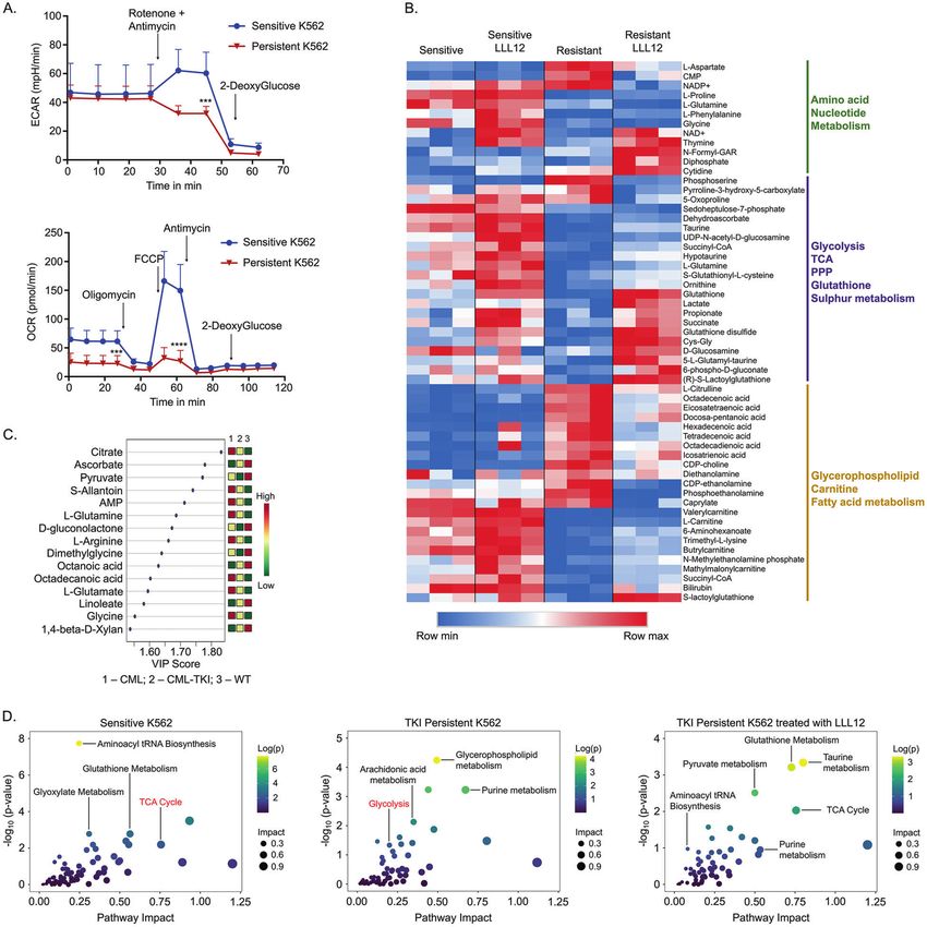
S.B. Patel et al.
                  2
                      [22, 23]. Significantly in CML, PKM2 deletion constrains disease                    RNAseq
                      progression and prolongs survival [24].                                            RNA was isolated using the RNeasy Plus mini kit (Qiagen). Libraries were
                         This study investigates the mechanism of STAT3-mediated TKI                     prepared with NexteraXT library construction (Illumina). The quality and size of
                      evasion in CML. We show that TKI-persistent LSCs undergo STAT3                     the indexed libraries were determined using the BioAnalyzer (Agilent) and
                      dependent transcriptional and metabolic changes from oxidative                     sequenced. Analysis was performed using Partek® Flow® software. Briefly,
                                                                                                         double-ended sequencing reads were aligned to the mouse (mm10) or human
                      phosphorylation to glycolysis. These insights led to the discovery                 (hg38) using Spliced Transcripts Alignment to a Reference (STAR 2.6.1d).
                      that TKI-persistent LSCs are susceptible to STAT3 inhibition and                   Aligned reads were then quantified to the transcriptome (RefSeq Transcripts
                      disruption of glycolysis. These findings reveal how STAT3 enables                   92) and normalized. Identified differentially expressed genes were used for
                      TKI-persistent CML cells to evade drug treatment in an oncogene-                   hierarchal clustering and pathway analysis.
                      independent manner and provides a therapeutic window to target
                      these cells specifically.                                                           Metabolomics and stable isotype tracing
                                                                                                         Metabolomics assays were performed via ultra-high pressure-liquid
                                                                                                         chromatography-mass spectrometry (Vanquish and Q Exactive, Thermo
                      MATERIAL AND METHODS                                                               Fisher) using the 3 min method [39]. For stable isotope tracing, K562 cells
                      Mouse models                                                                       were incubated with 13C3-1,2,3-glucose (Sigma Aldrich) or 13C515N2-L-
                      All mice are housed in UAB’s animal facility and experiments performed             Glutamine (Cambridge Isotope Laboratories) or U-13C16-Sodium Palmitate
                      under IACUC approved protocol. Double transgenic BCR-ABL×SCL-tTA                   (Sigma Aldrich) for 30 min to 72 h where indicated [20]. From each
                      mice were used as a CML model at ~60% myeloid cells in peripheral blood,           experiment, the labeled isotopologues as a percentage of the total
                      tested by flow cytometry as well as HemaVet [25, 26]. STAT3flox/flox mice             (labeled + unlabeled) were calculated, normalized to the relative abun-
                      (Jackson Laboratory) were bred with BCR-ABL×SCL-tTA mice and MxCre                 dance of each substrate. Data was analyzed using metaboanalyst [40].
                      mice to generate MxCre×STAT3flox/flox×CML mice (CML-STAT3 mice). H2b-
                      GFP-CML mice were generated by breeding BCR-ABL×SCL-tTA mice to
                      Rosa26+/rtTA Col1A1+/TetOP-H2B-GFP homozygous mice (Jackson Laboratory)            Statistical analysis
                                                                                                         Analyses were performed depending on the spread of the variable and
                      [27]. Males and females were equally distributed throughout the study.
                                                                                                         reported as standard deviation (SD). A Shapiro–Wilk test determined
                                                                                                         normal versus abnormal distributions, and all continuous variables were
                      Bone marrow transplant
1234567890();,:

                                                                                                         tested for mean differences. Depending on the spread of variable both
                      8-week-old CD45.1 mice were sub-lethally irradiated (450 rads), and retro-         nonparametric: Mann–Whitney U test, ANOVA Kruskal–Wallis test,
                      orbitally transplanted with 5 × 10⁶ bone marrow cells from CD45.2 CML or           Wilcoxon test, and parametric: Student’s t test and ANOVA were used.
                      control mice. Imatinib treatment was started ~4 weeks post-transplant [26]. For    For ANOVA, Tukey’s or Sidak post-test was used to compare groups
                      both the primary and transplanted mice, 200 or 400 mg/kg imatinib was              (GraphPad Prism version 7.0, La Jolla, CA).
                      administered by gavage every other day for 30 days. For STAT3 excision,
                      100 mg/kg pI:pC was administered i.p. every other day for 12 days after the
                      mice showed sign of disease (myeloid expansion detected in the peripheral          Data sharing statement
                      blood) [26]. For STAT3 small molecule inhibition studies, mice were treated        The data are deposited in NCBI’s Gene Expression Omnibus, accessible
                      with 25 mg/kg LLL12 i.p every alternate day in combination with imatinib for       through GEO series accession number GSE152713.
                      30 days.

                                                                                                         RESULTS
                      ChIP-seq                                                                           TKI-persistent CML cells have a distinct STAT3 mediated
                      Cells were cross-linked and sheared for ChIP with IgG and pSTAT3-Y705
                      (#9131, Cell Signaling). Double stranded cDNA libraries were constructed           transcriptional and proteomic profile
                      using Index Illumina library construction and sequenced [28]. Analysis of          TKIs reduce disease burden in patients, and yet, upon drug
                      ChIP-seq: Optical duplicates were removed from raw fastq files using                withdrawal, the disease recurs [41, 42] due to the persistence of
                      clumpify from BBMap (https://sourceforge.net/projects/bbmap/) with the             TKI-insensitive LSCs [4–7]. To mimic this phenotype, we used a
                      flags “dedupe spany addcount”. This was followed by, adapter trimming               previously described transgenic CML mouse model [25]. In brief,
                      using BBDuk (from BBMap) with the flags “ref = /…/bbmap/resources/                  once the mice are taken off tetracycline, the oncogene BCR-ABL
                      adapters.fa ktrim=l hdist=2”. Next, reads were trimmed using trimmomatic           gets expressed in the stem cells, giving rise to disease with
                      [29] with the flags “LEADING:20 SLIDINGWINDOW:4:20 TRAILING:20                      myeloid expansion detected after 4 weeks [25, 26]. TKI treatment,
                      MINLEN:20”. After cleanup, reads were aligned to mm10 using bwa mem                in this case Imatinib (IM), was carried out for 4-weeks after the
                      v0.7.17-r1188 [30] with default settings. Bam file sorting and indexing was
                      performed using samtools [31]. Bam coverage maps were generated using              mice had a leukemic burden of 40% [26]. Similar to patients, IM-
                      bamCoverage from deeptools [32] using default. These coverage maps are             treated CML mice had reduced disease burden with the
                      uploaded to GEO for access. Next, peak calling was performed on all                persistence of LSCs, which phenotypically resemble HSCs (Figs. 1A
                      samples using MACS2 [33], HOMER [34], Genrich (available at https://github.        and S1A) [43, 44]. One proposed mechanism for the survival of
                      com/jsh58/Genrich) and SICER2 [35, 36]. All peak callers were run in default       CML LSCs with TKI-treatment is quiescence [7]. To determine the
                      parameters for broad peaks as recommended by developers. Once peaks                quiescence of LSCs, we used a pulse and chase H2b-GFP mouse
                      were called, they were merged using HOMERs mergePeaks command with                 model, wherein GFP is lost with each cell division leaving dormant
                      the flag “-d 100.” Average fold change of each peak over input was                  cells marked over time [27]. H2b-GFP-CML mice revealed that IM-
                      calculated using a custom python script and considering only uniquely              persistent LSCs retained more GFP, which was more quiescent
                      mapped reads. Peaks were also annotated using HOMER’s annotatePeaks
                      command using the mm10 genome. To ascertain peak overlap between                   than untreated LSCs (Fig. S1B). Transcriptomic analysis of these
                      the 3 CML/TKI/WT samples, HOMERs mergePeaks command was used with                  untreated and IM-persistent cells in vivo and in vitro revealed
                      the flag “-d given”, and resultant sub-peak files annotated using HOMER’s            significant differences in gene expression as well as enriched
                      annotatePeaks command with default parameters. Genome coverage plots               pathways (Fig. S1C, D). We focused on signatures common
                      were generated using ngsplot [37] with the flags “-G mm10 -R tss -L 2000 -RB        between our in vitro and in vivo model by integrating our murine
                      0.05. The ChIP-seq enrichment plot was generated using plotFingerprint from        LSC and K562 RNA-seq data (Fig. 1B). Pathway analysis on these
                      the deepTools suite with the flags “-skipZeros -bs 25.” For generating              common, differentially upregulated genes revealed that the CML
                      coverage regions presented within the manuscript, normalized bam coverage          cells (126 genes) had enriched Rap1 and PI3-Akt signaling
                      maps were generated using bamCoverage using the flags “-outFileFormat
                                                                                                         pathway, while the IM-persistent cells (154 genes) had enrichment
                      bedgraph -normalizeUsing None -binSize 25 –effectiveGenomeSize
                      2652783500.” The generated bedgraphs were then imported into R v4.0.3
                                                                                                         in the central carbon metabolism, histidine metabolism and cell-
                      (available at https://www.r-project.org/index.html) and figures generated           cell interaction pathways (Fig. 1B). This is consistent with previous
                      using the package Sushi [38].                                                      findings demonstrating BCR-ABL mediated regulation of FOXO,

                                                                                                                                                Leukemia _#####################_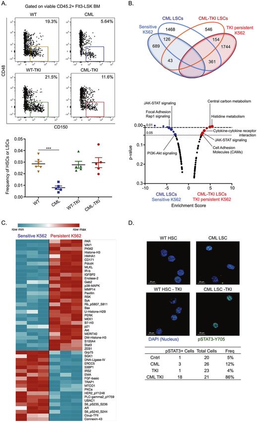
S.B. Patel et al.
3
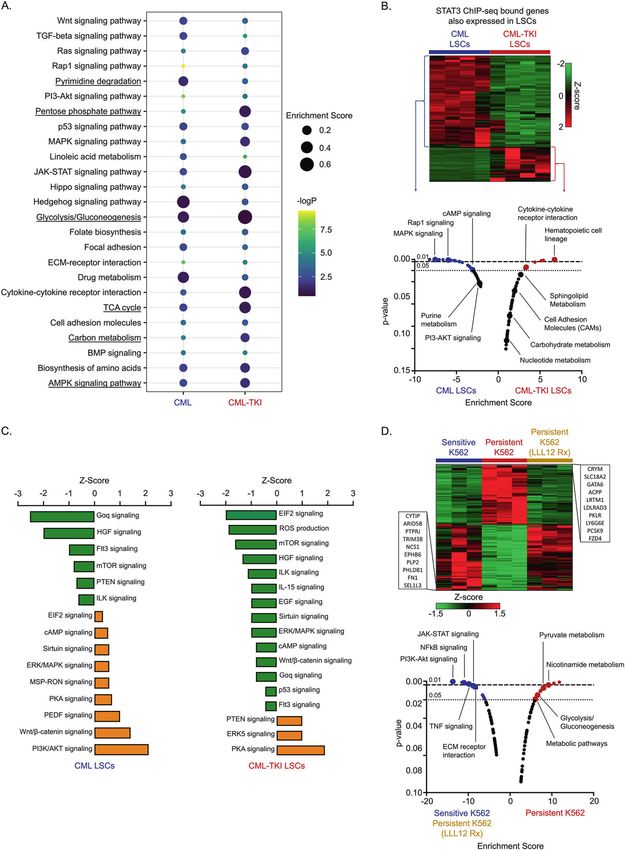
Ras/MAPK, PI3K/AKT/mTOR, Wnt, and STAT5 signaling [3, 43]. pharmacokinetics, and toxic effects [50]. These challenges highlight
However, targeting these pathways in combination with TKI, failed the need to identify the molecular mechanism of drug-insensitivity
to eradicate LSCs suggesting alternative mechanisms of cell downstream of STAT3 for alternative therapeutic targets. Since active
survival [3, 8]. Therefore, we sought to identify additional signaling STAT3 is a transcription factor [51], we performed pSTAT3-Y705 ChIP-
pathways contributing to transcriptional changes in IM-persistent seq using Lin−Sca1+cKit+ (LSK) cells to determine the STAT3
CML cells by performing reverse-phase protein array. Active transcriptional targets in IM-persistent CML stem and progenitors.
signaling proteins in the IM-persistent cells included STAT3, AKT1, We observed 13444 unique pSTAT3-Y705 localizations from IM-
MAP2K1, and PKM2 (Fig. 1C; Table S1). Pathway analysis of the top treated CML mice compared to 9931 in control and 10266 sites in
differentially activated proteins contributed to glycolysis, HIF and leukemic mice (Fig. S3A). Moreover, motif analysis revealed that
FoxO signaling in IM-persistent K562 (Fig. S1E, F). String analysis of unique sites of enrichment from IM-persistent CML LSK had a greater
the differentially active proteins within IM-persistent K562 percentage of peaks with the known STAT3 consensus sequence
revealed STAT3 and AKT1 as central nodes (Fig. S1G) [45]. compared to WT and CML (Fig. S3B). Pathway analysis on these
Increased STAT3 activation was also observed in our murine pSTAT3-Y705-bound sites revealed target genes enriched in
CML model, where 86% of IM-persistent LSCs having pSTAT3-Y705 glycolysis, carbohydrate metabolism, pentose phosphate pathway,
as opposed to 12% of untreated LSCs (Fig. 1D). Overall, these data AMPK signaling and cytokine-cytokine interaction in the CML-TKI LSK,
indicate that the IM-persistent CML LSCs and K562 are transcrip- while hedgehog signaling, Wnt/β-catenin signaling and TGFβ
tionally rewired and have distinct proteomic signatures suggestive signaling are enriched in the CML LSK (Fig. 3A), corroborating the
of metabolic changes. enriched pathways observed in the transcriptomic analysis. Integra-
tion of ChIP-seq and RNA-seq analysis revealed that genes bound by
STAT3 is essential for the leukemic potential of TKI-persistent pSTAT3-Y705 as well as those upregulated in CML-TKI LSCs were also
LSC found to be enriched in cytokine interaction and metabolism related
Since STAT3 and AKT were central nodes driving differential signatures (Fig. 3B). Moreover, ingenuity pathway analysis also
signaling in TKI-persistent K562, we evaluated their potential as disclosed significant positive correlation of PKA signaling in the CML-
therapeutic targets. Inhibition of AKT has previously been TKI LSCs, which is known to be activated by glycolysis [52] (Fig. 3C).
demonstrated to increase proliferation and apoptosis of CML Hence, to specifically define STAT3 mediated transcriptional changes,
LSCs but it also impairs maintenance, self-renewal and quies- we performed RNA-seq on sensitive, IM-persistent and LLL12 treated
cence of normal HSCs, making it a difficult target [46, 47]. IM-persistent K562. Since STAT3 inhibition sensitizes the IM-persistent
Alternatively, STAT3 has been essential for initiating disease and K562s (Fig. 2B), we focused on signatures shared between LLL12
TKI resistance in CML in an oncogene-independent manner treated persistent and sensitive cells (Fig. 3D). Pathway analysis
[10–12]. Consistently, we observed reduced pBCR and pSTAT5, revealed an enrichment of pyruvate metabolism and glycolysis in IM-
along with increased pSTAT3-Y705 but not pSTAT3-S727 and persistent K562 (Fig. 3E). Meanwhile, the sensitive and LLL12 treated
pCRKL in IM-persistent K562s (Fig. S2A) [10, 11]. STAT3 CRISPR cells were enriched in the NFkB, PI3-Akt, TNF, and JAK-STAT signaling
knock-out sensitive K562s had reduced cell growth and survival pathway known to be essential for CML maintenance (Fig. 3E) [3].
when treated with increasing doses of IM, indicating the Overall, these analyses reveal STAT3 mediated transcriptomic
importance of STAT3 for CML persistence during IM-treatment changes in TKI-persistent CML cells with enrichment of metabolic
(Fig. S2B, C). To test the impact of STAT3 KO on LSCs in vivo, we pathways.
generated MxCre×STAT3flox/flox × CML (CML-STAT3) mice. Condi-
tional deletion of STAT3 from hematopoietic cells of CML mice Drug insensitive CML cells have altered metabolism mediated
after disease progression significantly increased LSCs and by STAT3
reduced leukemic burden, suggesting the importance of STAT3 CML stem and progenitor cells rely on oxidative phosphoryla-
in CML maintenance (Fig. S2D–F). As STAT3 KO eliminates the tion compared to their non-transformed counterparts [44, 53].
pro-inflammatory IL-6 signature necessary for the maintenance of Moreover, short-term TKI-treatment of sensitive cells reduces
CML disease burden [26], it precluded our ability to use a genetic glucose uptake and its utilization for nucleotide and fatty acid
loss of STAT3 for studying IM-persistent LSCs. To circumvent this synthesis while increasing mitochondrial activity [54, 55], but
problem, we used a direct inhibitor of STAT3 phosphorylation at alterations in the metabolism of IM-persistent CML cells is yet to
the Y705 site, LLL12 (Fig. S2G), unlike the widely used JAK2- be studied. Thus, to examine metabolic differences between
related STAT3 inhibitors, WP1066 or Ruxolitinib [48, 49]. Pre- sensitive and IM-persistent K562, we measured cellular mito-
treatment of K562 cells with LLL12 followed by IM treatment led chondrial function and glycolysis using extracellular flux
to apoptosis of 80.4 ± 9.4% IM-persistent K562 (Fig. 2A). Moreover, analyzer. We observed similar basal extracellular acidification
pSTAT3-Y705 inhibition also significantly increased persistent rate (ECAR) but reduced reserve glycolytic capacity of the
cells’ susceptibility to IM in a dose-dependent manner with persistent K562s compared to sensitive cells indicating maximal
minimal effect on the sensitive K562 (Fig. 2B). Consistently, in vivo glycolysis usage in persistence (Fig. 4A). Moreover, oxygen
LLL12 treatment alone had no impact on LSC count; however, consumption rate (OCR) was significantly reduced in IM-
dual administration of LLL12 and IM significantly reduced LSC persistent cells (Fig. 4A), and hence we next evaluated
number in CML mice (Fig. 2C). This reduction was associated with mitochondrial damage. We observed three times more total
significantly increased apoptosis and proliferation of dual treated mitochondrial DNA in IM-persistent K562 (Fig. S4A); however,
LSCs compared to LSCs from mice treated with IM or LLL12 alone there was no difference in mitochondrial counts or area
(Fig. 2C). Furthermore, LLL12/IM dual treatment extended survival (Fig. S4B). Furthermore, we noted reduced protein abundance
of CML mice compared to single-agent treated and untreated of complex III and IV, Cytochrome C reductase and oxidase
CML mice (Fig. 2D). Moreover, the dual treated LSCs had reduced components of the electron transport chain (ETC) (Fig. S4C) [56].
engraftment upon transplantation (Fig. 2E). These data thus These data indicate loss of mitochondrial metabolism and
demonstrate that STAT3 is essential for the maintenance and function with a maintained glycolytic rate in IM-persistent K562,
leukemogenic potential of TKI-persistent LSCs. which suggests reduced energy requirement observed with
dormancy (Fig. S1B).
TKI-persistent LSCs are transcriptionally rewired with STAT3, mostly pSTAT3-S727, acSTAT3-K685 and unphosphory-
differential STAT3 localization to metabolic genes lated STAT3 play an essential role in maintaining mitochondrial
STAT3 targeting agents have not yet progressed beyond clinical function by localizing to the inner mitochondrial membrane
trials’ initial stages due to lack of specificity, unpredictable [16, 17, 57]. Mitochondrial STAT3 (mitoSTAT3) interacts with
Leukemia _#####################_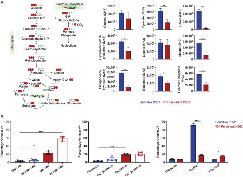
S.B. Patel et al.
4

    complex I, II, and IV of the ETC and also transcriptionally regulates      persistent CML cells, we generated metabolic profiles with STAT3
    mitochondrial DNA [58, 59]. We observed an accumulation of                 inhibition. We noted an accumulation of nucleosides and
    tSTAT3, pSTAT3-S727, and acSTAT3-K685 in the mitochondria of               unsaturated fatty acids in IM-persistent K562, which reduced with
    IM-persistent K562 compared to sensitive K562s (Fig. S4D). Hence,          STAT3 inhibition (Fig. 4B). In addition, stable metabolic profiles on
    to determine STAT3 mediated alteration of metabolism in IM-                murine LSCs and control HSCs with and without IM-treatment also

                                                                                                                Leukemia _#####################_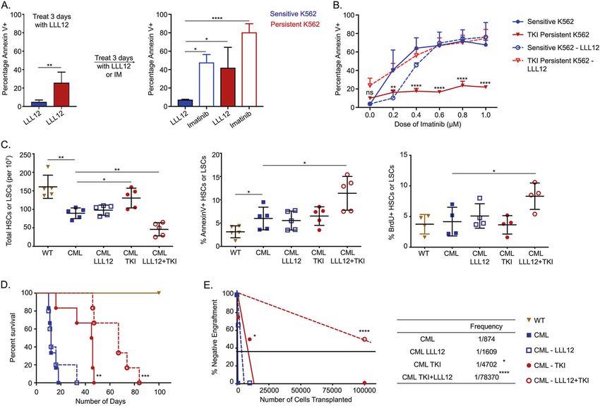
S.B. Patel et al.
                                                                                                                                                  5
Fig. 1 TKI-persistent CML LSCs and K562s have a distinct transcriptional and proteomic signature. A Representative FACS plot (left) and
scatter plot (right) for frequency of HSC/LSC (LSK Flt3−CD48−CD150+) off the stem and progenitor (LSK Flt3−) cells in the BM of CML, CML
mice treated with imatinib (200 mg/kg) for 4 weeks and their respective controls. n = 5. B Venn diagram (top) and KEGG pathway analysis
(bottom) for common genes upregulated in LSCs obtained from CML mice and CML mice treated with imatinib (200 mg/kg) for 4 weeks and
sensitive and IM-persistent K562 cells, obtained from RNA-seq. C Heatmap depicting differentially expressed protein in sensitive and
persistent K562 with a fold change >1.4 and p value ≥ 0.01 when assayed by Reverse Phase Protein Array (RPPA). D Representative confocal
imaging of (HSC/LSC (Lin−cKit+Sca1+Flt3−CD150+CD48−) for pSTAT3-Y705 (Green) and nucleus (Blue). Frequency of cells having stat3
expression is shown in the table to the right. The transcriptomic and proteomic data was carried out in triplicates. For scatter plot, two-way
ANOVA was used to determine statistical significance with Tukey’s multiple comparison. p values < 0.05 were considered statistically
significant. *p < 0.05, **p < 0.01, ***p < 0.001, ****p < 0.0001; ns = not significant.

Fig. 2 Active STAT3 is essential for survival and leukemic potential of TKI-persistent CML LSCs. A The sensitive and persistent K562 were
treated with LLL12 (1 µM) for 3 days. Treatment was switched to either imatinib (1 µM) or continued with LLL12 (1 µM) for additional 3 days.
Cells were analyzed for AnnexinV by flow cytometry. B Dose curve of imatinib for the sensitive and persistent K562 untreated or treated with
LLL12 (1 µM) for 3 days. The cells were stained with annexin V and analyzed by flow cytometry. C HSC or LSC count per 10 million BM cells
(left), percent AnnexinV stem cells (middle) and 24 hr BrDU incorporation in stem cells (right) of control and CML mice post 4 weeks of
imatinib (200 mg/kg) or LLL12 (25 mg/kg) treatment individually or in combination. n = 5. D Survival curves of control and CML mice post 4-
week imatinib (200 mg/kg) or LLL12 (25 mg/kg) treatment alone, or in combination. E Limiting dilution CRU (competitive repopulation unit)
assay is shown. The indicated numbers of control or leukemic-exposed HSCs were transplanted along with 2 × 10⁵ BM cells of a competitor
(CD45.2+) into lethally irradiated hosts (CD45.1+). Reconstitution was evaluated in the blood at 16 weeks post-transplantation (p = 0.0001).
Mice with CD45.1+ chimerism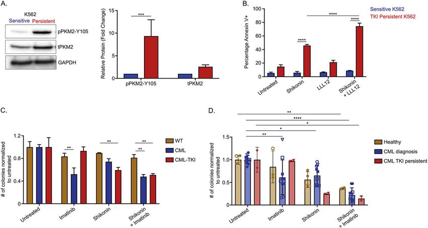
S.B. Patel et al.
6

    Fig. 3 STAT3 localizes to metabolic genes in transcriptionally altered TKI-persistent LSCs. A Kegg GO Pathway analysis depicting z score
    and p value for the genes differentially bound by pSTAT3-Y705 in the CML and IM-persistent CML LSK. B Heatmap and pathway analysis for
    integration of the pSTAT3-Y705 bound ChIP-seq peaks and the upregulated RNA-seq genes in the CML and IM-persistent LSK and LSCs
    respectively. C Ingenuity Pathway analysis bar chart depicting z scores of pathways correlated to overlapping genes of CML vs WT bulk-RNA
    sequencing and pSTAT3-Y705 ChIP-seq peaks unique to CML as well as overlapping genes of CML-TKI vs WT bulk-RNA sequencing and
    pSTAT3-Y705 ChIP-seq peaks unique to CML-TKI. D Heatmap and pathway analysis for upregulated genes common between sensitive K562
    and IM-persistent K562 treated with 1 µM LLL12 (1 µM) but different from IM-persistent K562. The transcriptomic data were carried out in
    triplicates.

                                                                                                           Leukemia _#####################_
S.B. Patel et al.
7
Fig. 4 STAT3 contributes to altered metabolism in TKI-persistent CML cells. A ECAR (top) from glycolysis rate assay (GRA) and OCR (bottom)
from mitochondrial stress test (MST) of 75,000 sensitive and IM-persistent K562 measured by seahorse. B Heatmap for the top 50 differentially
expressed metabolites between IM-persistent K562 and those treated with LLL12 (1 µM) with a p value ≤0.05. Raw values were used to
generate the heatmap. C VIP plot for component 1 of the PLS-DA comparing WT HSCs, CML LSCs, IM-persistent CML LSCs. The VIP score
indicates the contribution of the metabolite to the clustering pattern. D Scatter plots obtained from integration of the K562 metabolic profile
with the K562 RNA-seq data. All figures are mean ± SD of a representative data of at least 2–3 independent experiments. Unpaired student
t test. was used to determine statistical significance with Tukey’s multiple comparison. p values < 0.05 were considered statistically significant.
*p < 0.05, **p < 0.01, ***p < 0.001, ****p < 0.0001; ns = not significant.
non-significant increase in the basal ECAR, the compensatory oxidation or using glutamine as a carbon source [19–21]. To
glycolysis of IM-persistent K562 increased to be similar to the determine contributions of metabolic substrates in sensitive and
sensitive K562s (Fig. S4H). Overall, these data suggest that STAT3 IM-persistent K562, we carried out metabolic flux analysis using
leads to metabolic alterations in TKI-persistent LSCs towards a 1,2,3-13C3-Glucose, 13C515N2-Glutamine and U-13C16-Palmitate.
reduced mitochondrial activity state while maintaining energy Glucose tracing revealed reduced labeling of several glycolytic
production through glycolysis. metabolites in IM-persistent K562 (Fig. 5A). Despite having a
slower flux of 13C-Glucose through glycolysis in IM-persistent
TKI-insensitive CML cells are dependent on glycolysis K562, the intracellular level of labeled lactate was comparable
LSCs utilize multiple pathways to evade drug treatment, with the sensitive K562 (data not shown). Moreover, sensitive
including dependency on amino acid metabolism, fatty acid K562 had a higher flux of glucose-derived carbon atoms into the
Leukemia _#####################_S.B. Patel et al.
8
Fig. 5 TKI-persistent K562 have an active glycolysis and increased fatty acid accumulation. A Unit area plots for metabolites labeled in
glycolysis, citric acid cycle and PPP post 3 h incubation of 200,000 sensitive and IM-persistent K562 with isotopically labeled 13C1,2,3-glucose.
The metabolites were measured with UHPLC-MS. B Sensitive and IM-persistent K562 treated with 1 µM Imatinib were starved with glucose
(right) or glutamine (middle) for 48 h or treated with 10 µM Etomoxir for 3 days. Cells were analyzed by flow cytometry using annexinV
apoptosis assay. The data are representative mean ± SD from two independent experiments. Unpaired student t test or Two-way ANOVA was
used to determine statistical significance with Tukey’s multiple comparison. p values < 0.05 were considered statistically significant. *p < 0.05,
**p < 0.01, ***p < 0.001, ****p < 0.0001; ns = not significant.
TCA cycle (Fig. 5A), consistent with a higher OCR (Fig. 4A). (Fig. 4B). Moreover, palmitate tracing revealed palmitate
Furthermore, IM-persistent K562 had lower levels of glucose- oxidation as a source of acetyl-carnitine in IM-persistent K56s,
derived labeled carbons into pentose phosphate pathway, or further utilized to make significantly more citrate (Fig. S5E).
PPP-derived ATP and 13C-acetyl-carnitine, consistent with acetyl- Surprisingly, treatment with the FAO inhibitor, Etomoxir, had a
CoA (Fig. S5A), an entry metabolite of TCA cycle. These data negligible impact on cell survival of IM-persistent K562 (16.65 ±
combined with reduced levels of 13C-Glucose derived ATP in IM- 1.06%) (Fig. 5B). However, starving IM-persistent K562 of glucose
persistent K562 (Fig. S5A) suggest decreased oxidative phos- led to increased apoptosis, 58.37 ± 6.23% (Fig. 5B), indicating
phorylation. Importantly, stable metabolite profiles revealed an their dependence on glucose for survival but not fatty acids or
increase in α-Ketoglutarate to Citrate ratio (Fig. S5B), suggesting glutamine (Fig. 5B). Overall, these results indicate that TKI-
a block in the TCA cycle beyond α-Ketoglutarate and an increase persistent K562 accumulate fatty acids but depend on glucose
in reductive carboxylation as a result of the altered mitochon- for their survival.
drial function [60, 61]. To identify alternative carbon sources
preferentially utilized by IM-persistent K562, we carried out Glycolysis is important for survival of TKI-persistent CML cells
glutamine metabolic flux analysis. Consistent with glucose STAT3 activation leads to transcriptional and metabolic changes
tracing, labeled carbon incorporation from glutamine into TCA towards glycolysis and is critical for the survival of TKI-insensitive
cycle was reduced in IM-persistent K562 (Fig. S5C) along with cells. Additionally, glycolysis modulates quiescence (Fig. S1B) by
increase in citrate isotopologues M + 5 (reductive carboxylation) reducing energy demand [62]. From our LSC RNA-seq, we
to M + 4 (oxidative) ratio (Fig. S5D). In line with the role of observed that genes in the glycolysis/gluconeogenesis pathway
reductive carboxylation as fuel to fatty acid synthesis [60], we are more enriched in the IM-persistent LSCs (Fig. S6A). Treating IM-
observed an increase in glutamine-derived 13C-acetyl-carnitine persistent K562 with 2-Dexoyglucose, 2-DG, a competitive
in IM-persistent K562 (Fig. S5D). Palmitate tracing highlighted an glycolysis inhibitor, led to increased apoptosis (Fig. S6B), con-
increase in fatty acid elongation and desaturation in IM- sistent with glucose starvation (Fig. 5B). However, since 2-DG
persistent K562 (Fig. S5E), which corroborates the accumulation cannot be translated clinically, we looked at other glycolytic
of unsaturated fatty acids observed in steady-state analyses targets. Interestingly, we observed approximately threefold
Leukemia _#####################_S.B. Patel et al.
9
Fig. 6 Glycolysis is important for the survival of TKI-persistent CML stem and progenitor cells. A Western blot for PKM2 using sensitive and
IM-persistent K562 cell lysate. The bar plot represents density of the persistent protein bands relative to sensitive and normalized to the
housekeeping protein, GAPDH. B Sensitive and IM-persistent K562 analyzed for apoptosis via flow cytometry post 3-day treatment with IM
(1 µM), LLL12 (1 µM), or Shikonin (0.2 µM) alone, or in combination. C Colony-forming assay for 1000 LSK cells/mL sorted and plated from
control, CML, and imatinib (400 mg/kg) treated CML mice. The plates were treated with IM (1 µM) or Shikonin (0.5 µM) alone, or in
combination, incubated in hypoxia and colonies counted after 10 days. Data is represented as counts normalized to untreated. D Colony-
forming assay for 1000 Lin-CD34+ BM cells/mL sorted and plated from 2 healthy individuals, 5 CML patients at diagnosis and 1 IM treated
disease persistent patient. The plates were treated with Imatinib (1 µM) or Shikonin (0.5 µM) alone, or in combination, incubated in hypoxia
and colonies counted after 10 days. Data are represented as counts normalized to untreated. The data are representative or a pool of mean ±
SD from 2 to 3 independent experiments. Unpaired student t test or Two-way ANOVA was used to determine statistical significance with
Tukey’s multiple comparison. p values < 0.05 were considered statistically significant. *p < 0.05, **p < 0.01, ***p < 0.001, ****p < 0.0001; ns = not
significant.
increase in PKM2 and approximately tenfold increase in pPKM2- DISCUSSION
Y105 in IM-persistent K562s (Figs. 1C and 6A). PKM2 in previous The persistence of cancer stem cells after drug treatment and
studies has shown to slow CML progression; however, it has subsequent relapse is a significant barrier to cure. Apart from
importance in drug persistence is not known [24]. We hence used mutational causes of drug resistance, cells can persist in drug
pharmacological inhibitors of PKM2, compound 3 K or Shikonin treatment due to the survival of a drug-tolerant form [65]. In this
[63, 64], and observed selectively increased apoptosis of persistent study, we describe a non-mutational mechanism of STAT3-
K562s with dose escalation (Fig. S6C–F). Interestingly, dual mediated transcriptional and metabolic regulation in drug-
treatment of IM-persistent K562 with LLL12 and Shikonin had an insensitive LSCs. Active STAT3 localized to several metabolic
additive effect on apoptosis (Fig. 6B). Since in vivo shikonin genes while also regulating mitochondrial metabolism. Notably,
treatment proved toxic for healthy mice (data not shown), we TKI-insensitive cells are reliant on glycolysis and the rate-limiting,
used ex vivo treatment to validate the K562 data in our murine stress-related enzyme PKM2. Understanding the mechanism of
model. Shikonin treatment of LSK leads to apoptosis of CML and STAT3-mediated drug-insensitivity provides potential drug targets
IM-treated LSKs compared to the single and dual treated non- to eliminate these TKI-persistent cells and reduces the chances of
transformed LSK (Fig. S6G), consistent with PKM2 knock out of relapse.
untreated CML cells [24]. In addition, we observed reduced Given the importance of STAT3 in tumor growth, survival, and
colony-forming potential of IM-persistent LSK with Shikonin alone chemoresistance, several clinical trials have been carried out
or in combination with IM (Fig. 6C). Similar trends were observed with STAT3 inhibitors. However, STAT3 targeting agents yield
in colony-forming potential of shikonin-treated CD34+ cells from minimal success because they inhibit activators of STAT3,
CML patients at diagnosis and following IM-treatment (Fig. 6D). like JAK family members. Besides, STAT3 can be activated by
Although, dual treatment of Shikonin and IM also reduced colony other kinases explaining the survival of TKI-persistent CML cells
forming potential of non-transgenic CD34 + cells, it did not treated with JAK-mediated STAT3 inhibitors [66]. Furthermore,
significantly affect the viability of these cells (Figs. 6D and S6H). many drugs that inhibit STAT3 activation bind the SH2 domain
Moreover, similar to K562 and murine LSK, we also observed that shares homology to other STAT family members leading to
reduced cell viability and increased apoptosis of CD34+ cells from toxic effects [50]. Recent drug discovery has been towards
IM-persistent CML patient samples treated with both IM and tagging STAT3 for ubiquitin-dependent degradation [67]. These
shikonin (Fig. S6H–I). These data indicate that IM-persistent LSCs agents hold some promise in clinical trials, yet understanding
have altered metabolism dependent on glycolysis for their the downstream activity of STAT3 during TKI-persistence
survival, hence providing a target to eliminate the TKI-persistent provides an additional opportunity to find alternative drug
LSCs in CML. targets.
Leukemia _#####################_S.B. Patel et al.
10
Consequently, we sought to understand the mechanism of 6. Jorgensen HG, Allan EK, Graham SM, Godden JL, Richmond L, Elliott MA, et al.
STAT3 mediated drug persistence. We observed transcriptional Lonafarnib reduces the resistance of primitive quiescent CML cells to imatinib
adaptations of metabolic genes and found STAT3 localized to mesylate in vitro. Leukemia 2005;19:1184–91.
CPT1B and citrate synthase, critical enzymes for fatty acid 7. Zhang B, Strauss AC, Chu S, Li M, Ho Y, Shiang KD, et al. Effective targeting
of quiescent chronic myelogenous leukemia stem cells by histone
oxidation and TCA cycle [14]. Conversely, STAT3 induces glycolysis
deacetylase inhibitors in combination with imatinib mesylate. Cancer Cell. 2010;
in a hypoxic environment while reducing mitochondrial genes 17:427–42.
[68]. Interestingly, it can also enter mitochondria to regulate 8. Holyoake TL, Vetrie D. The chronic myeloid leukemia stem cell: stemming the tide
mitochondrial function [17, 58, 59], and cellular stress [15, 16]. of persistence. Blood. 2017;129:1595–606.
These observations likely explained the increased mitoSTAT3 in 9. Hoelbl A, Kovacic B, Kerenyi MA, Simma O, Warsch W, Cui Y, et al. Clarifying the
TKI-persistent K562 with minimal mitochondrial activity and role of Stat5 in lymphoid development and Abelson-induced transformation.
compensated by increased glycolysis. LSCs have an increased Blood. 2006;107:4898–906.
oxygen consumption rate upon initial drug treatment [53]; 10. Bewry NN, Nair RR, Emmons MF, Boulware D, Pinilla-Ibarz J, Hazlehurst LA. Stat3
however, our data suggest that with prolonged treatment, the contributes to resistance toward BCR-ABL inhibitors in a bone marrow micro-
environment model of drug resistance. Mol Cancer Ther. 2008;7:3169–75.
more quiescent, TKI-persistent cells are biased towards glycolysis,
11. Eiring AM, Page BD, Kraft IL, Mason CC, Vellore NA, Resetca D, et al. Combined
possibly due to reduced energy requirements. Additionally, STAT3 and BCR-ABL1 inhibition induces synthetic lethality in therapy-resistant
reductive carboxylation, a de novo lipogenesis pathway [69], chronic myeloid leukemia. Leukemia. 2015;29:586–97.
and elongation of fatty acids in TKI-persistent K562 are consistent 12. Hoelbl A, Schuster C, Kovacic B, Zhu B, Wickre M, Hoelzl MA, et al. Stat5 is
with utilization of carbon from glucose for nucleic acid and fatty indispensable for the maintenance of bcr/abl-positive leukaemia. EMBO Mol Med.
acid synthesis [54, 70]. Moreover, free fatty acids can form a feed- 2010;2:98–110.
forward loop with fatty acid oxidation, a reserve energy source 13. Poli V, Camporeale A. STAT3-mediated metabolic reprograming in cellular
[71]. These indicate metabolic reorganization of TKI-persistent transformation and implications for drug resistance. Front Oncol. 2015;5:121.
LSCs to aid survival under cellular stress. 14. Wang T, Fahrmann JF, Lee H, Li YJ, Tripathi SC, Yue C, et al. JAK/STAT3-regulated
fatty acid beta-oxidation is critical for breast cancer stem cell self-renewal and
The main regulator of cellular stress is PKM2, a key enzyme in
chemoresistance. Cell Metab. 2018;27:136–50. e5
glycolysis [63] that promotes reductive glutamine metabolism 15. Meier JA, Hyun M, Cantwell M, Raza A, Mertens C, Raje V, et al. Stress-induced
[61]. PKM2 knockout in healthy HSCs has shown no substantial dynamic regulation of mitochondrial STAT3 and its association with cyclophilin D
defect minus loss of reconstitution potential observed upon reduce mitochondrial ROS production. Sci Signal. 2017;10.
multiple serial transplants. Additionally, PKM2 knockout delays 16. Szczepanek K, Chen Q, Derecka M, Salloum FN, Zhang Q, Szelag M, et al.
CML onset and death [24], consistent with our data. Our findings Mitochondrial-targeted signal transducer and activator of transcription 3
uniquely reveal an increased dependence of TKI-persistent cells on (STAT3) protects against ischemia-induced changes in the electron transport
PKM2. Oxidative stress can lead to PKM2 dimerization and chain and the generation of reactive oxygen species. J Biol Chem. 2011;286:
phosphorylation to act as a transcription factor or kinase, with 29610–20.
17. Wegrzyn J, Potla R, Chwae YJ, Sepuri NB, Zhang Q, Koeck T, et al. Function of
evidence of activating STAT3 [23, 72]. This observation suggests a
mitochondrial Stat3 in cellular respiration. Science. 2009;323:793–7.
possible feed-forward loop of PKM2 and STAT3 in regulating TKI- 18. Lagadinou ED, Sach A, Callahan K, Rossi RM, Neering SJ, Minhajuddin M, et al.
persistence and provides a potential drug target to eradicate the BCL-2 inhibition targets oxidative phosphorylation and selectively eradicates
TKI-persistent LSCs. quiescent human leukemia stem cells. Cell Stem Cell. 2013;12:329–41.
These findings emphasize STAT3 regulation of gene expres- 19. Matre P, Velez J, Jacamo R, Qi Y, Su X, Cai T, et al. Inhibiting glutaminase in acute
sion and metabolism in TKI-persistent leukemic cells to a myeloid leukemia: metabolic dependency of selected AML subtypes. Oncotarget.
glycolytic state. Our study could be extrapolated to other drug- 2016;7:79722–35.
resistant diseases like Flt3-ITD AML, multiple myeloma or solid 20. Jones CL, Stevens BM, D’Alessandro A, Reisz JA, Culp-Hill R, Nemkov T, et al. Inhibition
tumors that acquire oncogene independent drug-insensitivity. of amino acid metabolism selectively targets human leukemia stem cells. Cancer Cell.
2018;34:724–40. e4
Additional work is needed to develop better therapeutic drugs
21. Samudio I, Harmancey R, Fiegl M, Kantarjian H, Konopleva M, Korchin B, et al.
for targeting STAT3, but we have characterized the downstream Pharmacologic inhibition of fatty acid oxidation sensitizes human leukemia cells
metabolic regulation as an alternative means to eliminate LSCs. to apoptosis induction. J Clin Investig. 2010;120:142–56.
Our findings implicate a therapeutic opportunity where inhibi- 22. Christofk HR, Vander Heiden MG, Harris MH, Ramanathan A, Gerszten RE, Wei R,
tion of glycolysis along with TKI-treatment preferentially targets et al. The M2 splice isoform of pyruvate kinase is important for cancer meta-
drug-insensitive LSCs. At the same time, non-transformed HSCs bolism and tumour growth. Nature. 2008;452:230–3.
can survive due to activating alternative metabolic pathways. 23. Li Q, Zhang D, Chen X, He L, Li T, Xu X, et al. Nuclear PKM2 contributes to gefitinib
However, there is still an ever-increasing need for developing resistance via upregulation of STAT3 activation in colorectal cancer. Sci Rep.
better and specific metabolic inhibitors, an impediment to 2015;5:16082.
24. Wang YH, Israelsen WJ, Lee D, Yu VWC, Jeanson NT, Clish CB, et al. Cell-state-
clinical translation in cancer. Importantly, we have shown that
specific metabolic dependency in hematopoiesis and leukemogenesis. Cell.
integrating signaling pathways, transcriptomics and metabolo- 2014;158:1309–23.
mics can discover drug targets to eliminate treatment persistent 25. Koschmieder S, Gottgens B, Zhang P, Iwasaki-Arai J, Akashi K, Kutok JL, et al.
cancer stem cells. Inducible chronic phase of myeloid leukemia with expansion of hematopoietic
stem cells in a transgenic model of BCR-ABL leukemogenesis. Blood.
2005;105:324–34.
REFERENCES 26. Welner RS, Amabile G, Bararia D, Czibere A, Yang H, Zhang H, et al. Treatment of
1. Dagogo-Jack I, Shaw AT. Tumour heterogeneity and resistance to cancer thera- chronic myelogenous leukemia by blocking cytokine alterations found in normal
pies. Nat Rev Clin Oncol. 2018;15:81–94. stem and progenitor cells. Cancer Cell. 2015;27:671–81.
2. Fialkow PJ, Jacobson RJ, Papayannopoulou T. Chronic myelocytic leukemia: clonal 27. Foudi A, Hochedlinger K, Van Buren D, Schindler JW, Jaenisch R, Carey V, et al.
origin in a stem cell common to the granulocyte, erythrocyte, platelet and Analysis of histone 2B-GFP retention reveals slowly cycling hematopoietic stem
monocyte/macrophage. Am J Med. 1977;63:125–30. cells. Nat Biotechnol. 2009;27:84–90.
3. Deininger MW, Goldman JM, Melo JV. The molecular biology of chronic myeloid 28. Bararia D, Kwok HS, Welner RS, Numata A, Sarosi MB, Yang H, et al. Acetylation of
leukemia. Blood. 2000;96:3343–56. C/EBPalpha inhibits its granulopoietic function. Nat Commun. 2016;7:10968.
4. Corbin AS, Agarwal A, Loriaux M, Cortes J, Deininger MW, Druker BJ. Human 29. Bolger AM, Lohse M, Usadel B. Trimmomatic: a flexible trimmer for Illumina
chronic myeloid leukemia stem cells are insensitive to imatinib despite inhibition sequence data. Bioinformatics. 2014;30:2114–20.
of BCR-ABL activity. J Clin Investig. 2011;121:396–409. 30. Li H, Durbin R. Fast and accurate short read alignment with Burrows-Wheeler
5. Heidel FH, Bullinger L, Feng Z, Wang Z, Neff TA, Stein L, et al. Genetic and transform. Bioinformatics. 2009;25:1754–60.
pharmacologic inhibition of beta-catenin targets imatinib-resistant leukemia 31. Li H, Handsaker B, Wysoker A, Fennell T, Ruan J, Homer N, et al. The Sequence
stem cells in CML. Cell Stem Cell. 2012;10:412–24. Alignment/Map format and SAM tools. Bioinformatics. 2009;25:2078–9.
Leukemia _#####################_S.B. Patel et al.
11
32. Ramirez F, Dundar F, Diehl S, Gruning BA, Manke T. deepTools: a flexible platform 57. Xu YS, Liang JJ, Wang Y, Zhao XJ, Xu L, Xu YY, et al. STAT3 undergoes acetylation-
for exploring deep-sequencing data. Nucleic acids Res. 2014;42:W187–91. Web dependent mitochondrial translocation to regulate pyruvate metabolism. Sci
Server issue Rep. 2016;6:39517.
33. Gaspar J Improved peak-calling with MACS2. bioRxiv; 2018. 58. Carbognin E, Betto RM, Soriano ME, Smith AG, Martello G. Stat3 promotes
34. Heinz S, Benner C, Spann N, Bertolino E, Lin YC, Laslo P, et al. Simple combina- mitochondrial transcription and oxidative respiration during maintenance and
tions of lineage-determining transcription factors prime cis-regulatory elements induction of naive pluripotency. EMBO J. 2016;35:618–34.
required for macrophage and B cell identities. Mol Cell. 2010;38:576–89. 59. Cui P, Wei F, Hou J, Su Y, Wang J, Wang S. STAT3 inhibition induced temozolomide-
35. Xu S, Grullon S, Ge K, Peng W. Spatial clustering for identification of ChIP- resistant glioblastoma apoptosis via triggering mitochondrial STAT3 translocation and
enriched regions (SICER) to map regions of histone methylation patterns in respiratory chain dysfunction. Cell Signal. 2020:109598.
embryonic stem cells. Methods Mol Biol. 2014;1150:97–111. 60. Fendt SM, Bell EL, Keibler MA, Olenchock BA, Mayers JR, Wasylenko TM, et al.
36. Zang C, Schones DE, Zeng C, Cui K, Zhao K, Peng W. A clustering approach for Reductive glutamine metabolism is a function of the alpha-ketoglutarate to
identification of enriched domains from histone modification ChIP-Seq data. citrate ratio in cells. Nat Commun. 2013;4:2236.
Bioinformatics. 2009;25:1952–8. 61. Liu M, Wang Y, Ruan Y, Bai C, Qiu L, Cui Y, et al. PKM2 promotes reductive
37. Shen L, Shao N, Liu X, Nestler E. ngs.plot: quick mining and visualization of next- glutamine metabolism. Cancer Biol Med. 2018;15:389–99.
generation sequencing data by integrating genomic databases. BMC Genom. 62. Takubo K, Nagamatsu G, Kobayashi CI, Nakamura-Ishizu A, Kobayashi H, Ikeda E,
2014;15:284. et al. Regulation of glycolysis by Pdk functions as a metabolic checkpoint for cell
38. Phanstiel DH, Boyle AP, Araya CL, Snyder MP. Sushi.R: flexible, quantitative and cycle quiescence in hematopoietic stem cells. Cell Stem Cell. 2013;12:49–61.
integrative genomic visualizations for publication-quality multi-panel figures. 63. Zhao X, Zhu Y, Hu J, Jiang L, Li L, Jia S, et al. Shikonin inhibits tumor growth in
Bioinformatics. 2014;30:2808–10. mice by suppressing pyruvate kinase M2-mediated aerobic glycolysis. Sci Rep.
39. Nemkov T, Hansen KC, D’Alessandro A. A three-minute method for high-throughput 2018;8:14517.
quantitative metabolomics and quantitative tracing experiments of central carbon 64. Ning X, Qi H, Li R, Li Y, Jin Y, McNutt MA, et al. Discovery of novel naphthoqui-
and nitrogen pathways. Rapid Commun mass Spectrom. 2017;31:663–73. none derivatives as inhibitors of the tumor cell specific M2 isoform of pyruvate
40. Chong J, Soufan O, Li C, Caraus I, Li S, Bourque G, et al. MetaboAnalyst 4.0: kinase. Eur J Med Chem. 2017;138:343–52.
towards more transparent and integrative metabolomics analysis. Nucleic Acids 65. Rambow F, Rogiers A, Marin-Bejar O, Aibar S, Femel J, Dewaele M, et al. Toward
Res. 2018;46:W486–W94. minimal residual disease-directed therapy in melanoma. Cell. 2018;174:843–55. e19
41. Mahon FX, Rea D, Guilhot J, Guilhot F, Huguet F, Nicolini F, et al. Discontinuation 66. Yang L, Lin S, Xu L, Lin J, Zhao C, Huang X. Novel activators and small-molecule
of imatinib in patients with chronic myeloid leukaemia who have maintained inhibitors of STAT3 in cancer. Cytokine Growth Factor Rev. 2019;49:10–22.
complete molecular remission for at least 2 years: the prospective, multicentre 67. Bai L, Zhou H, Xu R, Zhao Y, Chinnaswamy K, McEachern D, et al. A potent and
Stop Imatinib (STIM) trial. Lancet Oncol. 2010;11:1029–35. selective small-molecule degrader of STAT3 achieves complete tumor regression
42. Hochhaus A, Larson RA, Guilhot F, Radich JP, Branford S, Hughes TP, et al. Long- in vivo. Cancer Cell. 2019;36:498–511. e17
term outcomes of imatinib treatment for chronic myeloid leukemia. N Engl J 68. Demaria M, Giorgi C, Lebiedzinska M, Esposito G, D’Angeli L, Bartoli A, et al. A
Med. 2017;376:917–27. STAT3-mediated metabolic switch is involved in tumour transformation and
43. Hamilton A, Helgason GV, Schemionek M, Zhang B, Myssina S, Allan EK, et al. STAT3 addiction. Aging. 2010;2:823–42.
Chronic myeloid leukemia stem cells are not dependent on Bcr-Abl kinase 69. Metallo CM, Gameiro PA, Bell EL, Mattaini KR, Yang J, Hiller K, et al. Reductive
activity for their survival. Blood. 2012;119:1501–10. glutamine metabolism by IDH1 mediates lipogenesis under hypoxia. Nature.
44. Abraham A, Qiu S, Chacko BK, Li H, Paterson A, He J, et al. SIRT1 regulates metabolism 2011;481:380–4.
and leukemogenic potential in CML stem cells. J Clin Investig. 2019;129:2685–701. 70. Kominsky DJ, Klawitter J, Brown JL, Boros LG, Melo JV, Eckhardt SG, et al.
45. Szklarczyk D, Gable AL, Lyon D, Junge A, Wyder S, Huerta-Cepas J, et al. STRING v11: Abnormalities in glucose uptake and metabolism in imatinib-resistant human
protein-protein association networks with increased coverage, supporting functional BCR-ABL-positive cells. Clin Cancer Res. 2009;15:3442–50.
discovery in genome-wide experimental datasets. Nucleic Acids Res. 2019;47: 71. Kuo CY, Ann DK. When fats commit crimes: fatty acid metabolism, cancer
D607–D13. stemness and therapeutic resistance. Cancer Commun. 2018;38:47.
46. Juntilla MM, Patil VD, Calamito M, Joshi RP, Birnbaum MJ, Koretzky GA. AKT1 and 72. Pucino V, Certo M, Bulusu V, Cucchi D, Goldmann K, Pontarini E, et al. Lactate
AKT2 maintain hematopoietic stem cell function by regulating reactive oxygen buildup at the site of chronic inflammation promotes disease by inducing CD4(+)
species. Blood. 2010;115:4030–8. T cell metabolic rewiring. Cell Metab. 2019;30:1055–74. e8
47. Nie ZY, Yang L, Liu XJ, Yang Z, Yang GS, Zhou J, et al. Morin inhibits proliferation
and induces apoptosis by modulating the miR-188-5p/PTEN/AKT regulatory
pathway in CML cells. Mol Cancer Ther. 2019;18:2296–307.
ACKNOWLEDGEMENTS
48. Iwamaru A, Szymanski S, Iwado E, Aoki H, Yokoyama T, Fokt I, et al. A novel
We would like to thank Drs. Paul Kincade, Deepak Bararia, Allon Klein, and Robert
inhibitor of the STAT3 pathway induces apoptosis in malignant glioma cells both
Signer for their support and feedback. This project was supported by NIH grants
in vitro and in vivo. Oncogene. 2007;26:2435–44.
1PO1HL131477; startup funds from the Division of Hematology/Oncology at the
49. Lin L, Hutzen B, Li PK, Ball S, Zuo M, DeAngelis S, et al. A novel small molecule,
University of Alabama at Birmingham (UAB); American Cancer Society-IRG Junior
LLL12, inhibits STAT3 phosphorylation and activities and exhibits potent growth-
Faculty Development Grant (2019); the American Society of Hematology Bridge Grant
suppressive activity in human cancer cells. Neoplasia. 2010;12:39–50.
(2018); and the Leukemia Research Funding (2019). TEM imaging was supported by
50. Wong ALA, Hirpara JL, Pervaiz S, Eu JQ, Sethi G, Goh BC. Do STAT3 inhibitors have
the National Cancer Institute Cancer Center Support Grant P30 CA013148 and used
potential in the future for cancer therapy? Exp Opin Investig Drugs. 2017;26:883–7.
the UAB High Resolution Imaging Facility. Flow cytometry was supported by the
51. Wingelhofer B, Neubauer HA, Valent P, Han X, Constantinescu SN, Gunning PT, et al.
University of Alabama at Birmingham (UAB) Center for AIDS Research CFAR, an NIH
Implications of STAT3 and STAT5 signaling on gene regulation and chromatin
funded program (P30 AI027767-31).
remodeling in hematopoietic cancer. Leukemia. 2018;32:1713–26.
52. Caza M, Kronstad JW. The cAMP/protein kinase a pathway regulates virulence
and adaptation to host conditions in cryptococcus neoformans. Front Cell Infect
Microbiol. 2019;9:212. AUTHOR CONTRIBUTIONS
53. Kuntz EM, Baquero P, Michie AM, Dunn K, Tardito S, Holyoake TL, et al. Targeting SBP designed, planned, and performed experiments, analyzed data and wrote the
mitochondrial oxidative phosphorylation eradicates therapy-resistant chronic manuscript; BLC, TN, DS, GAB, VRM, VC, ATH, DET, and JP helped perform experiments;
myeloid leukemia stem cells. Nat Med. 2017;23:1234–40. SLW, LY, ZY, MAB, HY, JYC analyzed data; TN, GAB, VDU, and AA assisted with data
54. Boren J, Cascante M, Marin S, Comin-Anduix B, Centelles JJ, Lim S, et al. Gleevec interpretation, experimental design, and edited the manuscript; RB, DGT, PBF, and RL
(STI571) influences metabolic enzyme activities and glucose carbon flow toward supervised the study and edited the manuscript; RSW performed experiments, supervised
nucleic acid and fatty acid synthesis in myeloid tumor cells. J Biol Chem. the study and assisted with data interpretation and manuscript writing.
2001;276:37747–53.
55. Gottschalk S, Anderson N, Hainz C, Eckhardt SG, Serkova NJ. Imatinib (STI571)-
mediated changes in glucose metabolism in human leukemia BCR-ABL-positive COMPETING INTERESTS
cells. Clin Cancer Res. 2004;10:6661–8. None of the material has been published or is under consideration for publication
56. Divakaruni AS, Rogers GW, Murphy AN. Measuring mitochondrial function in elsewhere. The authors have no financial conflicts of interest that might influence our
permeabilized cells using the seahorse XF analyzer or a clark-type oxygen elec- results or their interpretation, and all authors have reviewed and agree to submit this
trode. Curr Protoc Toxicol. 2014;60:1–16. 25 2 manuscript.
Leukemia _#####################_S.B. Patel et al.
12
ADDITIONAL INFORMATION Open Access This article is licensed under a Creative Commons
Supplementary information The online version contains supplementary material Attribution 4.0 International License, which permits use, sharing,
available at https://doi.org/10.1038/s41375-021-01315-0. adaptation, distribution and reproduction in any medium or format, as long as you give
appropriate credit to the original author(s) and the source, provide a link to the Creative
Correspondence and requests for materials should be addressed to R.S.W. Commons license, and indicate if changes were made. The images or other third party
material in this article are included in the article’s Creative Commons license, unless
Reprints and permission information is available at http://www.nature.com/ indicated otherwise in a credit line to the material. If material is not included in the article’s
reprints Creative Commons license and your intended use is not permitted by statutory regulation or
exceeds the permitted use, you will need to obtain permission directly from the copyright
Publisher’s note Springer Nature remains neutral with regard to jurisdictional claims holder. To view a copy of this license, visit http://creativecommons.org/licenses/by/4.0/.
in published maps and institutional affiliations.
© The Author(s) 2021
Leukemia _#####################_You can also read