MCC950/CRID3 potently targets the NACHT domain of wildtype NLRP3 but not disease-associated mutants for inflammasome inhibition - bioRxiv
←
→
Page content transcription
If your browser does not render page correctly, please read the page content below

bioRxiv preprint first posted online May. 11, 2019; doi: http://dx.doi.org/10.1101/634493. The copyright holder for this preprint
 (which was not peer-reviewed) is the author/funder, who has granted bioRxiv a license to display the preprint in perpetuity.
                              It is made available under a CC-BY-NC-ND 4.0 International license.

   MCC950/CRID3 potently targets the NACHT domain of wildtype NLRP3
          but not disease-associated mutants for inflammasome inhibition

     Lieselotte Vande Walle1,2,3, Irma B. Stowe4, Pavel Šácha5, Bettina L. Lee4, Dieter Demon1,2, Amelie
     Fossoul1,2, Filip Van Hauwermeiren1,2,6, Pedro H. V. Saavedra1,2, Petr Šimon5, Vladimír Šubrt7, Libor
     Kostka7, Craig E. Stivala8, Victoria C. Pham8, Steven T. Staben8, Sayumi Yamazoe8, Jan Konvalinka5,
     Nobuhiko Kayagaki4, Mohamed Lamkanfi1,2,6

     1
         Inflammation Research Center, VIB, Ghent, B-9000, Belgium;
     2
      Department of Internal Medicine and Pediatrics, Ghent University, Ghent, B-9000, Belgium;
     3
      Discovery Sciences, Janssen Research & Development, Pharmaceutical Companies of Johnson &
     Johnson, Beerse, B-2340, Belgium;
     4
         Department of Physiological Chemistry, Genentech, South San Francisco, CA, 94080, USA;
     5
      Institute of Organic Chemistry and Biochemistry of The Czech Academy of Sciences, Prague, 16610,
     Czech Republic;
     6
      Janssen Immunosciences, World Without Disease Accelerator, Pharmaceutical Companies of Johnson
     & Johnson, Beerse, B-2340, Belgium;
     7
      Institute of Macromolecular Chemistry, Academy of Science of the Czech Republic, Prague, 16206,
     Czech Republic;
     8
      Department of Discovery Chemistry, Genentech, South San Francisco, CA, 94080, USA.

     *Correspondence should be addressed to:

     Nobuhiko Kayagaki, Department of Physiological Chemistry, Genentech, South San Francisco, CA,
     94080, USA; Email: kayagaki.nobuhiko@gene.com;
     Or
     Mohamed Lamkanfi, Janssen Immunosciences, Janssen Pharmaceutica, Turnhoutseweg 30, B-2340
     Beerse, Belgium; Email: mlamkanf@its.jnj.com

     Keywords: MCC950, CRID3, Nlrp3, inflammasome, IL-1, pyroptosis, inflammation, inhibitor

     Summary

                                                                                                                                    1
bioRxiv preprint first posted online May. 11, 2019; doi: http://dx.doi.org/10.1101/634493. The copyright holder for this preprint
 (which was not peer-reviewed) is the author/funder, who has granted bioRxiv a license to display the preprint in perpetuity.
                              It is made available under a CC-BY-NC-ND 4.0 International license.

     The NLRP3 inflammasome drives pathological inflammation in a suite of autoimmune,
     metabolic, malignant and neurodegenerative diseases. Additionally, NLRP3 gain-of-function
     point mutations cause systemic periodic fever syndromes that are collectively known as
     cryopyrin-associated periodic syndromes (CAPS). There is significant interest in the discovery
     and development of diarylsulfonylurea Cytokine Release Inhibitory Drugs (CRIDs) such as
     MCC950/CRID3, a potent and selective inhibitor of the NLRP3 inflammasome, for the
     treatment of CAPS and other diseases. However, drug discovery efforts have been constrained
     by the lack of insight in the molecular target and mechanism by which these CRIDs inhibit the
     NLRP3 inflammasome. Here, we show that the NACHT domain of NLRP3 is the molecular
     target of diarylsulfonylurea inhibitors. Interestingly, we find photoaffinity labelling of the
     NACHT domain requires an intact (d)ATP-binding pocket and is substantially reduced for most
     CAPS-associated NLRP3 mutants. In concordance, MCC950/CRID3 failed to inhibit NLRP3-
     driven inflammatory pathology in two mouse models of CAPS. Moreover, it abolished
     circulating levels of interleukin (IL)-1β and IL-18 in LPS-challenged wildtype mice but not in
     Nlrp3L351P knock-in mice and ex vivo-stimulated mutant macrophages. These results identify
     wildtype NLRP3 as the molecular target of MCC950/CRID3, and show that CAPS-related NLRP3
     mutants escape efficient MCC950/CRID3 inhibition. Collectively, this work suggests that
     MCC950/CRID3-based therapies may effectively treat inflammation driven by wildtype NLRP3,
     but not CAPS-associated mutants.

                                                                                                                                    2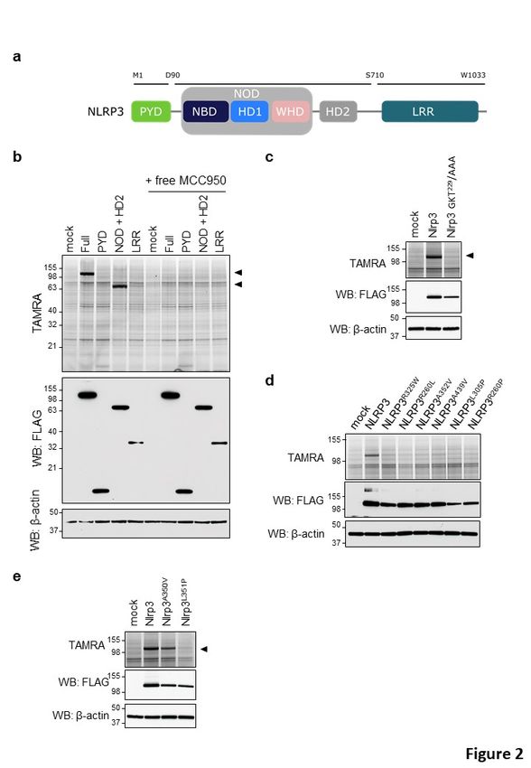
bioRxiv preprint first posted online May. 11, 2019; doi: http://dx.doi.org/10.1101/634493. The copyright holder for this preprint
 (which was not peer-reviewed) is the author/funder, who has granted bioRxiv a license to display the preprint in perpetuity.
                              It is made available under a CC-BY-NC-ND 4.0 International license.

     Introduction

     Inflammasomes are a suite of multi-protein complexes that play central roles in innate
     immune responses through their ability to recruit and activate caspase-1 (Broz and Dixit, 2016;
     Lamkanfi and Dixit, 2014). This cysteine protease cleaves the cytokines interleukin (IL)-1β and
     IL-18 and drives pyroptosis, a highly inflammatory regulated cell death mode that is induced
     by cleavage of gasdermin D (GSDMD) (Kayagaki et al., 2015; Shi et al., 2015). Amongst the
     different inflammasome pathways, the NLRP3 inflammasome responds to the broadest suite
     of inflammasome agonists that includes diverse pathogen-associated molecular patterns
     (PAMPs), host-derived danger-associated molecular patterns (DAMPs) like ATP, protein
     aggregates and β-fibrils such as β-amyloid, a broad range of environmental insults and
     ionophores such as nigericin and medically relevant crystals such as alum, CCPD, MSU, silica
     and asbestos (Broz and Dixit, 2016; Lamkanfi and Dixit, 2014). Moreover, the NLRP3
     inflammasome is engaged by Gram-negative pathogens, lipopolysaccharides (LPS) of which
     are sensed in the cytosol by the non-canonical NLRP3 inflammasome pathway (Hagar et al.,
     2013; Kayagaki et al., 2011; Kayagaki et al., 2013; Shi et al., 2014). Through the latter
     mechanism, cleavage of GSDMD by caspase-11 – and its human orthologs caspases 4 and 5 –
     promotes assembly of cytolytic GSDMD pores in the plasma membrane that also activate the
     NLRP3 inflammasome to drive caspase-1-dependent IL-1β and IL-18 maturation (Kayagaki et
     al., 2015; Kayagaki et al., 2011). Consistent with the chemical diversity of NLRP3 stimuli,
     activation of the NLRP3 inflammasome is thought to converge on sensing of a secondary
     messenger or cellular state that is universally induced by NLRP3-activating agents (Munoz-
     Planillo et al., 2013).

     Aberrant NLRP3 inflammasome activity is thought to contribute to the pathogenesis of many
     chronic diseases, including inflammatory diseases such as gout and pseudogout, metabolic
     diseases like atherosclerosis and NAFLD/NASH, and neurodegenerative diseases like
     Alzheimer’s disease, Parkinson’s disease and multiple sclerosis (Mangan et al., 2018; Voet et
     al., 2019). Moreover, gain-of-function mutations in and around the central NACHT domain of
     NLRP3 cause three autosomal dominantly inherited periodic fever syndromes that together
     are known as cryopyrin-associated periodic syndrome (CAPS). Symptoms span a clinical
     spectrum with Familial Cold Autoinflammatory Syndrome (FCAS) being the mildest; Muckle-
     well syndrome (MWS) being of moderate severity; and Neonatal Onset Multisystem

                                                                                                                                    3
bioRxiv preprint first posted online May. 11, 2019; doi: http://dx.doi.org/10.1101/634493. The copyright holder for this preprint
(which was not peer-reviewed) is the author/funder, who has granted bioRxiv a license to display the preprint in perpetuity.
It is made available under a CC-BY-NC-ND 4.0 International license.
Inflammatory Disease (NOMID)/Chronic Infantile Neurological, Cutaneous and Articular
Syndrome (CINCA) being the most severe form of CAPS, featuring systemic inflammation,
neurological and sensory impairment and deforming arthropathy (Van Gorp et al., 2019).
Selective and potent inhibitors of the NLRP3 inflammasome may have broad therapeutic
potential in CAPS and other diseases (Mangan et al., 2018; Voet et al., 2019). Early studies
with the sulfonylurea drug glyburide provided proof-of-concept that PAMP-, DAMP-, and
crystal-induced activation of the NLRP3 inflammasome pathway may be selectively targeted
without interfering with other inflammasome pathways (Lamkanfi et al., 2009). The related
diarylsulfonylurea compound MCC950/CRID3 was originally reported as an inhibitor of IL-1ß
secretion (Laliberte et al., 2003), and subsequently shown to potently and selectively inhibit
the NLRP3 inflammasome pathway in murine and human macrophages and monocytes with
IC50 values in the low nM range (Coll et al., 2015; Primiano et al., 2016). There is significant
interest in the discovery and development of diarylsulfonylurea CRIDs such as MCC950/CRID3
for the treatment of CAPS and other diseases based on its ability to curb inflammatory
pathology in mouse models of CAPS, the experimental autoimmune encephalomyelitis mouse
model of multiple sclerosis, NAFLD/NASH, and many other inflammatory disease models
(Mangan et al., 2018; Voet et al., 2019). However, the direct molecular target of
MCC950/CRID3 in the NLRP3 inflammasome pathway has remains elusive, hampering the
rational optimization and development of MCC950/CRID3-based therapies.
Utilizing two different chemoproteomic strategies, we here demonstrate that the NACHT
domain of NLRP3 is the molecular target of diarylsulfonylurea CRIDs. Interestingly, we find
photoaffinity labelling (PAL) of the NACHT domain of NLRP3 requires an intact (d)ATP-binding
pocket and is substantially reduced for most CAPS-associated NLRP3 mutants. In accordance,
NLRP3-driven inflammatory pathology in mouse models of CAPS was not efficiently curbed by
MCC950/CRID3. Consistently, MCC950/CRID3 abolished circulating levels of interleukin (IL)-1β
and IL-18 in LPS-challenged wildtype mice but not in CAPS mice and ex vivo-stimulated mutant
macrophages. These results identify the central NACHT domain of wildtype NLRP3 as the
molecular target of MCC950/CRID3 and show that CAPS-related NACHT mutations prevent
efficient MCC950/CRID3 inhibition. Collectively, this work suggests that MCC950/CRID3-based
therapies may effectively treat inflammation driven by wildtype NLRP3, but not CAPS-
associated mutants.
4bioRxiv preprint first posted online May. 11, 2019; doi: http://dx.doi.org/10.1101/634493. The copyright holder for this preprint
(which was not peer-reviewed) is the author/funder, who has granted bioRxiv a license to display the preprint in perpetuity.
It is made available under a CC-BY-NC-ND 4.0 International license.
Results
MCC950/CRID3 selectively binds to human and murine NLRP3
As a first approach to identify the molecular target of MCC950/CRID3, we made use of photo-
affinity labeling (PAL) in combination with click chemistry (Smith and Collins, 2015). Guided by
limited structure-activity studies, a cell-permeable photo-affinity probe was synthesized (Fig.
1A, compound PAL-CRID3) that contains a photo-reactive benzophenone group to enable
direct covalent labeling of MCC950/CRID3 targets upon exposure to UV light. The alkyne
functionality in PAL-CRID3 allowed in situ click reaction with a 5-carboxytetramethylrhodamine
(TAMRA) fluorescent reporter to support in-gel fluorescence detection of the covalent
MCC950/CRID3-protein adduct by SDS-PAGE. A dose-response analysis confirmed that PAL-
CRID3 retained the ability to potently inhibit NLRP3 inflammasome activation with sub-
micromolar IC50 values. PAL-CRID3 inhibited nigericin-induced IL-1β secretion from
Pam3CSK4-primed primary bone marrow-derived macrophages (BMDMs) (Fig. 1b; IC50 = 731
nM) and ER-Hoxb8-immortalized macrophages (iMac) (Fig. 1c; IC50 = 453 nM). As a reference,
MCC950/CRID3 inhibited nigericin-induced IL-1β secretion with approximately 8- and 11-fold
lower IC50 values of 90 and 40 nM, respectively (Fig. 1b, c). We hypothesize somewhat lesser
activity of PAL-CRID3 compared to MCC950/CRID3 is a combination of physical property and
binding differences that decrease target occupancy.
To screen candidate targets of MCC950/CRID3, HEK293T cells overexpressing FLAG-epitope
tagged fusions of the human inflammasome sensor proteins NLRP3, NLRP1, NLRC4, NLRP6 and
MEFV were incubated with PAL-CRID3 and exposed to UV light to allow covalent binding of
PAL-CRID3 to potential targets. Following cell lysis, the probe was conjugated to the TAMRA
reporter by click chemistry, and protein lysates were separated by SDS-PAGE. Notably, in-gel
fluorescence imaging showed significant TAMRA labelling of ectopically expressed NLRP3, but
not other inflammasome sensors in the panel (Fig. 1d). To further extend these findings, we
next confirmed binding to murine Nlrp3 (Fig. 1e). As controls, protein phosphatase PP1Cb and
glutathione S-transferase GSTO1, which has been proposed as the target of CRID compounds
(Laliberte et al., 2003), were not labelled by PAL-CRID3 (Fig. 1e). Moreover, binding of PAL-CRID3
to human NLRP3 and murine Nlrp3 only was observed following UV crosslinking and labelling
was rescued by competition with excess MCC950/CRID3 (Fig. 1e), thus ruling out non-specific
cross-linking and validating specificity of these findings. To further characterize the interaction
5bioRxiv preprint first posted online May. 11, 2019; doi: http://dx.doi.org/10.1101/634493. The copyright holder for this preprint
(which was not peer-reviewed) is the author/funder, who has granted bioRxiv a license to display the preprint in perpetuity.
It is made available under a CC-BY-NC-ND 4.0 International license.
of PAL-CRID3 with components of the NLRP3 inflammasome, we assessed binding to human
NEK7, ASC, caspase-1 and GSDMD. PAL-CRID3 failed to label the NLRP3 inflammasome
components in the panel apart from NLRP3 (Fig. 1f). Collectively, these results suggest that
CRID compounds, including MCC950/CRID3, inhibit NLRP3 inflammasome signalling by directly
binding to NLRP3.
To confirm and extend these findings to endogenous NLRP3, we made use of recently
described iBody technology to immobilize MCC950/CRID3 on polymers that enabled
immunoprecipitation of MCC950/CRID3 targets (Simon et al., 2018). In brief, MCC950/CRID3
was stochastically modified with a photo-activatable phenyldiazirine linker, and the resulting
isomeric mixture was conjugated to a hydrophilic N-(2-hydroxypropyl)methacrylamide
(HPMA) polymer backbone that is decorated with a biotin affinity tag (for details please refer
to the Materials and Methods section and (Simon et al., 2018)). The MCC950/CRID3 iBody
conjugate is further referred to as iBody U121. The corresponding iBody conjugate lacking
MCC950/CRID3 served as a negative control and is referred to as iBody U126.
BMDMs of wildtype (C57BL/6J) and Nlrp3-/- mice were primed with LPS to transcriptionally
upregulate NLRP3 inflammasome components (Bauernfeind et al., 2009), and cell lysates were
subsequently incubated with iBody U121 (MCC950/CRID3) or iBody U126 (control) followed
by immunoprecipitation with streptavidin-coupled beads. Contrary to control iBody U126,
iBody U121 immunoprecipitated endogenous Nlrp3 from wildtype BMDMs (Fig. 1g). iBody
U121 failed to pulldown inflammasome components ASC and caspase-1 (Fig. 1g). As expected,
the immunoreactive band was absent from immunoprecipitates and lysates of LPS-primed
Nlrp3-deficient macrophages (Fig. 1g). Given the lack of suitable antibodies to detect
endogenous NLRC4, we made use of reported Nlrc43xFlag/3xFlag knock-in mice (Matusiak et al.,
2015; Qu et al., 2012) to further validate selective targeting of NLRP3. Consistent with our
earlier results, only iBody U121 precipitated endogenous Nlrp3 from LPS-primed
Nlrc43xFlag/3xFlag BMDMs (Fig. 1h). Comparable background binding of Nek7 was observed with
iBody U121 and control iBody U126, whereas neither Nlrc4 (detected using FLAG antibodies),
Nek7 nor Pyrin were retrieved in iBody U121 immunoprecipates of LPS-primed Nlrc4Flag/Flag
BMDMs (Fig. 1h). Collectively, these results show that MCC950/CRID3 selectively binds to
NLRP3 in LPS-primed macrophages.
6bioRxiv preprint first posted online May. 11, 2019; doi: http://dx.doi.org/10.1101/634493. The copyright holder for this preprint
(which was not peer-reviewed) is the author/funder, who has granted bioRxiv a license to display the preprint in perpetuity.
It is made available under a CC-BY-NC-ND 4.0 International license.
PAL-CRID3 targets the central NACHT domain of wildtype NLRP3, but not CAPS mutants
NLRP3 consists of an amino-terminal Pyrin domain (PYD), a central nucleotide binding and
oligomerization (NACHT) domain and carboxy-terminal leucine-rich repeats (LRRs) that are
thought to lock NLRP3 in an inactive conformation. To map the NLRP3 region(s) to which
MCC950/CRID3 binds, we generated deletion mutants that cover the following three regions
of human NLRP3: (i) the N-terminal PYD; (ii) the central NACHT domain (comprising the NOD
domain and helical domain 2 (HD2)); and (iii) the carboxy-terminal LRR region (Fig. 2a).
Consistent with our previous results (Fig. 1), TAMRA analysis showed significant binding of
PAL-CRID3 to ectopically expressed full-length NLRP3 in HEK293T cells (Fig. 2b). In addition,
we observed binding of PAL-CRID3 to the isolated NACHT (NOD + HD2) region of NLRP3, but
not to the PYD and LRR domains (Fig. 2b). Labelling of both full-length NLRP3 and the isolated
NACHT region by PAL-CRID3 was rescued by treatment with an excess amount of free
MCC950/CRID3 (Fig. 2b), confirming specificity and suggesting PAL-CRID3 and MCC950/CRID3
compete for the same binding site. Binding of ATP/dATP to the Walker A pocket in the NACHT
domain is essential for NLRP3 inflammasome assembly and function (Duncan et al., 2007).
Interestingly, mutation of the Walker A motif (GKT229/AAA) in full-length Nlrp3 abolished PAL-
CRID3 binding (Fig. 2c), suggesting that an intact ATP binding pocket is required for strongest
binding of diarylsulfonylurea CRID compounds.
The central NACHT domain of NLRP3 also contains most reported gain-of-function mutations
that cause CAPS disease (https://infevers.umai-montpellier.fr/web/index.php). This
prompted us to explore PAL-CRID3 binding to CAPS-associated NLRP3 mutants. Unexpectedly,
introducing a random selection of 6 different CAPS mutations in the NACHT domain of human
NLRP3 that are associated with respectively MWS (NLRP3R325W, NLRP3R260L, NLRP3A352V), FCAS
(NLRP3A439V, NLRP3L305P) or NOMID (NLRP3R260P) all blunted binding of PAL-CRID3 to full-length
human NLRP3 (Fig. 2d). The classical MWS A352V and FCAS L353P mutations in human NLRP3
correspond to the A350V and L351P mutations that have been knocked into the murine Nlrp3
gene to model CAPS disease in mice (Brydges et al., 2009). Notably, the Nlrp3A350V mutant did
not significantly affect labelling by PAL-CRID3, whereas the Nlrp3L351P mutation abolished
labelling by PAL-CRID3 (Fig. 2e). Together, these results suggest that photoaffinity labelling of
the NACHT domain requires an intact (d)ATP-binding pocket and is substantially reduced for
most CAPS-associated NLRP3 mutants.
7bioRxiv preprint first posted online May. 11, 2019; doi: http://dx.doi.org/10.1101/634493. The copyright holder for this preprint
(which was not peer-reviewed) is the author/funder, who has granted bioRxiv a license to display the preprint in perpetuity.
It is made available under a CC-BY-NC-ND 4.0 International license.
MCC950/CRID3 inhibits the inflammasome in wildtype but not Nlrp3L351P macrophages
Considering that our observation that CAPS-associated Nlrp3 mutants escape PAL-CRID3
binding may have potential implications for treating CAPS with MCC950/CRID3, we sought to
functionally validate these results with MCC950/CRID3 in the reported Nlrp3L351P CAPS model
(Brydges et al., 2009). To this end, mice that were homozygous for the Nlrp3L351P allele were
bred to transgenic mice that hemizygously expressed the tamoxifen-inducible Cre-ERT2 fusion
gene (CreT) (Ventura et al., 2007). Following tamoxifen treatment and excision of the floxed
neomycin resistance cassette, BMDMs of the resulting Nlrp3L351P/+CreT+ mice expressed Nlrp3
from both the wildtype and Nlrp3L351P alleles. Macrophages from Cre-ERT2-negative
littermates (Nlrp3L351P/+CreT-), which only express Nlrp3 from the wildtype allele, were used
as controls in these experiments.
Culture media of LPS-stimulated Nlrp3L351P/+CreT+ macrophages contained significant levels of
IL-1β (Fig. 3a) and IL-18 (Fig. 3b) that were associated with marked maturation of caspase-1
and IL-1β in cell lysates (Fig. 3c). As reported (Brydges et al., 2009), these LPS-induced
inflammasome responses were driven by the CAPS-associated Nlrp3L351P allele because
Nlrp3L351P/+CreT- BMDMs failed to secrete detectable levels of IL-1β (Fig. 3a) and IL-18 (Fig.
3b), and did not contain mature caspase-1 and IL-1β in their cell lysates (Fig. 3c). Consistent
with our previous results that CAPS-associated Nlrp3L351P escaped PAL-CRID3 binding, we
found that MCC950/CRID3 failed to inhibit the above inflammasome responses driven by the
Nlrp3L351P allele. We confirmed that MCC950/CRID3 was active against wildtype NLRP3
because it abolished levels of LPS-ATP-induced secretion of IL-1β (Fig. 3d) and IL-18 (Fig. 3e)
in culture media of Nlrp3L351P/+CreT- BMDMs (that are solely driven by wildtype Nlrp3 in this
genotype given the absence of a Cre-ERT2 transgene), as well as the concomitant maturation
of caspase-1 and IL-1β in the corresponding cell lysates (Fig. 3f). Similarly, MCC950/CRID3
abolished LPS+nigericin-induced secretion of IL-1β (Fig. 3g) and IL-18 (Fig. 3h), and maturation
of caspase-1 and IL-1β by Nlrp3L351P/+CreT- macrophages (Fig. 3i). In marked contrast, LPS+ATP-
and LPS+nigericin-induced inflammasome responses were insensitive to MCC950/CRID3
blockade in Nlrp3L351P/+CreT+ BMDMs that express both the Nlrp3L351P allele and wildtype Nlrp3
(Fig. 3d-i). Together, these results establish that the CAPS-associated Nlrp3L351P allele is
insensitive to MCC950/CRID3 inhibition.
8bioRxiv preprint first posted online May. 11, 2019; doi: http://dx.doi.org/10.1101/634493. The copyright holder for this preprint
(which was not peer-reviewed) is the author/funder, who has granted bioRxiv a license to display the preprint in perpetuity.
It is made available under a CC-BY-NC-ND 4.0 International license.
MCC950/CRID3 inhibition of inflammasome responses in Nlrp3A350V macrophages
Unlike for Nlrp3L351P, labelling of Nlrp3A350V by G03086997 was not significantly impacted by
the mutation (Fig. 2e). To address whether this was mirrored by potent inhibition of
Nlrp3A350V-driven inflammasome responses by MCC950/CRID3, mice that were homozygous
for the previously reported Nlrp3A350V allele were bred to the Cre-ERT2 (CreT) transgenic mice
described above. Following tamoxifen treatment and excision of the floxed neomycin
resistance cassette, BMDMs of the resulting Nlrp3A350V/+CreT+ mice expressed Nlrp3 from both
the wildtype and Nlrp3A350V alleles. BMDMs from CreT-negative littermates (Nlrp3A350V/+CreT-
), which only express Nlrp3 from the wildtype allele, were used as controls in these
experiments.
Like Nlrp3L351P/+CreT+ BMDMs (Fig. 3a-c), Nlrp3A350V/+CreT+ macrophages that expressed
Nlrp3A350V secreted high levels of secreted IL-1β (Fig. 4a) and IL-18 (Fig. 4b) in response to LPS
stimulation alone. This was accompanied by proteolytic maturation of caspase-1 and proIL-1β
as demonstrated by immunoblot analysis (Fig. 4c). As expected, these responses were absent
from LPS-stimulated Nlrp3A350V/+CreT- BMDMs that expressed wildtype Nlrp3 only (Fig. 4a-c).
MCC950/CRID3 potently inhibited LPS-induced IL-1β (Fig. 4a) and IL-18 (Fig. 4b) secretion, and
Nlrp3A350V-driven cleavage of caspase-1 and IL-1β (Fig. 4c) in marked contrast to
Nlrp3L351P/+CreT+ macrophages that proved insensitive to MCC950/CRID3 blockade (Fig. 3a-c).
LPS+ATP and LPS+nigericin potently triggered IL-1β and IL-18 secretion, and maturation of
caspase-1 and proIL-1β from both Nlrp3A350V/+CreT+ and Nlrp3A350V/+CreT- control BMDMs (Fig.
4d-i). However, whereas MCC950/CRID3 abolished LPS+ATP- and LPS+nigericin-induced IL-1β
secretion from Nlrp3A350V/+CreT- control BMDMs, it failed to alter secretion of IL-1β from
parallelly-treated Nlrp3A350V/+CreT+ BMDMs (Fig. 4d, g). MCC950/CRID3 also abrogated
LPS+ATP- and LPS+nigericin-induced IL-18 secretion from control Nlrp3A350V/+CreT- BMDMs,
but only reduced IL-18 secretion from parallelly stimulated Nlrp3A350V/+CreT+ macrophages by
about 50% (Fig. 4e, h). Aligned with these results, MCC950/CRID3 inhibited LPS+ATP- and
LPS+nigericin-induced cleavage of caspase-1 and proIL-1β in control Nlrp3A350V/+CreT- BMDMs
but not in Nlrp3A350V/+CreT+ macrophages (Fig. 4f, i). Together, these results show that
although binding of PAL-CRID3 to Nlrp3A350V was not significantly compromised, the mutation
subtly alters the ability of MCC950/CRID3 to inhibit inflammasome activation in primary
9bioRxiv preprint first posted online May. 11, 2019; doi: http://dx.doi.org/10.1101/634493. The copyright holder for this preprint
(which was not peer-reviewed) is the author/funder, who has granted bioRxiv a license to display the preprint in perpetuity.
It is made available under a CC-BY-NC-ND 4.0 International license.
macrophages, with potent inhibition seen only in response to LPS but not following ‘signal 2’
triggers such as ATP and nigericin.
Inflammasome inhibition in homozygous mutant macrophages
The studies described above were performed in heterozygous macrophages that express both
wildtype and CAPS-associated Nlrp3 mutants. Considering that the Nlrp3 NACHT region
facilitates Nlrp3 oligomerization, we decided to further assess MCC950/CRID3 responses in
macrophages that uniquely express the CAPS-associated Nlrp3 mutants in the absence of
wildtype Nlrp3. To do so, we transduced wildtype, Nlrp3A350V/A350V and Nlrp3L351P/L351P BMDMs
with Cre recombinase-expressing lentiviruses to excise the neomycin resistance cassette that
is placed upstream of the Nlrp3 mutation and allow expression of the CAPS-associated Nlrp3
mutants.
As expected, wildtype macrophages failed to secrete IL-1β and IL-18 in response to LPS alone,
and MCC950/CRID3 abolished secretion of IL-1β and IL-18 when wildtype BMDM were
stimulated with LPS+ATP or LPS+nigericin (Fig. 5a, b). LPS stimulation alone was sufficient to
induce extracellular release of IL-1β and IL-18 in homozygous Nlrp3L351P/ L351P and Nlrp3A350V/
A350V macrophages (Fig. 5c-f). MCC950/CRID3 inhibited Nlrp3L351P-induced secretion of IL-1β
and IL-18 neither in response to LPS alone, nor when combined with ‘signal 2’ agents ATP or
nigericin (Fig. 5c, d), unequivocally establishing that Nlrp3L351P-induced inflammasome
activation is insensitive to MCC950/CRID3 blockade. Consistent with our previous results in
heterozygous Nlrp3A350V mutant macrophages, MCC950/CRID3 partially inhibited LPS-induced
IL-1β and IL-18 levels in culture supernatants of homozygous Nlrp3A350V/ A350V macrophages,
but this was not observed in response to LPS+ATP and LPS+nigericin (Fig. 5e, f).
In vivo MCC950/CRID3 inhibition of inflammasome activation in CAPS disease models
Myeloid-specific expression of the Nlrp3L351P and Nlrp3A350V alleles in knock-in mice was shown
to drive systemic inflammation accompanied by respectively embryonic and perinatal lethality
that in both cases required Nlrp3 inflammasome activation (Brydges et al., 2009). To seek
further validation of the notion that the Nlrp3L351P mutations escapes MCC950/CRID3
inhibition, we next investigated how MCC950/CRID3 treatment impacts on the CAPS
phenotype of Nlrp3L351P/+CreT+ mice. As expected, serum levels of IL-1β, IL-18 and IL-6 were
significantly increased in Nlrp3L351P/+CreT+ mice three days after tamoxifen dosing relative to
10bioRxiv preprint first posted online May. 11, 2019; doi: http://dx.doi.org/10.1101/634493. The copyright holder for this preprint
(which was not peer-reviewed) is the author/funder, who has granted bioRxiv a license to display the preprint in perpetuity.
It is made available under a CC-BY-NC-ND 4.0 International license.
the basal levels of tamoxifen-treated Nlrp3L351P/+CreT- littermate mice (Supplementary Fig. 1a-
c). Moreover, tamoxifen treatment resulted in Nlrp3L351P/+CreT+ mice presenting with
substantial weight loss and mortality, with all mice being lost or requiring termination because
of humane endpoints within 5 days after commencing tamoxifen treatment (Fig. 6a, b). As a
control, tamoxifen administration did not alter body weight or survival of Nlrp3L351P/+CreT-
mice (Fig. 6a, b). Daily i.p. injection of MCC950/CRID3 did not rescue body weight loss or
mortality rates of Nlrp3L351P/+CreT+ mice, suggesting that MCC950/CRID3 failed to inhibit in
vivo Nlrp3L351P-induced inflammasome activation (Fig. 6a, b). In agreement, i.p. dosing of
MCC950/CRID3 failed to reduce tamoxifen-induced levels of IL-1β, IL-18 and IL-6 in serum of
Nlrp3L351P/+ CreT+ mice (Fig. 6c-e). Thus, the CAPS-associated L351P mutation renders Nlrp3
insensitive to MCC950/CRID3 inhibition.
Our results in Nlrp3A350V macrophages showed that this mutation subtly altered the ability of
MCC950/CRID3 to inhibit inflammasome activation with potent inhibition seen only in
response to LPS but not following ‘signal 2’ triggers such as ATP and nigericin (Fig. 4). To
determine how this translates to the in vivo disease setting, we analyzed circulating cytokine
levels and body weight loss of Nlrp3A350V/+CreT+ mice following tamoxifen administration.
Although there was a trend towards increased serum levels of IL-1β, the low measured
concentrations did not reach statistical significance compared to tamoxifen-treated
Nlrp3A350V/+CreT- littermate mice (Supplementary Fig. 1d). However, serum concentrations of
IL-18 and IL-6 were signficantly elevated in tamoxifen-treated Nlrp3A350V/+CreT+ mice relative
to Nlrp3A350V/+CreT- littermates (Supplementary Fig. 1e, f). Nevertheless, levels of the latter
cytokines were on average 10 to 20-fold lower than seen in tamoxifen-treated
Nlrp3L351P/+CreT+ mice, a finding that is consistent with the milder pathology and the lack of
mortality associated with the Nlrp3A350V/+CreT+ CAPS model. Consistent with published
findings (McGeough et al., 2012), Nlrp3A350V/+CreT+ mice developed an inflammatory
phenotype characterized by a steady weight loss of up to 20 % within 27 days (Fig. 6e).
Notably, daily i.p. dosing of MCC950/CRID3 stabilized Nlrp3A350V-mediated body weight loss in
Nlrp3A350V/+CreT+ mice relative to PBS-treated controls, although differences were small and
MCC950/CRID3-treated mice failed to thrive and gain weight like the Nlrp3A350V/+CreT- control
group (Fig. 6e). In agreement, MCC950/CRID3 had a mild or no effect on circulating levels of
the systemic inflammatory markers IL-18 (Fig. 6f) and IL-6 (Fig. 6g), respectively. Together,
11bioRxiv preprint first posted online May. 11, 2019; doi: http://dx.doi.org/10.1101/634493. The copyright holder for this preprint
(which was not peer-reviewed) is the author/funder, who has granted bioRxiv a license to display the preprint in perpetuity.
It is made available under a CC-BY-NC-ND 4.0 International license.
these results establish that MCC950/CRID3 has a weak, but measurable effect on Nlrp3A350V-
induced inflammasomopathy in adult mice.
MCC950/CRID3 inhibition of LPS-induced cytokines in CAPS mutant mice
To complement the chronic CAPS disease models described above, we next evaluated the
potency of MCC950/CRID3 in inhibiting acute Nlrp3-dependent inflammasome responses by
subjecting wildtype and CAPS mutant mice to LPS-induced endotoxemia and probing the
effect of MCC950/CRID3 on well-documented Nlrp3-dependent readouts such as LPS-induced
elevation of serum levels of IL-1β and IL-18 (He et al., 2013).
As expected, Nlrp3L351P/+CreT- control mice presented with increased serum levels of IL-1β and
IL-18 3 hours after LPS challenge (Fig. 7a, b). MCC950/CRID3 substantially curbed circulating
IL-1β and IL-18 levels in this control group (Fig. 7a, b), consistent with reported findings in LPS-
challenged C57BL/6 mice (Coll et al., 2015). Contrastingly, serum levels of IL-1β and IL-18 in
LPS-dosed Nlrp3L351P/+CreT+ mice were not significantly impacted by MCC950/CRID3 relative
to PBS-treated Nlrp3L351P/+CreT+ littermates (Fig. 7c, d).
LPS challenge increased serum concentrations of IL-1β and IL-18 in Nlrp3A350V/+CreT- control
mice, which were inhibited by MCC950/CRID3 (Fig. 7e, f) similarly to its effect on LPS-treated
Nlrp3L351P/+CreT- control mice (Fig. 7a, b). This is not unexpected since both genotypes express
solely wildtype Nlrp3 in the absence of Cre-recombinase-mediated excision of the neomycin
resistance cassette that disrupts expression of the respective CAPS-associated Nlrp3 mutants.
However, in marked contrast to Nlrp3L351P-expressing CreT+ mice (Fig. 7c, d), MCC950/CRID3
markedly curbed circulating concentrations of IL-1β and IL-18 in Nlrp3A350V-expressing CreT+
mice (Fig. 7g, h). These results confirm that MCC950/CRID3 potently inhibits inflammasome
activation by wildtype Nlrp3 and the MWS-associated Nlrp3A350V mutant, but not the FCAS-
associated Nlrp3L351P mutant.
12bioRxiv preprint first posted online May. 11, 2019; doi: http://dx.doi.org/10.1101/634493. The copyright holder for this preprint
(which was not peer-reviewed) is the author/funder, who has granted bioRxiv a license to display the preprint in perpetuity.
It is made available under a CC-BY-NC-ND 4.0 International license.
Discussion
Activation of the NLRP3 inflammasome has been observed in many diseases, and its central
role in driving pathological inflammation renders it an attractive target for therapeutic
intervention (Mangan et al., 2018). Gain-of-function mutations in NLRP3 cause hereditary
periodic fever syndromes that are collectively referred to as CAPS, and these patients are
currently treated with biologics that target secreted IL-1 (Van Gorp et al., 2019). As reported
(Ridker et al., 2017), chronic IL-1-blockade increases risk for fatal infections and sepsis,
suggesting that selective targeting of the NLRP3 inflammasome may potentially be a safer and
more efficacious therapeutic strategy as it would block production of the central inflammatory
mechanisms that contribute to inflammatory pathology while at the same time keeping non-
targeted inflammasomes available to produce IL-1β to cope with infections.
Early studies with the sulfonylurea compound glyburide provided proof-of-concept that small
molecules may selectively inhibit the NLRP3 inflammasome pathway without interfering with
other inflammasomes (Lamkanfi et al., 2009). Subsequently, several additional compounds
have been reported to specifically inhibit the NLRP3 inflammasome pathway, but the majority
of these agents has weak activity against the NLRP3 inflammasome pathway (μM IC50
concentrations) and may target NF-κB signaling and other immune pathways (reviewed in
(Mangan et al., 2018)). MCC950/CRID3 is structurally related to glyburide (Coll et al., 2015;
Laliberte et al., 2003) and is considered the most potent and selective inhibitor of NLRP3
inflammasome signaling reported to date. There is substantial interest in developing this
chemical scaffold for the treatment of CAPS and other diseases. However, these efforts are
constrained by the lack on insight in the molecular target and mechanism by which
MCC950/CRID3 and related sulfonylurea molecules inhibit activation of the NLRP3
inflammasome pathway.
Making use of photoaffinity labeling and iBody technology as complementary chemical
biology approaches, we here identified NLRP3 as the physical target of MCC950/CRID3. We
further mapped the binding pocket to the central NACHT domain of NLRP3 and showed that
photoaffinity labelling required an intact ATP/dATP binding pocket. This suggests that binding
may occur at the nucleotide-binding pocket of NLRP3, although it cannot be excluded that
mutations in the Walker A motif may cause long-distance conformational changes that distort
a remote MCC950/CRID3 binding pocket elsewhere in the NACHT domain. Notable in this
regard is our observation that a randomly selected panel of 6 CAPS-associated gain-of-
13bioRxiv preprint first posted online May. 11, 2019; doi: http://dx.doi.org/10.1101/634493. The copyright holder for this preprint
(which was not peer-reviewed) is the author/funder, who has granted bioRxiv a license to display the preprint in perpetuity.
It is made available under a CC-BY-NC-ND 4.0 International license.
function mutations in human NLRP3 all failed to be labelled by our PAL probe, further
supporting the notion that the sulfonylurea CRID binding pocket is highly sensitive for
conformational changes in the protein that are imposed by mutations. Further insight in the
conformational requirements and the molecular mechanism by which MCC950/CRID3 inhibits
NLRP3 activation awaits a high-resolution structural analysis of the MCC950/CRID3 binding
pocket.
Considering the potential implications of these findings for treating CAPS with MCC950/CRID3-
based therapies, we evaluated the functional impact of MCC950/CRID3 in two reported CAPS
models (Brydges et al., 2009). When knocked into the murine Nlrp3 sequence, the FCAS-
associated Nlrp3L351P mutant (corresponding to human L353P) also failed to bind to PAL-CRID3.
Consistent with these results, our analysis of both ex vivo-stimulated mutant macrophages
and in vivo CAPS and endotoxemia models unequivocally established that MCC950/CRID3
potently inhibits inflammasome activation by wildtype Nlrp3, but not the FCAS-associated
Nlrp3L351P mutant. Surprisingly, however, we found that labelling by PAL-CRID3 to the MWS-
linked Nlrp3A350V mutant (corresponding to adjacent residue A352V in human NLRP3) was not
significantly impacted by the mutation. MCC950/CRID3 partially inhibited LPS-induced IL-1β
and IL-18 secretion from macrophages that homozygously or heterozygously expressed the
Nlrp3A350V mutant, but inhibition of the Nlrp3A350V inflammasome was completely lost in
response to ‘signal 2’ agents ATP and nigericin. Consequently, MCC950/CRID3 significantly
lowered IL-1β and IL-18 levels in serum of LPS-challenged Nlrp3A350V knock-in mice, whereas it
provided only limited protection against chronic CAPS mutation-driven body weight loss. The
CreERT2 recombinase expression system used with our CAPS disease models allows for
controlled ubiquitious tissue expression of the mutant Nlrp3 knock-in allele upon tamoxifen
treatment in adult animals. Expression of the Nlrp3A350V allele does not induce lethality in adult
mice under these conditions. However, another study (Coll et al., 2015) that relied on
Lysosome M-Cre-driven expression of the Nlrp3A350V allele in cells of the myeloid lineage
observed that MCC950/CRID3 rescued neonatal lethality, consistent with our observation that
the Nlrp3A350V mutant retained sensitivity to MCC950/CRID3 inhibition. This report also
suggested MCC950/CRID3 inhibits LPS-induced IL-1β processing in peripheral blood
mononuclear cells (PBMCs) from MWS patients. Another study showed that unlike samples
from healthy controls, LPS- and LPS+ATP-induced IL-1β secretion from whole blood samples
of genetically defined CAPS patients resisted MCC950/CRID3 inhibition (Grinstein et al., 2018),
14bioRxiv preprint first posted online May. 11, 2019; doi: http://dx.doi.org/10.1101/634493. The copyright holder for this preprint
(which was not peer-reviewed) is the author/funder, who has granted bioRxiv a license to display the preprint in perpetuity.
It is made available under a CC-BY-NC-ND 4.0 International license.
which is consistent with our results suggesting that MCC950/CRID3-based therapies may
effectively treat inflammation driven by wildtype NLRP3, but may be less effective in CAPS
patients. To conclude, by identifying the molecular target of MCC950/CRID3 in the NLRP3
inflammasome pathway, and by evaluating its ability to inhibit CAPS mutatant variants, the
findings presented here provide a mechanistic framework for advancing therapeutic
development of this chemical scaffold and for understanding its therapeutic potential in
patients.
15bioRxiv preprint first posted online May. 11, 2019; doi: http://dx.doi.org/10.1101/634493. The copyright holder for this preprint
(which was not peer-reviewed) is the author/funder, who has granted bioRxiv a license to display the preprint in perpetuity.
It is made available under a CC-BY-NC-ND 4.0 International license.
Material and methods
Synthesis of PAL-CRID3. Detailed experimental procedures for the synthesis of PAL-CRID3 are
provided in the Supplementary Data section.
Synthesis of iBody conjugates. Experimental procedures for the preparation of the iBody
conjugates have been described by (Simon et al., 2018) and details for the production of
MCC950/CRID3 iBody conjugates are provided in the Supplementary Data section.
iMac and BMDM culture. Bone marrow cells from C57BL/6 mice were immortalized by ER-
Hoxb8 (iMac) as described previously (Wang et al., 2006). Primary bone marrow or iMac
progenitor cells were differentiated in DMEM or IMDM supplemented with 10% endotoxin-
free heat-inactivated fetal bovine serum, 20-30% L929-conditioned medium, 100 U ml-1
penicillin, and 100 mg ml-1 streptomycin for 5-6 days at 37°C in a humidified atmosphere
containing 5% CO2. 6 days later, cells were collected and seeded at a density of 8.5 x 10⁵ cells
per well in 12-well plates in IMDM containing 10% heat-inactivated FBS and 1% non-essential
amino acids in the presence of antibiotics. The next day BMDMs were either left untreated or
treated with 1 μM MCC950/CRID3 (S7809, Selleckchem) and then stimulated with 0.5 μg ml-1
ultrapure LPS from Salmonella minnesota (tlrl-smlps, Invivogen) for 3 h followed by 5 mM ATP
(10519987001, Roche) or 20 μM nigericin (N-7143, Sigma-Aldrich) for 45 min. For the dose-
response analysis of PAL-CRID3 and MCC950/CRID3, adherent BMDMs or differentiated iMac
were seeded at 1 x 10⁵ cells per well in 96-well plates and cultured overnight. The following
day, medium was removed and replaced with OPTI-MEM I (Thermo Fisher Scientific)
containing 1 μg mL-1 Pam3CSK4 (Invivogen). Post-priming (5-6 h later), cells were incubated
with DMSO (1:1,000), MCC950/CRID3 (0.001-100 µM), or PAL-CRID3 (0.001-100 µM) for 30
min and then stimulated with 5 μg mL-1 nigericin (Invivogen) for 30 min.
Transfections. HEK293T cells were maintained in DMEM media supplemented with 10% fetal
bovine serum. HEK293T cells were transfected in 12-well plates with Lipofectamine 2000
(Thermo Fisher Scientific) according to manufacturer’s instructions. All constructs, including
NLRP3 cDNAs were synthesized and subcloned into pCDNA3 (+) Zeo (Thermo Fisher Scientific).
Photo-labeling. Transfected HEK293T were incubated with DMSO or photo probe PAL-CRID3
(1 μM) for 30 min at 37°C in 500 μL OPTI-MEM. For competition experiments with free
16bioRxiv preprint first posted online May. 11, 2019; doi: http://dx.doi.org/10.1101/634493. The copyright holder for this preprint
(which was not peer-reviewed) is the author/funder, who has granted bioRxiv a license to display the preprint in perpetuity.
It is made available under a CC-BY-NC-ND 4.0 International license.
MCC950/CRID3, 10 μM MCC950/CRID3 was added 30 min prior. Photo-labeling and click
chemistry experiments were performed as described with slight modification (Mackinnon and
Taunton, 2009). Briefly, cells were washed once with ice-cold PBS and UV irradiated at 365 nm
(100 W) on ice for 10 min. Cells were washed once with ice-cold PBS and frozen at -80°C. Lysis
buffer was added (40 mM HEPES, 140 mM NaCl, 0.1% Triton-X-100, Roche EDTA-free complete
protease inhibitor) and cells were detached with a cell scraper. Crude lysates were clarified by
centrifugation (30 min at 14,000 rpm, 4°C). For click chemistry, lysates (20 μL) were incubated
with 5 μL freshly mixed click cocktail comprised of: 1.7 mM TBTA in 1:4 DMSO/t-BuOH (1.5
μL); 5 mM TAMRA-N3 (0.3 μL); 50 mM TCEP (0.5 μL, freshly prepared); 10 % SDS (2.7 μL). Then,
50 mM CuSO4 (0.5 μL), was added and reactions were incubated for 1 hr at RT with gentle
mixing. Reactions were quenched with 4X LDS buffer and separated by SDS-PAGE. Gels were
scanned for TAMRA fluorescence on a Typhoon Trio scanner (GE Life Sciences).
Mice. Nlrp3-/- (Mariathasan et al., 2006), NLRC43xFlag (Qu et al., 2012) were described. The CAPS
models Nlrp3A350VneoR and Nlrp3L351PneoR (Brydges et al., 2009), and R26-CreERT2 mice (Ventura
et al., 2007) (B6.129-Gt(ROSA)26Sortm1(cre/ERT2)Tyj/J, Jax stock number: 008463; here
abbreviated as CreT) were originally obtained from The Jackson Laboratories and colonies
were further maintained at animal facilities of Ghent University. Mice were housed in
individually ventilated cages and kept under pathogen-free conditions at animal facilities of
Ghent University and Genentech. All animal experiments were conducted with permission of
the Ethical committees on laboratory animal welfare of Ghent University. Nlrp3A350VneoR and
Nlrp3L351PneoR that are homozygous for the mutated Nlrp3 gene were bred to the tamoxifen-
inducible Cre line R26-CreERT2 mice to generate Nlrp3A350neoR/+ R26-CreERT2 Tg+ (herein referred
to as Nlrp3A350V/+CreT+) and Nlrp3L351PneoR/+R26-CreERT2 Tg+ mice (here referred to as
Nlrp3L351P/+CreT+), in which expression of mutant Nlrp3 is induced through administration of
tamoxifen. 4-5 week old mice received at two consecutive days tamoxifen (T5648, Sigma,
dissolved in 1:9 ethanol:corn oil (C-8267, Sigma) at 50 mg ml-1) through oral gavage at a dose
of 5 mg tamoxifen per mouse per day. On day 3, tamoxifen was administered through diet
(Teklad Global 16% Rodent Diet met 400 ppm Tamoxifen per kg, Harlan).
in vivo LPS challenge. 6-12 weeks old mice were intraperitoneally injected with PBS or 50 mg
kg-1 MCC950/CRID3, 30 minutes before being challenged with 40 mg kg-1 LPS (E. coli, serotype
0111:B4, L-2630, Sigma). Mice were euthanized 3 hours after LPS challenge for blood
17bioRxiv preprint first posted online May. 11, 2019; doi: http://dx.doi.org/10.1101/634493. The copyright holder for this preprint
(which was not peer-reviewed) is the author/funder, who has granted bioRxiv a license to display the preprint in perpetuity.
It is made available under a CC-BY-NC-ND 4.0 International license.
collection. At the 0 h timepoint before LPS challenge, blood was collected by retro-orbital
bleeding.
iBody immunoprecipitation. 12 x 106 BMDMs were harvested and cells were washed in PBS
and lysed by three cycles of freeze/thawing in PBS with 0.09% NP40 supplemented with
Complete Protease Inhibitor cocktail (4693159001, Roche Applied Science). Cell lysates were
clarified by centrifugation at 14.000 rpm for 20 minutes and supernatants was subsequently
incubated with 1 μM of the indicated iBody at RT for 10 minutes. Then prewashed streptavidin
conjugated beads were added, followed by overnight incubation at 4°C. The next day, beads
were washed 3 times in PBS with 0.09% NP40 buffer and biotinylated proteins were eluted in
Laemmli buffer and analyzed by Western blot.
Lentiviral Cre transduction. To induce lentivirus production, the lentiviral GFP.Cre empty
vector (20781, Addgene) together with 2nd generation packaging vector psPAX2 (12260,
Addgene) and VSV-G-expressing envelope plasmid pCMV-VSV-G (8454, Addgene) were
transfected into HEK293T cells using jetPRIME transfection reagent (114-15, PolyPlus
Transfect). Lentiviral particle-containing medium was collected 48 hours after transfection,
filtered using a 0.45 μm filter and incubated with harvested bone marrow. 6 days later,
differentiated macrophages were collected, washed and seeded into 12-well cell culture
plates prior to stimulation.
Cytokine analysis. Cytokine levels in culture medium and serum were determined by magnetic
bead-based multiplex assay using Luminex technology (Bio-Rad), IL-1β ELISA (R&D Systems)
and mouse IL-1β tissue culture kit (Meso Scale Discovery) according to the manufacturers’
instructions.
Western blotting. Cell lysates were prepared using lysis buffer containing 20 mM Tris HCl pH
7.4, 200 mM NaCl and 1% NP-40. Samples for detection of caspase-1 and IL-1β processing
were prepared by combining cell lysates with culture supernatants. Samples were denatured
in Laemlli buffer and boiled at 95°C for 10 min. SDS-PAGE-separated proteins were transferred
to PVDF membranes and immunoblotted with primary antibodies against caspase-1 (AG-20B-
0042-C100, Adipogen), IL-1β (GTX74034, Genetex), Flag-tag (Flag M2-Peroxidase, Sigma-
Aldrich) and actin (AC15, Novus Biologicals). Horseradish peroxidase-conjugated goat anti-
mouse (115-035-146, Jackson Immunoresearch Laboratories) or anti–rabbit secondary
18bioRxiv preprint first posted online May. 11, 2019; doi: http://dx.doi.org/10.1101/634493. The copyright holder for this preprint
(which was not peer-reviewed) is the author/funder, who has granted bioRxiv a license to display the preprint in perpetuity.
It is made available under a CC-BY-NC-ND 4.0 International license.
antibody (111-035-144, Jackson Immunoresearch Laboratories) was used to detect proteins
by enhanced chemiluminescence (Thermo Scientific).
Statistical analysis. GraphPad Prism 5.0 software was used for data analysis. For survival
studies, data were compared by log rank (Mantel-Cox) test. Two-way ANOVA test were used
to assess body weight differences between groups. Unpaired 2-tailed Student’s t-test was
applied to compare cytokine serum levels. Data are shown as mean with standard
deviation. P < 0.05 was considered to indicate statistical significance.
19bioRxiv preprint first posted online May. 11, 2019; doi: http://dx.doi.org/10.1101/634493. The copyright holder for this preprint
(which was not peer-reviewed) is the author/funder, who has granted bioRxiv a license to display the preprint in perpetuity.
It is made available under a CC-BY-NC-ND 4.0 International license.
References
Bauernfeind, F.G., Horvath, G., Stutz, A., Alnemri, E.S., MacDonald, K., Speert, D., Fernandes-Alnemri,
T., Wu, J., Monks, B.G., Fitzgerald, K.A., et al. (2009). Cutting edge: NF-kappaB activating
pattern recognition and cytokine receptors license NLRP3 inflammasome activation by
regulating NLRP3 expression. Journal of immunology 183, 787-791.
Broz, P., and Dixit, V.M. (2016). Inflammasomes: mechanism of assembly, regulation and signalling.
Nat Rev Immunol 16, 407-420.
Brydges, S.D., Mueller, J.L., McGeough, M.D., Pena, C.A., Misaghi, A., Gandhi, C., Putnam, C.D., Boyle,
D.L., Firestein, G.S., Horner, A.A., et al. (2009). Inflammasome-mediated disease animal models
reveal roles for innate but not adaptive immunity. Immunity 30, 875-887.
Coll, R.C., Robertson, A.A., Chae, J.J., Higgins, S.C., Munoz-Planillo, R., Inserra, M.C., Vetter, I., Dungan,
L.S., Monks, B.G., Stutz, A., et al. (2015). A small-molecule inhibitor of the NLRP3
inflammasome for the treatment of inflammatory diseases. Nature medicine 21, 248-255.
Duncan, J.A., Bergstralh, D.T., Wang, Y., Willingham, S.B., Ye, Z., Zimmermann, A.G., and Ting, J.P.
(2007). Cryopyrin/NALP3 binds ATP/dATP, is an ATPase, and requires ATP binding to mediate
inflammatory signaling. Proc Natl Acad Sci U S A 104, 8041-8046.
Grinstein, L., Endter, K., Hedrich, C.M., Reinke, S., Luksch, H., Schulze, F., Robertson, A.A.B., Cooper,
M.A., Rosen-Wolff, A., and Winkler, S. (2018). An optimized whole blood assay measuring
expression and activity of NLRP3, NLRC4 and AIM2 inflammasomes. Clin Immunol 191, 100-
109.
Hagar, J.A., Powell, D.A., Aachoui, Y., Ernst, R.K., and Miao, E.A. (2013). Cytoplasmic LPS activates
caspase-11: implications in TLR4-independent endotoxic shock. Science 341, 1250-1253.
He, Y., Franchi, L., and Nunez, G. (2013). TLR agonists stimulate Nlrp3-dependent IL-1beta production
independently of the purinergic P2X7 receptor in dendritic cells and in vivo. Journal of
immunology 190, 334-339.
Kayagaki, N., Stowe, I.B., Lee, B.L., O'Rourke, K., Anderson, K., Warming, S., Cuellar, T., Haley, B., Roose-
Girma, M., Phung, Q.T., et al. (2015). Caspase-11 cleaves gasdermin D for non-canonical
inflammasome signalling. Nature 526, 666-671.
Kayagaki, N., Warming, S., Lamkanfi, M., Vande Walle, L., Louie, S., Dong, J., Newton, K., Qu, Y., Liu, J.,
Heldens, S., et al. (2011). Non-canonical inflammasome activation targets caspase-11. Nature
479, 117-121.
Kayagaki, N., Wong, M.T., Stowe, I.B., Ramani, S.R., Gonzalez, L.C., Akashi-Takamura, S., Miyake, K.,
Zhang, J., Lee, W.P., Muszynski, A., et al. (2013). Noncanonical inflammasome activation by
intracellular LPS independent of TLR4. Science 341, 1246-1249.
20bioRxiv preprint first posted online May. 11, 2019; doi: http://dx.doi.org/10.1101/634493. The copyright holder for this preprint
(which was not peer-reviewed) is the author/funder, who has granted bioRxiv a license to display the preprint in perpetuity.
It is made available under a CC-BY-NC-ND 4.0 International license.
Laliberte, R.E., Perregaux, D.G., Hoth, L.R., Rosner, P.J., Jordan, C.K., Peese, K.M., Eggler, J.F.,
Dombroski, M.A., Geoghegan, K.F., and Gabel, C.A. (2003). Glutathione s-transferase omega 1-
1 is a target of cytokine release inhibitory drugs and may be responsible for their effect on
interleukin-1beta posttranslational processing. J Biol Chem 278, 16567-16578.
Lamkanfi, M., and Dixit, V.M. (2014). Mechanisms and functions of inflammasomes. Cell 157, 1013-
1022.
Lamkanfi, M., Mueller, J.L., Vitari, A.C., Misaghi, S., Fedorova, A., Deshayes, K., Lee, W.P., Hoffman,
H.M., and Dixit, V.M. (2009). Glyburide inhibits the Cryopyrin/Nalp3 inflammasome. J Cell Biol
187, 61-70.
Mackinnon, A.L., and Taunton, J. (2009). Target Identification by Diazirine Photo-Cross-linking and Click
Chemistry. Curr Protoc Chem Biol 1, 55-73.
Mangan, M.S.J., Olhava, E.J., Roush, W.R., Seidel, H.M., Glick, G.D., and Latz, E. (2018). Targeting the
NLRP3 inflammasome in inflammatory diseases. Nat Rev Drug Discov 17, 688.
Mariathasan, S., Weiss, D.S., Newton, K., McBride, J., O'Rourke, K., Roose-Girma, M., Lee, W.P.,
Weinrauch, Y., Monack, D.M., and Dixit, V.M. (2006). Cryopyrin activates the inflammasome in
response to toxins and ATP. Nature 440, 228-232.
Matusiak, M., Van Opdenbosch, N., Vande Walle, L., Sirard, J.C., Kanneganti, T.D., and Lamkanfi, M.
(2015). Flagellin-induced NLRC4 phosphorylation primes the inflammasome for activation by
NAIP5. Proc Natl Acad Sci U S A 112, 1541-1546.
McGeough, M.D., Pena, C.A., Mueller, J.L., Pociask, D.A., Broderick, L., Hoffman, H.M., and Brydges,
S.D. (2012). Cutting edge: IL-6 is a marker of inflammation with no direct role in inflammasome-
mediated mouse models. Journal of immunology 189, 2707-2711.
Munoz-Planillo, R., Kuffa, P., Martinez-Colon, G., Smith, B.L., Rajendiran, T.M., and Nunez, G. (2013).
K(+) efflux is the common trigger of NLRP3 inflammasome activation by bacterial toxins and
particulate matter. Immunity 38, 1142-1153.
Primiano, M.J., Lefker, B.A., Bowman, M.R., Bree, A.G., Hubeau, C., Bonin, P.D., Mangan, M., Dower,
K., Monks, B.G., Cushing, L., et al. (2016). Efficacy and Pharmacology of the NLRP3
Inflammasome Inhibitor CP-456,773 (CRID3) in Murine Models of Dermal and Pulmonary
Inflammation. Journal of immunology 197, 2421-2433.
Qu, Y., Misaghi, S., Izrael-Tomasevic, A., Newton, K., Gilmour, L.L., Lamkanfi, M., Louie, S., Kayagaki, N.,
Liu, J., Komuves, L., et al. (2012). Phosphorylation of NLRC4 is critical for inflammasome
activation. Nature 490, 539-542.
Ridker, P.M., Everett, B.M., Thuren, T., MacFadyen, J.G., Chang, W.H., Ballantyne, C., Fonseca, F.,
Nicolau, J., Koenig, W., Anker, S.D., et al. (2017). Antiinflammatory Therapy with Canakinumab
for Atherosclerotic Disease. N Engl J Med 377, 1119-1131.
21bioRxiv preprint first posted online May. 11, 2019; doi: http://dx.doi.org/10.1101/634493. The copyright holder for this preprint
(which was not peer-reviewed) is the author/funder, who has granted bioRxiv a license to display the preprint in perpetuity.
It is made available under a CC-BY-NC-ND 4.0 International license.
Shi, J., Zhao, Y., Wang, K., Shi, X., Wang, Y., Huang, H., Zhuang, Y., Cai, T., Wang, F., and Shao, F. (2015).
Cleavage of GSDMD by inflammatory caspases determines pyroptotic cell death. Nature 526,
660-665.
Shi, J., Zhao, Y., Wang, Y., Gao, W., Ding, J., Li, P., Hu, L., and Shao, F. (2014). Inflammatory caspases
are innate immune receptors for intracellular LPS. Nature 514, 187-192.
Simon, P., Knedlik, T., Blazkova, K., Dvorakova, P., Brezinova, A., Kostka, L., Subr, V., Konvalinka, J., and
Sacha, P. (2018). Identification of Protein Targets of Bioactive Small Molecules Using Randomly
Photomodified Probes. ACS Chem Biol 13, 3333-3342.
Smith, E., and Collins, I. (2015). Photoaffinity labeling in target- and binding-site identification. Future
Med Chem 7, 159-183.
Van Gorp, H., Van Opdenbosch, N., and Lamkanfi, M. (2019). Inflammasome-Dependent Cytokines at
the Crossroads of Health and Autoinflammatory Disease. Cold Spring Harb Perspect Biol 11.
Ventura, A., Kirsch, D.G., McLaughlin, M.E., Tuveson, D.A., Grimm, J., Lintault, L., Newman, J., Reczek,
E.E., Weissleder, R., and Jacks, T. (2007). Restoration of p53 function leads to tumour
regression in vivo. Nature 445, 661-665.
Voet, S., Srinivasan, S., Lamkanfi, M., and van Loo, G. (2019). Inflammasomes in neuroinflammatory
and neurodegenerative diseases. EMBO Mol Med.
Wang, G.G., Calvo, K.R., Pasillas, M.P., Sykes, D.B., Hacker, H., and Kamps, M.P. (2006). Quantitative
production of macrophages or neutrophils ex vivo using conditional Hoxb8. Nat Methods 3,
287-293.
22You can also read