BIMA82 - HT2018 Early development in vertebrates I: Amphibians (Anamniotes)
←
→
Page content transcription
If your browser does not render page correctly, please read the page content below
BIMA82

Early development in vertebrates I:
            Amphibians
           (Anamniotes)

             HT2018
                                      1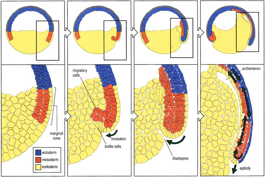
BIMA82

         2
3
Animal Architecture

 Organization of the
vertebrate body plan

                       4
Animals are made of repeating units

 Lobopodian 500 mio år
                         Salamander 150 mio år

                          Dinosaur 150 mio år    5
  Trilobite 500 mio år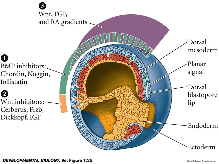
Diversity arises from variations in the
   numbers and kinds of repeating units

All vertebrate limbs are variations on a
common design in which the number,
size and shape of elements differ

                                             6
             Human hand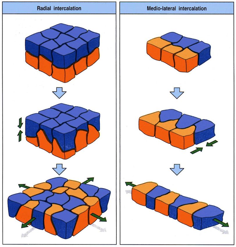
Evolution as variation in number and kind

Samuel Williston’s Law (1914): In the course of evolution the
parts in an organism tend toward a reduction in number and the
fewer parts showing a great specialization in function.

                                                                 7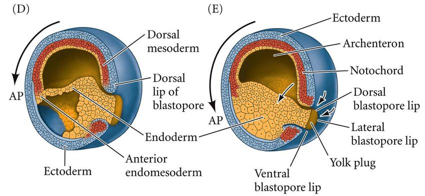
How is form encoded in the genome?

Modularity, symmetry and
  polarity are universal
features of animal design

•   bilateral symmetry
•   anterior-posterior axis
•   proximal-distal axis
•   dorsal-ventral axis

                                         8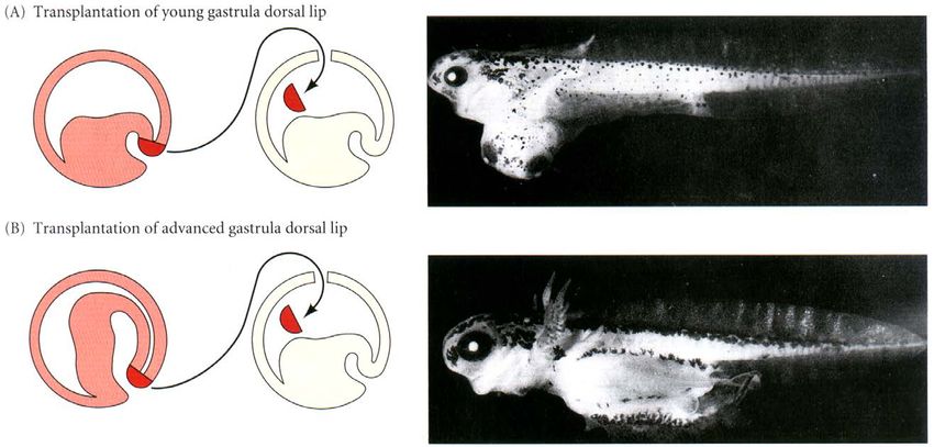
Early asymmetry in the amphibian egg

Are factors required for
development asymmetrically
localized in the fertilized egg?

Hans Spemann and Hilde Mangold
        Nobel prize 1935
                                       9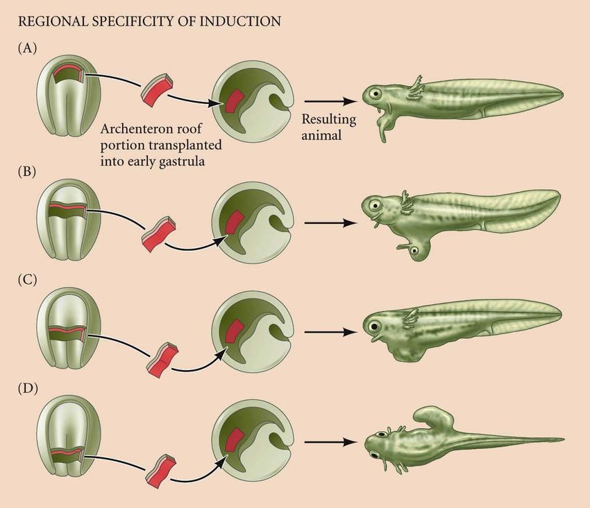
Induction
   Spemann organizer              Polydactyly

Zone of polarizing activity

                                                10
Homeotic Selector Genes
Wild type
Wild type
Ultrabithorax
Antennapedia
Antennapedia
11Drosophila Homeotic (Hox) Genes
• Specify identity of body
region
• Located in homeotic
complexes: Antennapedia
complex, Bithorax complex
• Complexes are conserved
throughout the animal
kingdom
12
Adapted from: Carroll et al., From DNA to Diversity , Blackwell Science, 2001 and S.F. Gilbert,
SinauerDrosophila Homeotic and Vertebrate Hox Genes
Control Anterior-Posterior Identity
13Pax6: A Master Control Gene for Eye Development
Come back to
polydactyly,
cancer and
hedgehog
14The genetic toolkit
So what’s in the toolkit?
Transcription Factors
15Cyclopia, Polydactyly, Cancer and Hedgehog
Drosophila hedgehog ZPA & AER Polydactyly
Cyclopia Basal Cell Carcinoma
in Sheep
16The toolkit paradox
• Humans and mice share nearly identical sets of
25000 genes, and humans and chimps are 99%
identical at the DNA level?
• How can the same set of tool kit genes sculpt
the different anatomies of arthropods and
vertebrates?
• At what point in development is a cells fate
sealed?
17Fate Maps
The fate map reveals that at some
point in development cells know
where they are in the embryo and to
what tissue or structure they belong.
How do cells learn their position or
identity?
18The Coordinate system
Define identities of modules
Defining the poles
The third axis is defined
Subdivide into smaller regions
Form new worlds at specific
coordinates
Initially similar modules are
distinguished according to
Within the worlds polarity is
their position 19
Refine into finer modules establishedThe Making of a Fly
Subdividing the Drosophila embryo
Wing
primordia
Leg primordia
marked by
Distalless
Subdivision of the D-V axis 20The Making of a Vertebrate
k-l: chordin and Frzb marking the early axes in the frog
m-n: subdivision of the mouse brain by hox genes
o-p: toolkit genes marking the segmented
organization of the somites
q: toolkit genes marking the position where
the limbs will form
21The Making of a Vertebrate
w: BMP4 marking tissue between the digits that will die
x: Patched receptor marking feather buds in the chicken
u: Gdf5 marking the future position
of joints in the digits
x: Scleraxis gene marking future
position of tendons of the limbs
22Genetic Switches
The positioning of toolkit genes determines the fate of cells and
builds tissues. But where are the operating instructions for the
toolkit?
How do toolkit genes know in what order to act or where to act
in the embryo?
What is the mechanism that positions toolkit genes?
Genetic switches are the key link between toolkit genes that
build animal complexity and diversity.
23Regulatory Sequences: Genetic “Switches”
One gene can have multiple “switches”: enhancers
The physical integrity of switches is very
important for normal development!
24How do Genetic Switches Work? A is an activator B and C are repressors 25
Changing Switches and Evolution

          Solving the toolkit
               paradox

A, B, U: enhancer binding proteins (U= ubx)
Gene 1: required for forewing development
Gene 2: required for fore- and hindwing
Gene 3: required for hindwing development     26
Summary § Animals are built of repeating units and have a modular design. § Diversity is created by evolutionary change of individual modules. § There is a universal gene toolkit used to build all animals. § Expression of toolkit genes foreshadows the development of tissues and organs § Evolutionary change is created by modulation of gene regulatory switches. § Every animal form is the product of two processes: development from an egg and evolution from its ancestors. 27
Axis formation in vertebrates

In vertebrates differences in axis formation are mainly due to
           differences in the mode of reproduction               28
The Xenopus life cycle
29The Xenopus body plan
30The Xenopus fate map
• Regulative development: The individual cell’s potential is greater than it’s normal
embryonic fate.
31
• Cell fate is determined by the interaction with neighboring cells: called induction!Fate Map of a Frog Blastula
32Early asymmetry in the amphibian egg
Are factors required for
development asymmetrically
localized in the fertilized egg?
Hans Spemann and Hilde Mangold
Nobel prize 1935
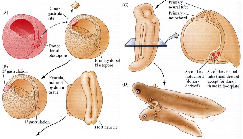
33Hans Spemann and Hilde Mangold:
Primary embryonic induction
34The dorsal blastopore lip is the
Spemann organizer
The Spemann organizer:
• initiates gastrulation
• induces dorsal mesoderm (notochord) and neural tube formation
• organizes tissues into an embryo with anterior-posterior polarity 35How does the organizer work?
- How is the organizer itself specified
- How do cells in the dorsal blastopore lip become
different from other cells?
- What factors are secreted by the organizer?
- How is anterior-posterior polarity established?
36The Spemann organizer is induced by the
Nieuwkoop center
37Transplantation experiments localize
the Nieuwkoop center
38The Nieuwkoop center is induced
by cortical rotation of the egg cytoplasm
+
- + -
39Experimental evidence: Cortical rotation
untreated UV irradiated
40Dishevelled stabilizes β-catenin on
the dorsal side
41Various factors can rescue axis formation in UV-
irradiated embryos
Injection of blastocysts at the 2-cell
stage with dominant-inactive GSK3.
42β-Catenin, VegT and Vg-1 induce
the Nieuwkoop center
Oocyte β-Catenin
VegT, Vg-1
Vg-1 (TGF-β family)
is localized during oogenesis
43The Spemann organizer is induced by the
Nieuwkoop center
Nieuwkoop center
Nodal-related molecules
(Xnr, TGF-β family)
44Mechanism inducing the Spemann organizer
VegT in endoderm
45Model for mesoderm induction and organizer
formation by β-catenin and TGFβ family molecules
46Transplantation
47Functions of the Spemann organizer
1. Differentiation of dorsal mesoderm (prechordal plate, axial
mesoderm)
2. Dorsalization of surrounding mesoderm into paraxial
mesoderm (somites)
3. Dorsalization of the ectoderm inducing neural tube formation
4. Initiation of gastrulation movements
48The homeodomain transcription factor Goosecoid
is expressed in the organizer
- activates the migration properties of dorsal blastopore lip cells.
- autonomously determines the fate of dorsal mesoderm cells.
- enables goosecoid-expressing cells to recruit neighboring cells
into the dorsal axis.
goosecoid encodes a transcription factor and must therefore
activate diffusible factors for its non-cell autonomous effects!
49The Spemann organizer secretes growth
factor antagonists
Noggin, Chordin, Follistatin BMP-4 (induces ventral structures)
Frzb-1, Dickkopf-1, Cerberus Wnts (prevent head formation)
Lefty, Activin TGF-β
BMP-4 Chordin
50Signals
① From vegetal cells to induce mesoderm in marginal cells of
the animal hemisphere (Vg1, low Xnr)!
② In dorsal vegetal cells Vg1 and β-catenin induce high
concentrations of Xnr (Nieuwkoop center).
③ From Nieuwkoop center cells (high Xnr) to marginal cells ( β-
catenin) to dorsalize the mesoderm (Spemann organizer)!
④ From Spemann organizer to ventrally adjacent marginal cells
to induce mediolateral mesoderm (noggin, chordin)!
⑤ From ventral mesodermal cells to oppose the dorsalizing
signal emanating from the Spemann organizer (BMP4
gradient)!
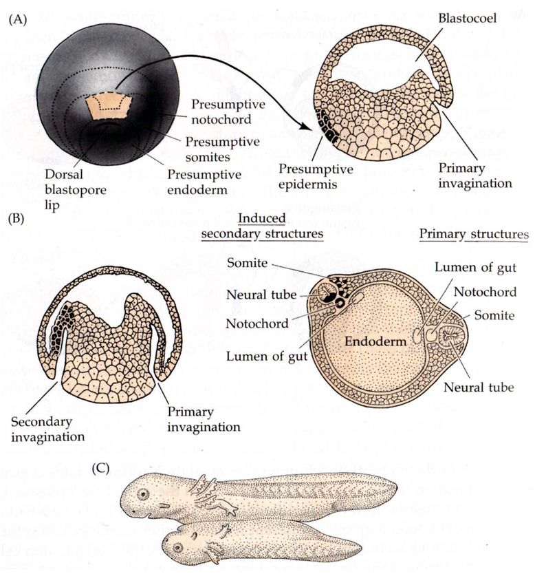
51Gastrulation in Amphibians
52Gastrulation in the Frog Embryo
53Migration of the germlayers in Xenopus
54Cell intercalation
brachyury
Epiboly Convergent extension 55Summary: Gastrulation in Xenopus
• Combination of involution, convergent extension and epiboly.
• Starts below the equator in the marginal zone on the dorsal side.
• Endodermal cells invaginate to form the blastopore. Cells change
their shape by apical constriction to form bottle cells.
• Mesoderm starts to involute across the dorsal blastopore lip
migrating towards the animal pole.
• Region of invagination (and the blastopore) widens laterally and
ventrally and more ventral cells involute.
• At the same time the ectoderm expands towards the vegetal pole
by epiboly (convergent extension + division).
• Finally the blastopore contracts and closes. The germlayers have
been internalized.
56Determination of the germlayers
• Closely linked to axis formation
w Animal vegetal axis formed by maternal
factors. Animal pole: ectoderm, vegetal pole:
endoderm.
w Mesoderm formation by “embryonic
induction”: ability of cells to determine the
embryonic fate of other neighboring cells by
cell communication.
57Temporal specificity of induction
58
Transplantation of the dorsal blastopore lip at different times during gastrulationRegional specificity of induction
59
Transplantation of tissue from different regions of the neural plateSignals from the mesoderm regionalize the
anterior-posterior axis
60Signals from the mesoderm regionalize the
anterior-posterior axis
61Wnt inhibitors regionalize the anterior-posterior axis
Frzb
chordin 62
Developmental Biology, 9e, Figure 7.32Double gradient model of patterning
the Xenopus body plan
β-catenin
63Sequence of events
• Four stages of specification in Xenopus:
① Dorsal ventral axis is determined by the point of sperm entry. Radial symmetry is
broken by cortical rotation. Opposite to the sperm entry will be dorsal.
② Vegetal cells (Nieuwkoop center) induce cells above them to become the Spemann
organizer (mesoderm).
③ Organizer converts neighboring mesoderm into dorsal mesoderm. Invagination
through the blastopore establishes the anterior-posterior axis (anterior structures
invaginate first).
④ Regional specificities are induced in the neural ectoderm.
Fertilization Cortical Rotation Late blastula Tailbud stage
64Literature
• Gilbert/Barresi, Developmental Biology, chapter 11 (11th edition),
chapter 8 (10th edition).
• Planar Cell Polarity, Vladar EK. & JD. Axelrod, Cold Spring Harbor
Perspectives Biol. 2009
• Carroll S., Endless Forms Most Beautiful, Quercus, 2011
65You can also read