Blood Oxygen Conservation in Diving Sea Lions: How Low Does Oxygen Really Go?
←
→
Page content transcription
If your browser does not render page correctly, please read the page content below
DISTRIBUTION STATEMENT A. Approved for public release; distribution is unlimited.

                     Blood Oxygen Conservation in Diving Sea Lions:
                           How Low Does Oxygen Really Go?
                                           Paul J. Ponganis
                          Center for Marine Biotechnology and Biomedicine
                                 Scripps Institution of Oceanography
                                        8655 Discovery Way
                                         215 Scholander Hall
                                      La Jolla, CA 92093-0204
             phone: (858) 822-0792 fax: (858) 534-1305 email: pponganis@ucsd.edu

                                       Award Number: N000141410404

LONG-TERM GOALS

California sea lions (Zalophus californianus) from San Nicolas Island regularly perform 350-meter
deep dives during maternal foraging trips to sea. The physiology of these extreme dives is relevant to
the development of the sea lion as a model to investigate deep diving physiology and the avoidance of
decompression sickness in a marine mammal. Such a model is essential to better understand the
potential role of decompression sickness in the etiology of the stranding of beaked whales after
exposure to naval sonar as well as to evaluate the value and accuracy of the many numerical models of
nitrogen uptake and distribution in these animals. This project continues prior physiological
investigations with these animals and focuses on the relationship of blood oxygen depletion patterns
during dives to heart rate and muscle workload.

OBJECTIVES

With techniques and backpack data recorders developed in prior ONR-funded research, this project
will a) compare blood oxygen depletion profiles in the anterior and posterior venae cavae of diving sea
lions, and b) investigate the effects of heart rate and flipper stroke rate on simultaneously recorded
venous oxygen depletion patterns during dives. The goals of this research are to a) document the
magnitude and pattern of depletion of the entire venous oxygen store during dives, b) examine the
effect of muscle work load (flipper stroke rate) and potential blood oxygen extraction by muscle on the
simultaneously recorded venous oxygen depletion profiles in both the anterior and posterior venae
cavae, and c) determine the effect of heart rate and potential perfusion-related blood oxygen extraction
by tissues on the simultaneously recorded venous oxygen depletion profiles in both the anterior and
posterior venae cavae.

APPROACH

Objective 1: In order to calculate the rate and magnitude of depletion of the blood O2 store during
dives, PO2 profiles will be obtained from a PO2 recorder and intravascular electrode deployed on sea
lions (McDonald and Ponganis 2012). The PO2 electrode will be placed in either the anterior or
posterior vena cava. As in previous research by the PI with California sea lions and other species

                                                             1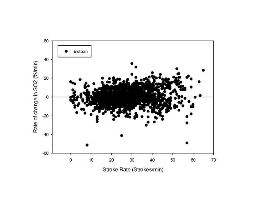
(McDonald and Ponganis 2013; Meir et al. 2009; Meir and Ponganis 2009), the PO2 profiles will be
converted to Hb saturation profiles with the use of the sea lion O2-Hb dissociation curve. In addition
to the PO2 and Hb saturation profile during a dive, the start-of-dive and end-of-dive % Hb saturations
can then be used to calculate the magnitude of blood O2 depletion during dives based on the net change
in % Hb saturation, and the known Hb concentration and blood volume.
Objective 2: ECG profiles will be simultaneously collected from freely diving sea lions equipped with
a PO2 electrode in either the anterior or posterior vena cava in order to assess the rate of venous O2
desaturation to heart rate throughout dives.
Objective 3: Accelerometers will be deployed on freely diving sea lions equipped with a PO2 electrode
in either the anterior or posterior vena cava in order to assess the rate of venous O2 desaturation to
work load (stroke rate or MSA) throughout dives.
WORK COMPLETED
The first field season of this grant has just been completed. Analysis of these data and pilot studies
from the previous field season are underway. Initial results will be presented at the upcoming Bio-
logging Conference.
RESULTS
As in a pilot study from the prior ONR grant, the PO2 and Hb desaturation profiles in the anterior and
posterior venae cavae are different (Fig. 1), but end-of-dive saturations approximate the same 20-30%
region of saturation.
100
80
% Hb Saturation (SO2)
AntVenaCava SO2
60
Arterial SO2
PostVenaCava SO2
40
20
0
0 1 2 3 4 5 6 7
Time into Dive (min)
Figure 1. Arterial, anterior vena caval, and posterior vena caval hemoglobin (Hb) saturation
proflies during 380-m deep dives of California sea lion. Each profile is from a different sea lion.
Analyses of venous hemoglobin saturation profiles and simultaneously paired heart rate or stroke rate
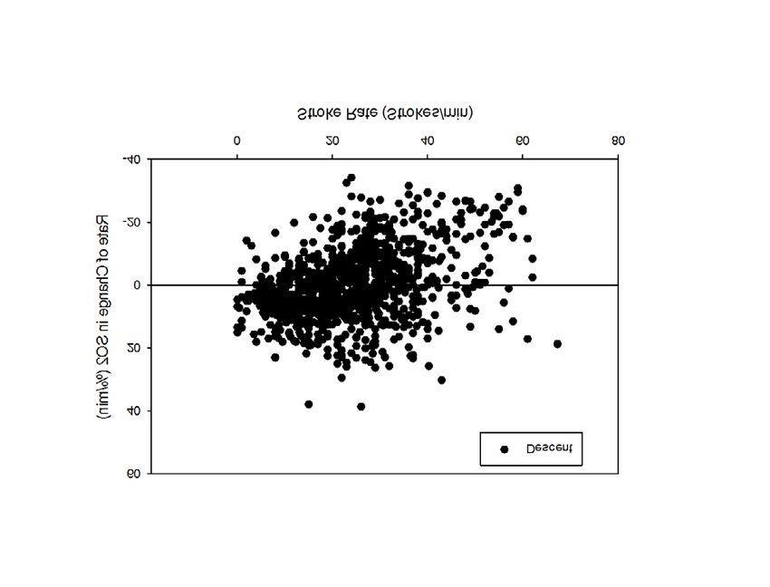
profiles have just begun. Review of posterior vena caval desaturation rates relative to stroke rate in all
dives (Fig. 2) reveals a highly variable relationship, suggesting that posterior vena caval desaturation
is not related to muscle workload.
2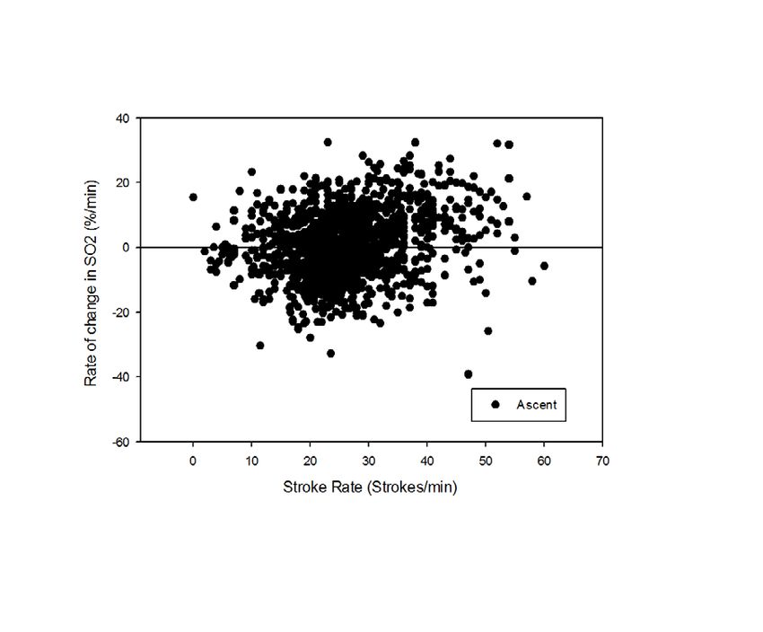
Figure 2. Rate of change in posterior venacaval hemoglobin saturation (SO2) in relation to stroke
              rate during descent, bottom phase, and ascent of all dives of sea lions.

IMPACT/APPLICATIONS

In prior ONR-funded research, partial pressure of oxygen (PO2) profiles provided evidence that lung
collapse occurred near 200-m depth in diving sea lions (McDonald and Ponganis 2012). This
impairment of gas exchange limits nitrogen uptake at depth and preserves lung oxygen for later use
during ascent. More recent research has revealed that heart rate rapidly declines during descent of deep
dives to values less than 10 beats min-1 (McDonald and Ponganis 2014). Such a low heart rate also
limits the absorption and distribution of both nitrogen and oxygen at depth (through reductions in
pulmonary and aortic blood flow). As a result of these physiological processes, the sea lion can
maintain arterial hemoglobin saturation above 90% during deep dives as long as 7 minutes (McDonald
and Ponganis 2012; McDonald and Ponganis 2013). In contrast, the elephant seal (Mirounga
angustirostris), which dives on expiration and has less than 5% of total body O2 stores in the
respiratory system, experiences significant hypoxemia with routine arterial hemoglobin desaturation to
10 to 20% (Meir et al. 2009). However, similar to the sea lion, the emperor penguin (Aptenodytes
forsteri), another animal that dives on inspiration with a large respiratory O2 store, also can maintain
arterial saturations during dives as long as 10 min (Meir and Ponganis 2009). It is also notable that a
severe bradycardia during descent occurs in deep-diving emperor penguins (Ponganis, unpublished
data), and in deep-diving bottlenose dolphins (Tursiops truncatus), which also dive on inspiration
(Houser et al. 2010; Williams et al. 1999). For these reasons, it is hypothesized that the heart rate
profile during deep dives of California sea lions is universal among higher vertebrates that dive on
inspiration. Hence, both lung collapse and the heart rate profile make the California sea lion a valuable
model to investigate physiological responses and gas uptake / distribution during deep dives.

The lower heart rates during deeper, longer dives observed in this study and the lack of complete blood
oxygen depletion during these deep dives that were documented in our prior ONR study (McDonald
and Ponganis 2012; McDonald and Ponganis 2013) also have implications for the management of
oxygen stores and the physiological basis of the ADL. The concept that most dives are aerobic in
nature and do not exceed an aerobic dive limit (ADL - dive duration associated with the onset of post-
dive blood lactate accumulation) has dominated the interpretation of dive behavior and foraging
ecology over the past 30 years (Costa et al. 2001; Kooyman et al. 1980). However, because of
technical difficulties, the ADL has rarely been measured. Instead, researchers have had to resort to

                                                    3
estimations of total O2 store depletion, i.e., calculated ADLs (cADLs) (Costa et al. 2001; Weise and
Costa 2007). Our findings in sea lions have supported the concept that the physiological basis of the
ADL is muscle oxygen depletion and subsequent glycolysis. The lung and blood oxygen stores are not
completely depleted in even the longest of sea lion dives. The severe bradycardia during deep dives
contributes to the preservation of the blood and lung oxygen for use during ascent, and it also creates
greater reliance of muscle metabolism on the myoglobin-bound muscle oxygen store. In addition, the
lack of correlation between heart rate and stroke rate during deeper dives (Figs. 1 and 3) suggests that
muscle blood flow and oxygen delivery are not coupled with stroke effort. These findings reinforce our
hypothesis that depletion of the muscle oxygen store with subsequent glycolysis underlies the ADL.
This current project will address the linkage of venous blood O2 depletion to heart rate and to muscle
work load (stroke rate). Furthermore, it will examine blood O2 depletion in both the anterior and
posterior vena cavae. These analyses will allow documentation of the degree of depletion of the entire
venous O2 store, and reveal which factor (heart rate or stroke rate) that depletion is most dependent
upon. Such analysis will further document the potential relation of heart rate and muscle blood flow to
muscle workload during dives, and to the physiological model of the ADL discussed in the prior
paragraph.
RELATED PROJECTS
This project is building on our findings from our previous ONR funded projects: “Blood oxygen
depletion in California sea lions: How close to the limit?” (award #: N000141010514), and “Deep
diving California sea lions: Are they pushing their physiological limits?” (award #: N000141210633).
REFERENCES
Costa, D.P., N.J. Gales and M.E. Goebel. 2001. Aerobic dive limit: how often does it occur in nature?
Comparative Biochemistry and Physiology A 129: 771-783.
Houser, D.S., L.A. Dankiewicz-Talmadge, T.K. Stockard and P.J. Ponganis. 2010. Investigation of the
potential for vascular bubble formation in a repetitively diving dolphin. J Exp Biol 213: 52-62.
Kooyman, G.L., E.A. Wahrenbrock, M.A. Castellini, R.W. Davis and E.E. Sinnett. 1980. Aerobic and
anaerobic metabolism during diving in Weddell seals: evidence of preferred pathways from
blood chemistry and behavior. Journal of Comparative Physiology 138: 335-346.
McDonald, B.I., and P.J. Ponganis. 2012. Lung collapse in the diving sea lion: hold the nitrogen and
save the oxygen. Biology Letters 8: 1047-1049.
McDonald, B.I., and P.J. Ponganis. 2013. Insights from venous oxygen profiles: oxygen utilization and
management in diving California sea lions. Journal of Experimental Biology 216: 3332-3341.
McDonald, B.I., and P.J. Ponganis. 2014. Deep-diving sea lions exhibit extreme bradycardia in long-
duration dives. Journal of Experimental Biology 217: 1525-1534.
Meir, J.U., C.D. Champagne, D.P. Costa, C.L. Williams and P.J. Ponganis. 2009. Extreme hypoxemic
tolerance and blood oxygen depletion in diving elephant seals. Am J Physiol Regul Integr Comp
Physiol 297: R927-939.
Meir, J.U., and P.J. Ponganis. 2009. High-affinity hemoglobin and blood oxygen saturation in diving
emperor penguins. Journal of Experimental Biology 212: 3330-3338.
4Weise, M.J., and D.P. Costa. 2007. Total body oxygen stores and physiological diving capacity of
California sea lions as a function of sex and age. Journal of Experimental Biology 210: 278-289.
Williams, T.M., J.E. Haun and W.A. Friedl. 1999. The diving physiology of bottlenose dolphins
(Tursiops truncatus). I. Balancing the demands of exercise for energy conservation at depth.
Journal of Experimental Biology 202: 2739-2748.
5You can also read