USING UNDERWATER PULSE OXIMETRY IN FREEDIVING TO EXTREME DEPTHS TO STUDY RISK OF HYPOXIC BLACKOUT AND DIVING RESPONSE PHASES - DIVA
←
→
Page content transcription
If your browser does not render page correctly, please read the page content below
ORIGINAL RESEARCH
                                                                                                                                                 published: 01 April 2021
                                                                                                                                        doi: 10.3389/fphys.2021.651128

                                                Using Underwater Pulse Oximetry in
                                                Freediving to Extreme Depths to
                                                Study Risk of Hypoxic Blackout and
                                                Diving Response Phases
                                                Eric Mulder 1* and Erika Schagatay 1,2
                                                1
                                                    Environmental Physiology Group, Department of Health Sciences, Mid Sweden University, Östersund, Sweden,
                                                2
                                                    Swedish Winter Sports Research Centre, Mid Sweden University, Östersund, Sweden

                                                Deep freediving exposes humans to hypoxia and dramatic changes in pressure. The effect
                                                of depth on gas exchange may enhance risk of hypoxic blackout (BO) during the last part
                                                of the ascent. Our aim was to investigate arterial oxygen saturation (SpO2) and heart rate
                          Edited by:            (HR) in shallow and deep freedives, central variables, which have rarely been studied
                Costantino Balestra,
                                                underwater in deep freediving. Four male elite competitive freedivers volunteered to wear
       Haute École Bruxelles-Brabant
                    (HE2B), Belgium             a newly developed underwater pulse oximeter for continuous monitoring of SpO2 and HR
                        Reviewed by:            during self-initiated training in the sea. Two probes were placed on the temples, connected
                  Neal William Pollock,         to a recording unit on the back of the freediver. Divers performed one “shallow” and one
              Laval University, Canada
                           Kay Tetzlaff,        “deep” constant weight dive with fins. Plethysmograms were recorded at 30 Hz, and
      University Hospital of Tübingen,          SpO2 and HR were extracted. Mean ± SD depth of shallow dives was 19 ± 3 m, and
                              Germany
                                                73 ± 12 m for deep dives. Duration was 82 ± 36 s in shallow and 150 ± 27 s in deep
                    Claus-Martin Muth,
  Universitaetsklinikum Ulm, Germany            dives. All divers desaturated more during deeper dives (nadir 55 ± 10%) compared to
                   *Correspondence:             shallow dives (nadir 80 ± 22%) with a lowest SpO2 of 44% in one deep dive. HR showed
                           Eric Mulder          a “diving response,” with similar lowest HR of 42 bpm in shallow and deep dives; the
                  eric.mulder@miun.se
                                                lowest value (28 bpm) was observed in one shallow dive. HR increased before dives,
                   Specialty section:           followed by a decline, and upon resurfacing a peak after which HR normalized. During
         This article was submitted to          deep dives, HR was influenced by the level of exertion across different diving phases;
   Environmental, Aviation and Space
                           Physiology,          after an initial drop, a second HR decline occurred during the passive “free fall” phase.
               a section of the journal         The underwater pulse oximeter allowed successful SpO2 and HR monitoring in freedives
               Frontiers in Physiology
                                                to 82 m depth – deeper than ever recorded before. Divers’ enhanced desaturation during
         Received: 08 January 2021
                                                deep dives was likely related to increased exertion and extended duration, but the rapid
          Accepted: 15 March 2021
           Published: 01 April 2021             extreme desaturation to below 50% near surfacing could result from the diminishing
                            Citation:           pressure, in line with the hypothesis that risk of hypoxic BO may increase during ascent.
   Mulder E and Schagatay E (2021)              Recordings also indicated that the diving response is not powerful enough to fully override
 Using Underwater Pulse Oximetry in
     Freediving to Extreme Depths to            the exercise-induced tachycardia during active swimming. Pulse oximetry monitoring of
 Study Risk of Hypoxic Blackout and             essential variables underwater may be an important step to increase freediving safety.
            Diving Response Phases.
          Front. Physiol. 12:651128.            Keywords: breath-hold diving, apnea, arterial oxygen saturation, syncope, heart rate, bradycardia, exercise,
   doi: 10.3389/fphys.2021.651128               oxygen conservation

Frontiers in Physiology | www.frontiersin.org                                           1                                           April 2021 | Volume 12 | Article 651128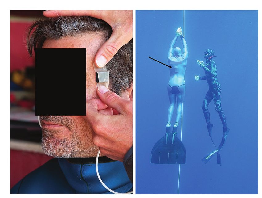
Mulder and Schagatay                                                                                               Pulse Oximetry in Deep Freediving

INTRODUCTION                                                                Only two previous studies have used pulse oximetry during
                                                                            freediving to shallow depth (Stanek et al., 1993; Kuch et al.,
Freediving is increasing in popularity both as a recreational               2010), and another few have measured heart rate (HR) during
and competitive sport (Divers Alert Network, 2017), where                   deep dives (Schagatay, 2011; Lemaitre et al., 2013).
all activities are performed on one breath of air. While most                  In order to further investigate the human physiological
freediving is done in the depth range down to 20 m, deep                    adaptations during actual freediving to great depth, new devices
freediving is an increasingly popular extreme sport where elite             must be developed for recording central variables remotely.
divers reach far beyond that depth, exposing themselves not                 But which variables are most central to determine a freediver’s
only to progressive hypoxia but also to dramatic changes in                 physiological performance limits and exposure to risks? While
pressure. These changes have several physiological effects, both            several factors determine the diver’s ultimate depth limits
by direct compression of air filled cavities (Boyles law) and               (Schagatay, 2011), maximal diving duration is set by the diver’s
on gas exchange, as partial pressure of oxygen changes                      total gas storage capacity, the lowest tolerable oxygen levels,
proportionally with ambient pressure (Dalton’s law). In                     and metabolic rate, i.e., the time it takes for the diver to use
competition freediving; “Apnea” there are four disciplines with             up the available oxygen stores (Schagatay, 2009). Individual
the aim to reach the greatest possible depth on one breath,                 oxygen storage capacity can be measured before the dive in
and to return back to the surface with one’s own muscular                   dry conditions, and a test determining the individual tolerance
effort, and to date competitive freedivers have reached 130 m               to hypoxia could also be done in a non-immersed diver.
using a monofin and 102 m swimming without fins (International              However, SpO2 measured during the dive would reveal remaining
Association for the Development of Apnea, 2021). A typical                  available oxygen stores and therefore be central to predicting
deep freedive is characterized by four different phases, with               diving duration, together with metabolic rate. Metabolic rate
different physiological demands, as buoyancy may help or                    relates to physical activity, but can be transiently limited by
counteract the swimming efforts (Schagatay, 2011).                          the cardiovascular “diving response,” which conserves oxygen
    The most obvious limitation to deep freediving is the                   by reducing peripheral circulation and myocardial oxygen
freediver’s maximal breath-holding capacity, as the diver cannot            consumption, leading to enhanced SpO2 and longer breath
immediately return to the surface once depth has been reached.              hold duration (Andersson and Schagatay, 1998; Schagatay and
Therefore, although all freedivers are exposed to risk of hypoxic           Andersson, 1998). Therefore, most central for determining
syncope (Craig, 1961; Lin et al., 1974), which is called “blackout”         metabolic rate and oxygen metabolism during freediving is
(BO) by the divers, its consequences may be more severe in                  monitoring of HR and SpO2 during the dive, two variables
deep diving. The effect of pressure on gas exchange may in                  that can be simultaneously measured via pulse oximetry.
addition expose deep divers to increased risk of blackout                      We therefore developed a pressure- and water-resistant
resulting from the air re-expansion during ascent, which can                pulse oximeter. Our immediate study aim was to monitor
dramatically reduce arterial oxygen saturation (SpO2), resulting            HR and SpO2 during freedives to great depth in order to
in a specific case of blackout often called “shallow water                  (1) determine if good quality data could be collected at 80 m
blackout” (Lanphier and Rahn, 1963; reviewed in Schagatay, 2011).           depth, and (2) to establish if HR and SpO2 patterns differ
    While during the descent, the increase in hydrostatic pressure          between shallow and deep dives, and whether HR differed
leads to an increment in alveolar partial pressure of oxygen,               between the different diving phases of deep dives. The long-
which may result in a temporary state of hyperoxia (Linér et al.,           term goal is to use this monitor to enhance the understanding
1993), during ascent the pressure diminishes with a concomitant             of the physiological events during deep freediving and to
decrease in alveolar oxygen pressure, which may invert the flow             be able to predict safe diving duration and thereby promote
of oxygen at the alveolar level. This situation may cause the               freediving safety.
SpO2 to drop very quickly, and cause an enhanced risk of hypoxic
blackout near the surface (Linér et al., 1993; Ferretti, 2001).
Although this situation is considered to be a major cause of                MATERIALS AND METHODS
deaths in freediving, continuous measurements of SpO2 during
deep dives are currently lacking. This is mainly because                    Participants
physiologists have up to now been highly dependent on access                Four healthy male elite competitive freedivers (mean ± SD
to the studied person to make direct measurements of physiological          age 38 ± 12 years, height 181 ± 10 cm and weight 76 ± 12 kg)
variables on the body using a range of methods and equipment.               volunteered to be part of this study during their regular
There exist few technological devices allowing recording of                 freediving training. They all had a minimum of 5 years of
physiological values underwater, and there is no possibility to             freediving experience, and qualified in category 5 in the 5-level
follow a diver underwater to extreme depths using SCUBA gear,               categorization system used for freedivers with respect to their
as the freedivers move so rapidly vertically across the water               training/experience (Schagatay and Andersson, 1998). The mean
column, that this could cause decompression illness in the SCUBA            personal best competition performance in constant weight
diver. Most research in freediving physiology to date is therefore          (CWT) deep diving with fins was 91 ± 10 m.
based on laboratory studies (Lin et al., 1974; Schagatay and                   The freedivers were well acquainted with and used the safety
Andersson, 1998; Bakovic et al., 2003; Lemaitre et al., 2007) or            procedures established in competitions, e.g., to be accompanied
at the water surface (Marongiu et al., 2015; Fernández et al., 2019).       during the last 20 m toward the surface by a safety diver.

Frontiers in Physiology | www.frontiersin.org                           2                                      April 2021 | Volume 12 | Article 651128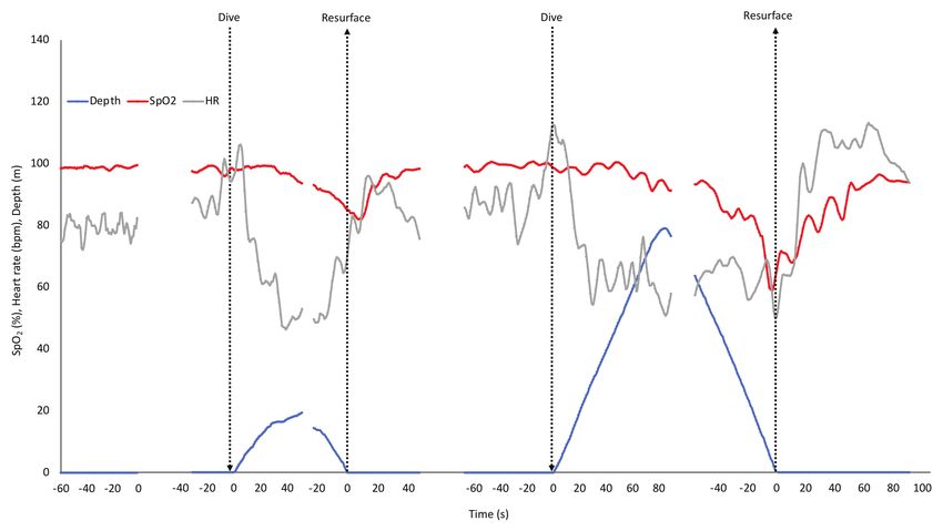
Mulder and Schagatay                                                                                                                Pulse Oximetry in Deep Freediving

All participants were given verbal information regarding the                         therefore optimized and included an auto-correlation algorithm
purpose of the study and signed an informed consent prior                            to filter motion artifacts. More specifically, the SpO2 algorithm
to engaging in their pre-diving routines. The study protocol                         was based on calculation of the Root Mean Square (RMS)
had been approved by the Regional Committee for Medical                              value of alternating current (AC) and direct current (DC) of
and Health Research Ethics in Umeå, Sweden, (Dnr 2019-                               the red and infrared channel as described by the manufacturer
05147) and tests were conducted in accordance with the 2004                          (Application note 6845, Maxim Integrated, CA, United States).
Declaration of Helsinki.                                                             An improved version of this algorithm was also performing
                                                                                     a correlation between infrared and red signal to calculate a
Diving Procedures and Data Recording                                                 measure of signal quality. In an undisturbed signal, infrared
When the freedivers had finished their pre-dive routines, they                       and red signals correlated well, and in case of bad correlation,
were equipped with a prototype pulse oximeter. The prototype                         the calculated values were discarded. The previous evaluation
pulse oximeter was evaluated prior to the current study and                          of the prototype showed that SpO2 and HR seemed in good
results will be presented elsewhere. Briefly, the prototype                          agreement with those measured of two conventional finger
consisted of a ruggedized universal datalogger platform based                        pulse oximeters during normoxic and hypoxic breathing, as
on a ST Microelectronics STM32L452 microprocessor                                    well as during body immersed static apnea.
(STMicroelectronics International N.V. Amsterdam, The                                    The sensor heads of the two probes were applied to the
Netherlands), with a 32 bit ARM Cortex M4 core, while a                              temples, and fixated with medical tape as well as by the pressure
32 GB micro-SD card was integrated for data storage.                                 from the hood (Figure 1A). If the hood was loose, an extra
   The prototype was equipped with two SpO2 probes based                             head band or swim cap was used to apply further fixation.
on the MAXIM MAX30102 chip (max 50 Hz sampling rate)                                 The quality of the plethysmograms signal was inspected and,
and an ambient pressure sensor. The MAX30102 (Maxim                                  if considered adequate, the data logger was inserted under the
Integrated, CA, United States) sensor frontend recorded                              wetsuit, and placed on the back of the freediver (Figure 1B).
plethysmograms and includes red and infra-red emitters, diode                        This ensured that no cables were outside of the wetsuit, thus
drivers, photodiode, photodiode amplifier, analog to digital                         not compromising the safety of the freediver.
converter, and controller. Distance between light/infra-red emitter                      To enable visual inspection of the quality of the
and photodiodes was 3 mm. A graphical user interface was                             plethysmograms prior to the diving session, the recording unit
developed in Labview (National Instruments Corporation, Austin,                      was connected to a computer through an optical fiber output
United States), which was used to show all parameters in real                        cable, which allowed real-time transmission of the data. The
time and display the recordings.                                                     cable was then removed and the recording was manually started.
   Sample algorithms for SpO2 calculations in low noise                              The prototype pulse oximeter registered plethysmograms at
environments were supplied by the manufacturer (Maxim                                30 Hz for continuous SpO2, HR, and depth recordings. The
Integrated, CA, United States); however, artifacts of different                      mean ambient air temperature was 26°C and sea surface
kinds may lead to incorrect SpO2 calculations. The algorithm was                     temperature was 22°C.

                                          A                                          B

 FIGURE 1 | (A) Sensor heads of the pulse oximeter probes were applied to the temple and fixated with medical tape, and thereafter by pressure from the hood of
 the wetsuit, (B) freedivers performed their training dives from a buoy and dove to their predetermined depth along a vertical line, and were accompanied up by a
 safety diver. Black arrow points at the recording unit of the pulse oximeter located underneath the wetsuit.

Frontiers in Physiology | www.frontiersin.org                                    3                                             April 2021 | Volume 12 | Article 651128
Mulder and Schagatay Pulse Oximetry in Deep Freediving
The freedivers entered the water and performed their planned current study was small and responses were in part determined
training dives from a buoy starting the dives on their own by direct inspection of individual and mean data graphs.
volition (Figure 1B). The divers did not follow an imposed
diving protocol; however, they all performed at least one shallower
freedive to approximately 20 m depth, and one deep freedive RESULTS
to at least 50 m. Dives were prepared according to each divers’
own preparatory routines, the last part of which involved lung Data Quality
packing to maximize lung volume (Örnhagen et al., 1998). There were periods of apparent motion artifacts in the recordings
After surfacing the divers used hook breathing for facilitated from one or both sensors, resulting in missing data points.
recovery (Fernández et al., 2019). The recording was manually Loss of data most often occurred at the start of the dive,
stopped and the equipment was removed once the divers had when the diver performed lung packing prior to the dive, and
completed their training session and exited the water. during the first phase of the descent, when middle ear equalization
needs to be done frequently. During the last phase of the
Data Analysis ascent near the end of dives, loss of data again occurred more
Arterial oxygen saturation and HR were extracted from the often, most likely linked to increased frequency and magnitude
plethysmograms at a window size of 6-s, and analyzed internally of involuntary breathing movements, and possibly as a result
every second. Since the pulse oximeter included two probes, of SpO2 being extremely low, which makes it harder for the
the mean was calculated from the results of both sensors to reflective probe to detect oxygen saturation. The dropout rate
obtain one single value for SpO2 and HR for each second. of 1 s values for all trials was 34% and after smoothing it
Dropout rate was presented and refers to interruptions in was 0.5%.
continuous SpO2 and/or HR data due to down time or machine-
probe unit nonfunction, and calculated as the percentage of Diving Patterns and Maximal Responses
time when SpO2 and/or HR data were not provided, as defined Depth of the shallow dives was 19 ± 3 m, while it was
by Barker and Shah (1997). The resulting time series for each 73 ± 12 m for the deep dives (p = 0.003; Table 1). The deep
dive was then smoothened using a 5-s moving median and dives performed were to 81 ± 10% of the diver’s personal
subsequently a 5-s moving average. best depth competition performance in the discipline CWT.
In order to follow the continuous events, a graph of HR Duration of the shallow dives was 82 ± 36 s and 150 ± 27 s
and SpO2 was drawn based on the three dives with the longest for the deep dives (NS).
durations for shallow and deep dives, respectively. The exclusion All divers desaturated more during the deeper dives with
of the two dives of shortest duration was done to provide a a mean nadir SpO2 of 55% compared to the shallow dives,
longer period of mean values. The presented data for the three when SpO2 dropped to a mean nadir SpO2 of 80% (p = 0.040;
included dives were synchronized at two points (1) at the dive Table 1). The lowest individual SpO2 observed was 44% after
initiation, and (2) at surfacing at the end of the dive, both a deep dive, and this diver had a low value, 48%, also after
marked as “0” on the time axis, with the middle part of the the shallower dive. The nadir SpO2 was observed near or just
longer dives omitted. after the end of the deep dives, and with some more delay
The nadir SpO2 and HR values for each dive and the time after surfacing from the shallower dives (Table 1).
of their occurrence were noted. Baseline values of SpO2 and The maximal heart rate reduction, characterizing the diving
HR were recorded during rest at the surface between 5 and response, was the same in shallower (42 bpm) and deep dives
15 min before the first dive. The maximal arterial oxygen (42 bpm; NS) and in both cases represented a mean HR
desaturation was calculated as the percent change from baseline reduction of 46% from the pre-dive level (Table 1). The lowest
for the lowest 1 s SpO2 value occurring between the start of HR observed was 28 bpm, and the time point of occurrence
the dive and 30 s after surfacing, to account for the circulatory of the lowest HR was similar at 52 s for shallow and 67 s
delay. The maximal magnitude of diving bradycardia was for deep dives (NS; Table 1).
evaluated as the percent change between the baseline HR and
the lowest 1 s HR value recorded during the dive. For each
diver, the different diving phases were identified through the
SpO2 and HR Patterns
Individual recording of data (after smoothing) of the shallow
depth profile and change of descent speed, and the mean HR
and deep dive for diver one is shown in Figure 2, with the
calculated for each phase.
phases of the deep dive marked. In the shallow dive, this
diver stayed at 20 m for a longer period, doing a training
Statistics “hang,” while in the deep dive the bottom turn only lasted
Calculated data are presented as mean ± SD for n = 4, unless for a few seconds (Figure 2). The individual dive data shown
otherwise stated. As subjects served as their own controls, represented the least interrupted data of the four deep dives.
paired Student’s t-test for selected data points, with Bonferroni The SpO2 in this diver was nearly unaffected in the shallow
correction for repeated tests, was used to compare diving dive, while it dropped toward the end of the deep dive,
phases and maximal responses between shallow and deep dives. with nadir SpO2 occurring just after surfacing due to the
Significance was accepted at p < 0.05. The sample size of the circulatory delay (Figure 2). HR showed a diving bradycardia of
Frontiers in Physiology | www.frontiersin.org 4 April 2021 | Volume 12 | Article 651128Mulder and Schagatay Pulse Oximetry in Deep Freediving
TABLE 1 | Individual and mean (SD) baseline reference values of SpO2 and HR during a period of immersed rest before dives, followed by depths, durations, and nadir
SpO2 (%) and HR (bpm) values during deep and shallow dives; time when nadir occurs is noted as seconds from start of the dive, as well as the maximal reduction from
pre-dive SpO2/HR reference values (%).
Shallow
Diver Pre SpO2 Depth Duration SpO2 nadir SpO2 nadir (s) SpO2 red (%) HR nadir HR nadir (s) HR red (%)
1 99.6 19 97 97 116 3 28 33 67
2 97.3 15 32 89 41 8 46 29 23
3 99.2 23 80 48 89 52 59 49 37
4 98.3 20 117 85 136 13 36 97 55
Mean 98.6 19.1 81.5 80.0 95.5 18.9 42.1 52.0 45.6
SD 1.0 3.1 36.3 21.9 41.1 22.3 13.4 31.2 19.1
p= 0.003 0.117 0.040 0.144 0.041 0.978 0.635 0.987
Diver Pre HR Deep
1 84.0 72 149 68 157 32 34 31 59
2 60.0 82 183 54 178 45 38 122 37
3 93.9 82 148 44 144 55 48 61 49
4 78.8 57 118 54 133 45 49 54 38
Mean 79.2 73.3 149.5 55.0 153.0 44.3 42.3 67.0 45.7
SD 1.0 11.8 26.6 9.7 19.3 9.6 7.2 38.8 10.6
p-values are shown for comparisons between deep and shallow dives.
FIGURE 2 | Individual continuous recordings from Diver 1 of arterial oxygen saturation (SpO2), heart rate (HR), and depth during immersion before starting to dive,
during the shallow and the deep dive followed by a recovery period. The interruptions represent about 5 min of cut out data. The typical phases of a deep dive are
indicated. In a deep dive, i.e., when lungs are compressed below residual volume which for most individuals occurs at greater depth than 30 m, four distinct phases
can be identified; (1) a phase of active swimming against positive buoyancy, (2) a passive “free fall” phase when the diver can relax and fall as a result of negative
buoyancy, which is after the turn followed by (3) a phase of intense swimming, when the diver actively swims upward against negative buoyancy and (4) the last
phase of the dive, when swimming is aided by positive buoyancy.
similar magnitude in both dives, but with a more variated reaching the target depth, which could be a result of
HR in the deeper dive. The nadir HR of approximately intense equalization.
30 bpm occurred at about 30 s into both dives. After In all divers HR varied more in the deep dives. For three
resurfacing, the HR increased above pre-dive levels, and of the four divers SpO2 traces remained stable and high during
returned to baseline after approximately 1 min, a recovery the descent, and dropped during ascent, while one diver started
increase which was greater after the deep dive (Figure 2). to desaturate already during the last part of the descent in
In the deep dive of this diver, HR suddenly increased before both the shallow and the deep dive.
Frontiers in Physiology | www.frontiersin.org 5 April 2021 | Volume 12 | Article 651128Mulder and Schagatay Pulse Oximetry in Deep Freediving
The mean HR and SpO2 response patterns for the shallow again when positive buoyancy reduced the energetic demands
and deep dives for three of the divers (with exclusion of the (Phase 4). During the initial 15 s after surfacing, when SpO2
two dives with shortest duration) is shown in Figure 3. The was still reduced, HR was low, but during the next 15 s it
SpO2 patterns revealed a moderate desaturation toward the increased to the pre-dive level (Figure 4).
end of the shallow dive, and a more pronounced desaturation
after the deeper dives (Figure 3). The rate of decline in SpO2
appeared to be increased in the deeper dive. DISCUSSION
Heart rate responses were similar in shallow and deep dives.
An initial increase at the start of the dive was followed by a The newly developed water and pressure-proof pulse oximeter
“diving response” with a progressive drop in HR during the allowed successful monitoring of both SpO2 and HR in freedivers
descent with nadir reached at or near the bottom. During down to 82 m depth – to our knowledge the greatest depth
ascent HR rose again, but dropped somewhat when divers where HR and SpO2 has been measured. There were short
were approaching the surface, and increased again during periods of data losses, but by using an averaging function,
recovery after which it normalized (Figure 3). continuous data could be obtained for the main portion of
the dives, revealing interesting response patterns of both
Diving Phases SpO2 and HR.
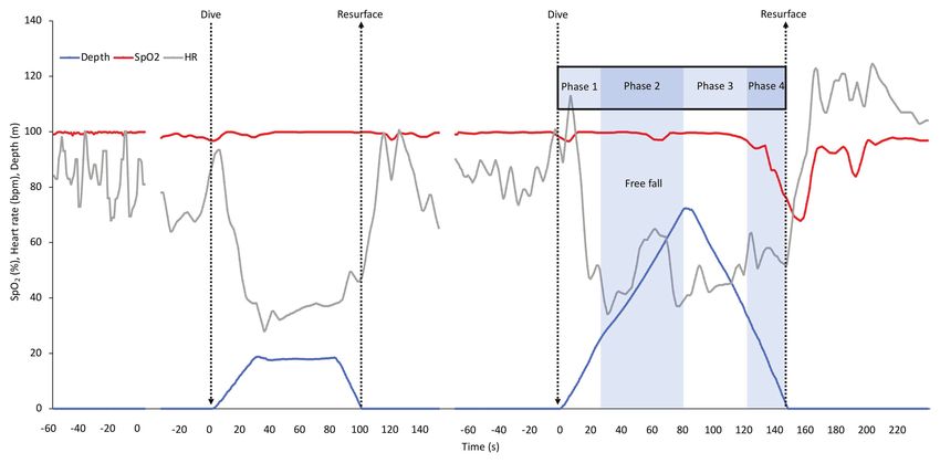
When HR responses across all four deep dives were analyzed
in detail, broken down to means for the separate phases related Arterial Oxygen Desaturation
to the required energetics for swimming, the response patterns While mean SpO2 was kept at pre-dive levels for the first third
became more evident (Figure 4). During preparation before of the dives, this was followed by a decline in both shallow
the dive, HR rose to above the pre-dive resting levels (Figure 4; and deep dives. SpO2 declined more and apparently at a faster
Table 1). During positive descent (Phase 1) HR declined rate during the latter part of ascent from the deep dives. Divers’
immediately from the pre-dive value, and declined further enhanced desaturation during the deep dives was likely in part
during the passive “free fall” (Phase 2), when divers stopped the result of an increased level of exertion and the extended
swimming, while at the bottom turn it remained on a similarly dive duration. However, the increased rate of desaturation
low level (Figure 4). When the diver started ascending by during ascent could be related to the rapidly diminishing
intense swimming (Phase 3) HR clearly increased but dropped pressure, with consequences for oxygen uptake (Muth et al., 2003).
FIGURE 3 | Mean depth, SpO2, and HR response pattern during rest before dives and in three of the four shallow and deep dives. SD has been left out for clarity.
The shallow and deep dives with the shortest durations were not included to allow longer means. Arrows indicate start and end of dives, and also marks the two
points, where dives were aligned for calculations. Gaps represent ommitted parts in the middle of the dives with the longest duration.
Frontiers in Physiology | www.frontiersin.org 6 April 2021 | Volume 12 | Article 651128Mulder and Schagatay Pulse Oximetry in Deep Freediving FIGURE 4 | Mean (SD) HR in each of the four deep diving phases, before (Pre) and at the turn of the dives, as well as immediately after surfacing (Post) and during recovery (Rec). ∗ indicates significant difference (p < 0.05) from the pre-dive value; “a” indicates difference from the previous phase. This situation could lead to a reduced or even reversed gas (Fernández et al., 2019), elevates SpO2, apparently in steps exchange during the ascent, when lung oxygen partial pressure with some drop back between individual hook maneuvers. may become lower than the corresponding pressure in the With the elevated HR reflecting a high cardiac output during blood, leading to transfer of oxygen from the blood back to the early part of recovery, transition time in the lungs could the lungs (Lanphier and Rahn, 1963). This observation is in perhaps be so high that this may not allow full saturation to line with the hypothesis that the risk of hypoxic blackout may occur, however, further investigation needs to confirm this increase during the ascent from deeper dives. hypothesis. The fact that SpO2 patterns describe similar events The extremely low SpO2 values observed at or near the end as those observed in the laboratory support that such data of the deep dives would be considered too low to support can be efficiently and correctly collected on depth. This is consciousness in non-divers (Lindholm and Lundgren, 2009). further supported by the similarity in our observed HR response, In experienced divers, apneic training has been suggested to compared to previous work. have resulted in increased hypoxia tolerance, also at the neural level (Lindholm and Lundgren, 2006), a conclusion which is Heart Rate Responses During Diving supported by our results. While three of our divers showed Phases marginal to moderate desaturation in the shallower dives, in The HR recordings clearly showed a “diving response” in both one diver, a shallow dive to 23 m resulted in a SpO2 of 48%, shallower and deep dives, in line with several studies in the nearly as low as the 44% observed after the deep dive to 82 m lab and a few done in the field (Ferrigno et al., 1997; Andersson lasting nearly twice as long. This illustrates that great individual et al., 2002, 2004; Lemaitre et al., 2008, 2013; Schagatay, 2011). differences in desaturation rate and most likely proneness to HR traces for all divers and dives had similar features: a rise hypoxic syncope may exist, and also shows that other dive in HR during preparation with lung packing prior to the characteristics than depth and duration determine oxygen cost. dive, followed by a decline in HR during the descent phase In line with this, the diver had a fairly high HR especially in and, upon resurfacing, a peak in HR during hook breathing the shallow dive, reflecting more intense exertion or a less and recovery, after which HR normalized. While response pronounced diving response suggesting a higher oxygen cost, patterns were similar, the levels of bradycardia differed which is supported by several previous studies done in the between individuals. laboratory (Andersson et al., 2002, 2004; Lemaitre et al., 2007). During the deeper dives, HR was more irregular, which During recovery after the dives, “hook breathing”, a breathing was likely a result of the larger variations in muscular work maneuver developed by the divers to overcome hypoxia and pressure. Four clear phases of the deep dives could Frontiers in Physiology | www.frontiersin.org 7 April 2021 | Volume 12 | Article 651128
Mulder and Schagatay                                                                                          Pulse Oximetry in Deep Freediving

be identified which involved different HR levels, showing that            Lemaitre et al. (2013) found a lowest HR of 23 bpm in
different energetic demands during these phases likely affected        one diver for a 10 s period, which is comparable to the lowest
HR despite an active diving response. The descent in the               value of 28 bpm we observed. The HR reduction is influenced
deep dives had two distinct phases, with a second drop in              by the difference between ambient air and water temperature
HR during free fall. When the four phases of the dive were             (Schagatay and Holm, 1996). In our study, there was a 4°C
analyzed separately it was clear that HR was significantly lower       difference between ambient air and sea surface temperature,
during the free fall, than during the phases involving active          but a lower temperature at depth may have enhanced the
swimming. When HR is less affected by the muscular work,               bradycardia. Our data further suggests that there may not
as during free fall, apparently a more pronounced bradycardia          be an effect of depth beyond 20 m on the diving response,
can develop, likely resulting in oxygen conservation, which            while comparing dives to 3 and 10 m in the study by Marabotti
may lead to a less reduced SpO2. Passive free fall has been            et al. (2008) revealed a larger HR reduction in the deeper
observed in deep diving seals, and is interpreted as a means           dives. The resulting HR during apnea is affected by lung volumes
to save energy during the descent by using the negative                in the upper range via stimulation of lung stretch receptors
buoyancy to their advantage for prolonged gliding and thus             (Andersson and Schagatay, 1998), but we think this effect may
minimizing active swimming (Williams et al., 2000).                    cease after the relatively deep “shallower” dive of our study,
    During ascent, there was a minor decrease in HR in Phase 4,        when lung volume approaches residual due to lung compression.
when the diver’s muscular efforts to reach the surface were
reduced by positive buoyancy. The lower HR in Phase 3                  Limitations
compared to Phase 1, despite similar swimming efforts, could           The main limitation of this study is the small sample size,
be due to the severe hypoxia during Phase 3. It has been               since only four divers were studied, which only allows preliminary
shown previously in a study involving static apneas, that elite        conclusions to be made. However, the fact that all divers showed
freedivers, but not non-divers, had a bi-phasic decrease in            similar general responses may allow some observations
HR, and the authors ascribed the second drop in HR to the              concerning general events that we intend to study further in
effects of their pronounced hypoxia (Lemaitre et al., 2008).           a larger group. The short periods with loss of data were handled
We also interpret the remaining low HR during the initial              by using an averaging method, which consequently showed
15 s of recovery to be a result of the prevailing hypoxia,             continuous changes in HR and SpO2 in line with previous
before the resumed breathing and oxygen uptake has resulted            observations in the laboratory, which is promising for future
in oxygen delivery to the tissues. These four phases are not           underwater studies in freediving and other diving physiology.
evident in the shallower dives, during which the diver                 However, due to the possible influence of disturbances like
constantly swims.                                                      involuntary breathing movements, equalizations, and hook
    The HR pattern found in our deep dives aligns with the             breathing on the quality of the plethysmograms, further
previous recording of HR in one diver during a dive to                 development is required to improve data quality. Another
44 m (Schagatay, 2011), where it also appeared that the                challenge is the reliability and precision of pulse oximetry
dive consists of different phases. Our present results contrast,       when values drop below 70% SpO2, which is a problem in
however, to the description of HR responses in the study               general but may be especially difficult to address when studying
by Lemaitre et al. (2013) who measured ECG in deep dives               deep freediving, where direct comparisons to other methods
of nine elite freedivers, diving to a depth of 70 ± 7 m,               may not be possible. Decreased oxygen saturation is central
similar to the depth reached by our divers. They found that            to studies on human hypoxia during freediving, and the new
HR increased abruptly at 2/3 of the ascent, despite the                underwater pulse oximeter must therefore be further tested
positive buoyancy and less muscular effort, and concluded              and validated both on land and underwater.
that diving HR is not directly affected by work intensity
(Lemaitre et al., 2013). Based on our observation of a slight
decrease in HR during the last phase, we find it more likely           CONCLUSION
that the resulting diving HR is a net-effect of both stimuli
where the opposite influences of the diving response                   Our study shows that monitoring SpO2 and HR with pulse
(bradycardia) and exercise (tachycardia) on HR balance oxygen          oximetry is possible in an extreme underwater hyperbaric
conservation with local energetic demands. In a study involving        environment, which may be an important step for future tele-
passive dives to 10 m in freedivers, Marabotti et al. (2009)           monitoring to enhance understanding of freediving physiology
showed that a reduction in both stroke volume and cardiac              and diving safety. The observed patterns of SpO2 and HR were
output accompanied the observed bradycardia, supporting                in line with previous observations done in the laboratory.
the conclusion of an oxygen conserving diving response in              Comparisons of our HR results to a few previous underwater
humans. The average of 46% HR reduction observed in our                studies also suggest that our recordings were successful. SpO2
study shows that the diving response in elite divers may               may apparently be reduced to levels below 50% during ascent
be of similar magnitude as the 40% bradycardia observed                from deep dives, values considered incompatible with
in Steller sea lions (Hindle et al., 2010), while in gray seals        consciousness in non-divers, confirming an enhanced hypoxia
average heart rate was shown to be reduced by nearly 90%               tolerance in elite divers, but also suggests a considerable
(Thompson and Fedak, 1993).                                            individual response. Our results support the hypothesis that

Frontiers in Physiology | www.frontiersin.org                      8                                      April 2021 | Volume 12 | Article 651128
Mulder and Schagatay Pulse Oximetry in Deep Freediving
effects of depth on gas exchange may enhance risk of hypoxic Research Ethics in Umeå, Sweden. The patients/participants
blackout during ascent. We further conclude that different provided their written informed consent to participate in
energetic demands, relating to the four phases of deep freediving, this study.
may result in a balance between the opposing sympathetic
exercise and the parasympathetic diving response stimuli, leading
to an optimization of the diving bradycardia. The diving response AUTHOR CONTRIBUTIONS
also seems to be independent of depth beyond 20 m. With
our results being based on only four divers with a total of EM contributed to the study design, planning and organization
eight dives, these findings need to be further investigated in of field study tests and procedures, data collection, data analysis,
a larger group. A next step could be to transfer data in real- and the manuscript writing. ES contributed with the original
time to the surface, which would not only allow future idea, planning and organization of field study tests and procedures,
physiological studies but could also enhance freediving safety; data analysis, and the manuscript writing. Both the authors
a diver’s low SpO2 or high HR could call for attention and contributed to the article and approved the submitted version.
preparation of rescue measures at the surface.
FUNDING
DATA AVAILABILITY STATEMENT
Funding was obtained by a donation from Peter and Jennifer
The datasets presented in this article are not readily available Francis and by a grant from the Swedish Research Council
as this conflicts with our ethics agreement with the participants. for Sport Science (CIF; P2019-0200).
Requests to access the datasets should be directed to eric.
mulder@miun.se.
ACKNOWLEDGMENTS
ETHICS STATEMENT We thank the participants for their kind participation in this
study. We also thank Mr. Boris Spajic for help with recruiting
The studies involving human participants were reviewed and participants and on-site organizational aspects and Dr. Frank
approved by Regional Committee for Medical and Health Pernett for helpful input on the manuscript.
REFERENCES Hindle, A. G., Young, B. L., Rosen, D. A. S., Haulena, M., and Trites, A. W.
(2010). Dive response differs between shallow- and deep-diving Stellar sea
Andersson, J., Linér, M., Fredsted, A., and Schagatay, E. (2004). Cardiovascular lions (Eutmetopias jubatus). J. Exp. Mar. Biol. Ecol. 394, 141–148. doi:
and respiratory responses to apneas with and without face immersion in 10.1016/j.jembe.2010.08.006
exercising humans. J. Appl. Physiol. 96, 1005–1010. doi: 10.1152/ International Association for the Development of Apnea (2021). World records.
japplphysiol.01057.2002 Available at: https://worldrecords.aidainternational.org/ (Accessed January 7, 2021).
Andersson, J. P. A., Linér, M. H., Rünow, E., and Schagatay, E. K. A. (2002). Kuch, B., Koss, B., Dujic, Z., Buttazzo, G., and Sieber, A. (2010). A novel
Diving response and arterial oxygen saturation during apnea and exercise wearable apnea dive computer for continuous plethysmographic monitoring
in breath-hold divers. J. Appl. Physiol. 93, 882–886. doi: 10.1152/ of oxygen saturation and heart rate. Diving Hyperb. Med. 40, 34–40.
japplphysiol.00863.2001 Lanphier, E. H., and Rahn, H. (1963). Alveolar gas exchange during breath-
Andersson, J., and Schagatay, E. (1998). Effects of lung volume and involuntary hold diving. J. Appl. Physiol. 18, 471–477. doi: 10.1152/jappl.1963.18.3.471
breathing movements on the human diving response. Eur. J. Appl. Physiol. Lemaitre, F., Buchheit, M., Joulia, F., Frontanari, P., and Tourny-Chollet, C.
77, 19–24. doi: 10.1007/s004210050294 (2008). Static apnea effect on heart rate and its variability in elite breath-
Bakovic, D., Valic, Z., Eterovic, D., Vukovic, I., Obad, A., Marinovic-Terzic, I., hold divers. Aviat. Space Environ. Med. 79, 99–104. doi: 10.3357/
et al. (2003). Spleen volume and blood flow response to repeated breath- ASEM.2142.2008
hold apneas. J. Appl. Physiol. 95, 1460–1466. doi: 10.1152/japplphysiol.00221.2003 Lemaitre, F., Lafay, V., Taylor, M., Costalat, G., and Gardette, B. (2013).
Barker, S. J., and Shah, N. K. (1997). The effects of motion on the performance Electrocardiographic aspects of deep dives in elite breath-hold divers. Undersea
of pulse oximeters in volunteers (revised publication). Anesthesiology 86, Hyperb. Med. 40, 145–154.
101–108. doi: 10.1097/00000542-199701000-00014 Lemaitre, F., Polin, D., Joulia, F., Boutry, A., and Le Pessot, D., Chollet., et al.
Craig, A. B. (1961). Underwater swimming and loss of consciousness. JAMA (2007). Physiological responses to repeated apneas in underwater hockey
176, 255–258. doi: 10.1001/jama.1961.03040170001001 players and controls. Undersea Hyperb. Med. 34, 407–414.
Divers Alert Network (2017). A report on 2015 diving fatalities, injuries and Lin, Y. C., Lally, D. A., Moore, T. O., and Hong, S. K. (1974). Physiological
incidents. Durham, NC: Divers Alert Network. 134. and conventional breath-hold breaking points. J. Appl. Physiol. 37, 291–296.
Fernández, F. A., Rodríguez-Zamora, L., and Schagatay, E. (2019). Hook breathing doi: 10.1152/jappl.1974.37.3.291
facilitates SaO2 recovery after deep dives in freedivers with slow recovery. Lindholm, P., and Lundgren, C. E. G. (2006). Alveolar gas composition before
Front. Physiol. 10:1076. doi: 10.3389/fphys.2019.01076 and after maximal breath-holds in competitive divers. Undersea Hyperb.
Ferretti, G. (2001). Extreme human breath-hold diving. Eur. J. Appl. Physiol. Med. 33, 463–467.
84, 254–271. doi: 10.1007/s004210000377 Lindholm, P., and Lundgren, C. E. (2009). The physiology and pathophysiology
Ferrigno, M., Ferretti, G., Ellis, A., Warkander, D., Costa, M., Cerretelli, P., of human breath-hold diving. J. Appl. Physiol. 106, 284–292. doi: 10.1152/
et al. (1997). Cardiovascular changes during deep breath-hold dives in a japplphysiol.90991.2008
pressure chamber. J. Appl. Physiol. 83, 1282–1290. doi: 10.1152/ Linér, M. H., Ferrigno, M., and Lundgren, C. E. G. (1993). Alveolar gas exchange
jappl.1997.83.4.1282 during simulated breath-hold diving to 20 m. Undersea Hyperb. Med. 20, 27–38.
Frontiers in Physiology | www.frontiersin.org 9 April 2021 | Volume 12 | Article 651128Mulder and Schagatay Pulse Oximetry in Deep Freediving Marabotti, C., Belardinelli, A., L’Abbate, A., Scalzini, A., Chiesa, F., Cialoni, D., Schagatay, E., and Holm, B. (1996). Effects of water and ambient air temperatures et al. (2008). Cardiac function during breath-hold diving in humans: an on human diving bradycardia. Eur. J. Appl. Physiol. 73, 1–6. doi: 10.1007/ echocardiographic study. Undersea Hyperb. Med. 35, 83–90. BF00262802 Marabotti, C., Scalzini, A., Cialoni, D., Passera, M., Ripoli, A., L’Abbate, A., et al. Stanek, K. S., Guyton, G. P., Hurford, W. E., Park, Y. S., Ahn, D. W., Qvist, J., (2009). Effects of depth and chest volume on cardiac function during breath- et al. (1993). Continuous pulse oximetry in the breath-hold diving women hold diving. Eur. J. Appl. Physiol. 106, 683–689. doi: 10.1007/s00421-009-1068-8 of Korea and Japan. Undersea Hyperb. Med. 20, 297–307. Marongiu, E., Crisafulli, A., Ghiani, G., Olla, S., Roberto, S., Pinna, M., et al. Thompson, D., and Fedak, M. A. (1993). Cardiac responses of grey seals during (2015). Cardiovascular responses during free-diving in the sea. Int. J. Sports diving at sea. J. Exp. Biol. 311, 788–796. Med. 36, 297–301. doi: 10.1055/s-0034-1389969 Williams, T. M., Davis, R. W., Fuiman, L. A., Francis, J., Le Boeuf, B. J., Muth, C. M., Radermacher, P., Pittner, A., Steinacker, J., Schabana, R., Hamich, S., Horning, M., et al. (2000). Sink or swim: strategies for cost- efficient diving et al. (2003). Arterial blood gases during diving in elite apnea divers. Int. by marine mammals. Science 288, 133–136. doi: 10.1126/science.288.5463.133 J. Sports Med. 24, 104–107. doi: 10.1055/s-2003-38401 Örnhagen, H., Schagatay, E., Andersson, J., Bergsten, E., Gustafsson, P., and Conflict of Interest: The authors declare that the research was conducted in SandstrÖm, S. (1998). “Mechanisms of ‘buccal pumping’ (‘lung packing’) and the absence of any commercial or financial relationships that could be construed its pulmonary effects” in 24th Annual Scientific Meeting, European Underwater as a potential conflict of interest. and Baromedical Society. August 12–15, Stockholm, Sweden, 80–83. Schagatay, E. (2009). Predicting performance in competitive apnea diving. Part Copyright © 2021 Mulder and Schagatay. This is an open-access article distributed I: static apnoea. Diving Hyperb. Med. 39, 88–99. under the terms of the Creative Commons Attribution License (CC BY). The use, Schagatay, E. (2011). Predicting performance in competitive apnea diving. Part distribution or reproduction in other forums is permitted, provided the original III: deep diving. Diving Hyperb. Med. 41, 216–228. author(s) and the copyright owner(s) are credited and that the original publication Schagatay, E., and Andersson, J. (1998). Diving response and apneic time in in this journal is cited, in accordance with accepted academic practice. No use, humans. Undersea Hyperb. Med. 25, 13–19. distribution or reproduction is permitted which does not comply with these terms. Frontiers in Physiology | www.frontiersin.org 10 April 2021 | Volume 12 | Article 651128
You can also read