Cancer Association of South Africa (CANSA)
←
→
Page content transcription
If your browser does not render page correctly, please read the page content below
Cancer Association of South Africa (CANSA)

                                                                                      Fact Sheet
                                                                                          on
                                                                                 Merkel Cell Carcinoma

Introduction
Merkel Cell Carcinoma (MCC), sometimes referred to as a neuroendocrine carcinoma of the skin,
arises from the uncontrolled growth of Merkel cells in the skin. It is a rare skin cancer with roughly
1 500 cases diagnosed per year in the United States of America. It is about 40 times less common
than melanoma. MCC has the potential
to be lethal, and thus prompt aggressive
treatment is warranted.

                              [Picture Credit: MCC]

MCC does not have a distinctive
appearance. It usually develops on sun-
exposed skin (e.g. head, neck, arms) as a
painless, firm, flesh-coloured to red or
blue bump (refer to photograph).
Frequently, patients seek advice from
their doctor because the bump grows
rapidly or the overlying skin breaks
down. Most MCCs are diagnosed when a
skin biopsy is performed to rule out
another sun-induced skin cancer or a
cyst. In the vast majority of cases, both
the doctor and the patient are surprised
by the diagnosis of MCC.

Merkel Cell Carcinoma
Merkel Cell Carcinoma is a rare but highly aggressive skin cancer, which, in most cases, is caused by
the Merkel cell polyomavirus (MCV) discovered by scientists at the University of Pittsburgh in
2008. It is also known as cutaneous APUDoma, primary neuroendocrine carcinoma of the skin,
primary small cell carcinoma of the skin, and trabecular carcinoma of the skin.

Researched and Authored by Prof Michael C Herbst
[D Litt et Phil (Health Studies); D N Ed; M Art et Scien; B A Cur; Dip Occupational Health; Dip Genetic Counselling; Dip
Audiometry and Noise Measurement; Diagnostic Radiographer; Medical Ethicist]
Approved by Ms Elize Joubert, Chief Executive Officer [BA Social Work (cum laude); MA Social Work]
March 2021                                                                                                               Page 1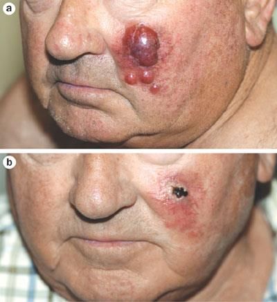
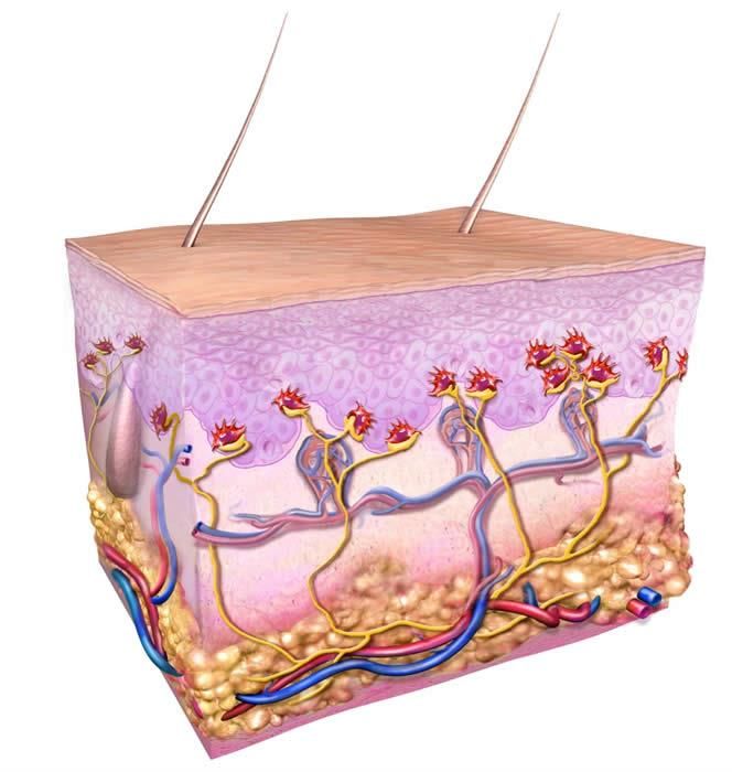
Normal Merkel cells in the skin: In
           this illustration of a cross-section of
          skin, normal Merkel cells are shown in
           red and connect to nerves shown in
           yellow. The structures drawn include
            the epidermis (upper third), dermis
            (middle), and deeper adipose layer
            containing the fatty tissue. Arteries
             are depicted as red and veins are
                             blue.

                                                                                      [Picture Credit: Merkel Cell Carcinoma]

This cancer is considered to be a form of neuroendocrine tumour. While patients with a small
tumour (less than 2 cm) that has not yet metastasised to regional lymph nodes have an expected 5-
year survival rate of more than 80 percent, once a lesion has metastasised regionally, the rate drops
to about 50 percent. Up to half of patients that have been seemingly treated successfully (i.e. that
initially appear cancer-free) subsequently suffer a recurrence of their disease. Recent reviews cite an
overall 5-year survival rate of about 60% for all MCC combined.

Merkel cell carcinoma (MCC) occurs most often on the sun-exposed face, head, and neck.

Walsh, N.M. & Cerronia, L. 2021.
“Merkel cell carcinoma has been a focus of active scientific investigation in recent years and new
information on the topic has emerged. Although uncommon, this primary cutaneous
neuroendocrine carcinoma, usually involving the head/neck of elderly individuals, has a poor
prognosis. Within the past two decades, an increase in the incidence of the tumor and the discovery
of its link to the Merkel cell polyomavirus have focused medical attention on the lesion. The
resulting studies have improved our understanding of the biology of the neoplasm and contributed
to clinical care. Specifically, two pathogenic subsets of the tumor have come to light, the majority
due to Merkel cell polyomavirus and the minority caused by ultraviolet radiation-induced genetic
damage. This dichotomy carries prognostic implications favoring the former subset. In addition,
having capitalized on the known susceptibility of the tumor to immune influences, investigators have
recently discovered its responsiveness to immune checkpoint inhibition. This revelation has
constituted a therapeutic milestone at the clinical level. Herein we provide an overview of the topic,
outline updates in the field and place an emphasis on dermatopathologic aspects of Merkel cell
carcinoma.”

Incidence of Merkel Cell Carcinoma (MCC) in South Africa
The National Cancer Registry (2017) does not make any mention of Merkel Cell Carcinoma.

Researched and Authored by Prof Michael C Herbst
[D Litt et Phil (Health Studies); D N Ed; M Art et Scien; B A Cur; Dip Occupational Health; Dip Genetic Counselling; Dip
Audiometry and Noise Measurement; Diagnostic Radiographer; Medical Ethicist]
Approved by Ms Elize Joubert, Chief Executive Officer [BA Social Work (cum laude); MA Social Work]
March 2021                                                                                                               Page 2
Cause of Merkel Cell Carcinoma (MCC)
A virus was discovered in 2008 to be frequently involved in MCC. This new virus is called Merkel Cell
Polyomavirus (MCPvV).The virus was found in 8 of 10 tumours tested, and it was associating with
the DNA of the tumour cells in such a way to suggest that it is involved in the development of MCC.
Several additional studies have validated this study, finding MCPvV in 43 of 53 patients.

Recently it was suggested that MCC also occurs more often in persons with HIV infection. In a search
of the Aids and cancer registers of the USA (1978–1996), ten MCC cases were identified as occurring
in both registers. In four of these cases, the MCC was diagnosed before the patient developed Aids.
In the remaining six cases, the MCC was diagnosed in persons with Aids, corresponding to a relative
risk of 13.4 compared with the general population.

Stages of Merkel Cell Carcinoma (MCC)
As of 2009 a new MCC staging system has been established. This new system is based on an analysis
of over 5,000 patients using the National Cancer Database as well as extensive review of the
literature. Stages I & II MCC are defined as disease that is localized to the skin at the primary site.
Stage I is for primary lesions less than or equal to 2 centimetres, and stage II is for primary lesions
greater than 2 cm. Stage III is defined as disease that involves nearby lymph nodes (regional lymph
nodes). Stage IV disease is found beyond regional lymph nodes.
          Stage Primary Tumour                       Lymph Node                                     Metastasis
             0     In situ primary tumour            No regional lymph node metastasis              No distant metastasis
                   Less than or equal to 2 cm
            IA     maximum tumour                    Nodes negative by pathologic exam              No distant metastasis
                   dimension
                   Less than or equal to 2 cm
                                                     Nodes negative by clinical exam* (no
            IB     maximum tumour                                                                   No distant metastasis
                                                     pathologic node exam performed)
                   dimension
                   Greater than 2 cm tumour
            IIA                                      Nodes negative by pathologic exam              No distant metastasis
                   dimension
                   Greater than 2 cm tumour          Nodes negative by clinical exam* (no
            IIB                                                                                     No distant metastasis
                   dimension                         pathologic node exam performed)
                   Primary tumour invades
            IIC    bone, muscle, fascia, or          No regional lymph node metastasis              No distant metastasis
                   cartilage
                   Any size tumour (includes
            IIIA                                     Micrometastasis**                              No distant metastasis
                   invading tumours)
                   Any size tumour (includes         Macrometastasis*** -OR-
            IIIB                                                                                    No distant metastasis
                   invading tumours)                 In transit metastasis****
                   Any size tumour (includes                                                        Metastasis beyond
             IV                                      Any lymph node metastasis
                   invading tumours)                                                                regional lymph nodes
         *Clinical detection of nodal disease may be via inspection, palpation, and/or imaging
         **Micrometastases are diagnosed after sentinel or elective lymphadenectomy
         ***Macrometastases are defined as clinically detectable nodal metastases confirmed by therapeutic
         lymphadenectomy or needle biopsy
         ****In transit metastasis: a tumour distinct from the primary lesion and located either (1) between the primary lesion
         and the draining regional lymph nodes or (2) distal to the primary lesion
         (Merkel Cell Carcinoma.Org).

Researched and Authored by Prof Michael C Herbst
[D Litt et Phil (Health Studies); D N Ed; M Art et Scien; B A Cur; Dip Occupational Health; Dip Genetic Counselling; Dip
Audiometry and Noise Measurement; Diagnostic Radiographer; Medical Ethicist]
Approved by Ms Elize Joubert, Chief Executive Officer [BA Social Work (cum laude); MA Social Work]
March 2021                                                                                                               Page 3
Risk Factors for Merkel Cell Carcinoma (MCC)
Factors that may increase your risk of Merkel cell carcinoma include:
• Excessive exposure to natural or artificial sunlight - Being exposed to ultraviolet light, such as the
light that comes from the sun or from tanning beds, increases one’s risk of Merkel cell
carcinoma. The majority of Merkel cell carcinomas appear on skin surfaces frequently exposed
to sun.
• A weakened immune system - People with weakened immune systems - including those with
HIV infection or those taking drugs that suppress the immune response - are more likely to
develop Merkel cell carcinoma.
• History of other skin cancers - Merkel cell carcinoma is associated with the development of
other skin cancers, such as basal cell or squamous cell carcinoma.
• Older age – One’s risk of Merkel cell carcinoma increases with age. This cancer is most common
in people older than age 50, though it can occur at any age.
• Light skin colour - Merkel cell carcinoma usually arises in people who have light-coloured skin.
Whites are much more likely to be affected by this skin cancer than are blacks.
Brady, M. & Spiker, A.M. 2020.
“Merkel cell carcinoma (MCC) is a rare, aggressive neuroendocrine tumor of the skin with increasing
incidence. It most frequently presents on the head and neck region of elderly, white males. Specific
risk factors include ultraviolet (UV) exposure, advancing age, and immunosuppression, and its
development is associated with Merkel cell polyomavirus (MCPyV) infection. Skin biopsy is
diagnostic, and sentinel lymph node evaluation should be performed in all patients who are
diagnosed with MCC, as the disease typically has a rapidly progressive course. Treatment consists of
wide local excision with or without adjuvant radiotherapy for local disease. New therapies for
metastatic MCC have shown promise and include immune-based therapies.”
Diagnosis of Merkel Cell Carcinoma (MCC)
Most MCCs are diagnosed when a skin biopsy is performed to rule out another sun-induced skin
cancer or a cyst.
Sachpekidis, C., Sidiropoulou, P., Hassel, J.C., Drakoulis, N. & Dimitrakopoulou-Strauss, A. 2020.
“Merkel cell carcinoma (MCC) is a rare neuroendocrine skin malignancy usually arising as a
nonspecific nodule on sun-exposed areas of the head and neck. Given the poor prognosis of this
aggressive tumor, assessment of disease burden in pre- and post-treatment care may ensure an
optimal management with significant implications for patient surveillance and prognosis. Although
imaging has established its role in locally advanced or distant metastatic MCC, a standard imaging
algorithm is yet to be determined and respective recommendations are mainly based on melanoma.
Positron emission tomography/computed tomography (PET/CT) is increasingly evolving as a valuable
imaging tool in metastatic or unresectable MCC, mostly utilizing the glucose analogue 18F-
fluorodeoxyglucose (18F-FDG) as a radiotracer. Despite being inferior in detecting the disease in its
early stages compared to the "gold standard" of sentinel lymph node biopsy, recent evidence
suggests an important role for 18F-FDG PET/CT in the routine workup of localized MCC.
Moreover, 68Ga-labeled somatostatin analogues have been employed as PET tracers in the field of
MCC with promising, yet comparable to 18F-FDG, results. This article provides a structured literature
Researched and Authored by Prof Michael C Herbst
[D Litt et Phil (Health Studies); D N Ed; M Art et Scien; B A Cur; Dip Occupational Health; Dip Genetic Counselling; Dip
Audiometry and Noise Measurement; Diagnostic Radiographer; Medical Ethicist]
Approved by Ms Elize Joubert, Chief Executive Officer [BA Social Work (cum laude); MA Social Work]
March 2021 Page 4review of the most important studies investigating the role of PET or PET/CT in the clinical practice of MCC.” Treatment of Merkel Cell Carcinoma (MCC) Merkel cell carcinoma is highly treatable with surgical and nonsurgical therapies, particularly if caught early. Treatments are often highly individualised, depending on a patient's general health, as well as the tumour's location, size, depth, and degree of spread. Patients with Merkel cell carcinoma are usually first treated with surgery. Patients with more advanced disease may receive adjuvant (additional) treatments such as radiation therapy and chemotherapy following, or instead of, surgery. Surgery - Surgery to remove the tumour is the most common treatment for Merkel cell carcinoma. A surgeon will also typically remove a safety margin of up to 2,5cm of normal skin around the tumour, and often underlying fatty and fibrous tissue as well, to ensure that all cancer cells have been removed. This is usually done in conjunction with a sentinel lymph node biopsy to determine if the cancer has spread to regional lymph nodes. Surgery may be the only treatment needed if the tumour is small and a wide margin of skin and soft tissue can be removed. Patients whose tumours have no lymph-node involvement have a greater than 60 percent chance of long-term survival or cure. Surgical removal of nearby lymph nodes, usually followed by radiation and chemotherapy, may also be required in patients whose tumours have spread regionally. Spread to lymph nodes is found in more than half of patients. Radiation Therapy and Chemotherapy - Localized radiation therapy is commonly used to destroy any remaining cancer cells following surgery to remove Merkel cell tumours. Radiation is also occasionally used to treat the area surrounding lymph nodes that have been surgically removed. Radiation therapy delivers penetrating beams of energy waves or streams of particles to the cancer cells and a small margin around the tumour. Radiation therapy can also be used to treat patients who are not candidates for surgery because of ill health or the location of their tumour, or to treat tumours that have returned after an initial round of treatment. Chemotherapy is another treatment option following surgery. The same platinum-based chemotherapy that is used for small cell lung cancer can be used against Merkel cell carcinoma that has spread to the lymph nodes. Patients whose tumours have spread to distant areas of the body or returned following initial treatment may also be treated with chemotherapy. Neoadjuvant chemotherapy (chemotherapy that is given before surgery) may be recommended for some patients with large Merkel cell tumours (greater than 2 centimetres) or lymph node involvement. Before this step is taken, however, consideration is needed to ensure that a patient treated with chemotherapy will still be healthy enough to subsequently undergo the surgery or radiation. Although the rarity of Merkel cell carcinoma has made it difficult to study, researchers continue to evaluate the best ways to use radiation therapy and chemotherapy in caring for patients with the disease. Researched and Authored by Prof Michael C Herbst [D Litt et Phil (Health Studies); D N Ed; M Art et Scien; B A Cur; Dip Occupational Health; Dip Genetic Counselling; Dip Audiometry and Noise Measurement; Diagnostic Radiographer; Medical Ethicist] Approved by Ms Elize Joubert, Chief Executive Officer [BA Social Work (cum laude); MA Social Work] March 2021 Page 5
Reconstruction After Surgery for Skin Cancer - Any form of surgery can leave a scar, some more
noticeable than others. When removal of a Merkel cell carcinoma leaves a wound that is too large to
close with simple sutures, surgeons can use skin grafts, flaps, and other reconstructive procedures to
help heal the skin and restore its appearance.

Follow-Up Care - Even after successful treatment, Merkel cell carcinomas can often come back. Also,
people who have one skin cancer are at higher-than-average risk for developing new skin cancers of
all types.

Individuals who have been treated for Merkel cell carcinoma should see their doctor immediately if
they find a growth, bump, spot, or any changes in their skin that could indicate a recurrence of
disease. Protection from sun exposure is also critical.

Patel, P. & Hussain, K. 2020.
“Merkel cell carcinoma (MCC) of the skin is a rare, aggressive form of skin cancer that metastasizes
to other parts of the body. This cutaneous neuroendocrine tumour mainly affects older people, with
most cases generally occurring over the age of 50 years. Merkel cell polyomavirus has been shown
to induce gene mutations resulting in this skin cancer, with immunosuppression and ultraviolet
radiation being other key risk factors in its pathogenesis. MCC is clinically seen as a rapidly enlarging,
isolated, irregular erythematous nodule typically found on sun-exposed sites. Diagnosis is through
clinical examination followed by tissue biopsy, which demonstrates characteristic histopathological
neuroendocrine features. Immunohistochemistry plays a crucial role in diagnosis with the
characteristic perinuclear staining with cytokeratin-20 helping to differentiate it from other
morphologically similar tumours. Sentinel lymph node biopsy and imaging is essential for staging and
determining prognosis. Surgical excision is the mainstay of treatment for localized disease although
adjuvant radiotherapy is often required. Metastatic disease involves a very poor prognosis, and
immune checkpoint inhibitors have recently shown promise in the treatment of metastatic disease.
Avelumab, a monoclonal antibody that binds to the programmed death-1 receptor, has been
approved by the National Institute for Health and Care Excellence and shown encouraging survival
outcomes. It provides an option for treating metastatic carcinoma in adults after they have failed ≥ 1
line of chemotherapy for metastatic disease.”

Topalian, S.L., Bhatia, S., Amin, A., Kudchadkar, R.R., Sharfman, W.H., Lebbé, C., Delord, J.P., Dunn,
L.A., Shinohara, M.M., Kulikauskas, R., Chung, C.H., Martens, U.M., Ferris, R.L., Stein, J.E., Engle,
E.L., Devriese, L.A., Lao, C.D., Gu, J., Li, B., Chen, T., Barrows, A., Horvath, A., Taube, J.M. &
Nghiem, P. 2020.
Purpose: Merkel cell carcinoma (MCC) is a rare, aggressive skin cancer commonly driven by the
Merkel cell polyomavirus (MCPyV). The programmed death-1 (PD-1)/programmed death-ligand 1
(PD-L1) immunosuppressive pathway is often upregulated in MCC, and advanced metastatic MCC
frequently responds to PD-1 blockade. We report what we believe to be the first trial of anti-PD-1 in
the neoadjuvant setting for resectable MCC.
Methods: In the phase I/II CheckMate 358 study of virus-associated cancer types, patients with
resectable MCC received nivolumab 240 mg intravenously on days 1 and 15. Surgery was planned on
day 29. Tumor regression was assessed radiographically and microscopically. Tumor MCPyV status,
PD-L1 expression, and tumor mutational burden (TMB) were assessed in pretreatment tumor
biopsies.
Results: Thirty-nine patients with American Joint Committee on Cancer stage IIA-IV resectable MCC
received ≥ 1 nivolumab dose. Three patients (7.7%) did not undergo surgery because of tumor
progression (n = 1) or adverse events (n = 2). Any-grade treatment-related adverse events occurred

Researched and Authored by Prof Michael C Herbst
[D Litt et Phil (Health Studies); D N Ed; M Art et Scien; B A Cur; Dip Occupational Health; Dip Genetic Counselling; Dip
Audiometry and Noise Measurement; Diagnostic Radiographer; Medical Ethicist]
Approved by Ms Elize Joubert, Chief Executive Officer [BA Social Work (cum laude); MA Social Work]
March 2021                                                                                                               Page 6
in 18 patients (46.2%), and grade 3-4 events in 3 patients (7.7%), with no unexpected toxicities.
Among 36 patients who underwent surgery, 17 (47.2%) achieved a pathologic complete response
(pCR). Among 33 radiographically evaluable patients who underwent surgery, 18 (54.5%) had tumor
reductions ≥ 30%. Responses were observed regardless of tumor MCPyV, PD-L1, or TMB status. At a
median follow-up of 20.3 months, median recurrence-free survival (RFS) and overall survival were
not reached. RFS significantly correlated with pCR and radiographic response at the time of surgery.
No patient with a pCR had tumor relapse during observation.
Conclusion: Nivolumab administered approximately 4 weeks before surgery in MCC was generally
tolerable and induced pCRs and radiographic tumor regressions in approximately one half of treated
patients. These early markers of response significantly predicted improved RFS. Additional
investigation of these promising findings is warranted.

Kwan, K., Ghazizadeh, S., Moon, A.S., Rünger, D., Sajed, D., Elashoff, D. & St John, M. 2020.
Objective: To evaluate the management and recurrence outcomes of head and neck Merkel cell
carcinoma (HN-MCC) at a single institution.
Study design: A retrospective review of outcomes in patients with HN-MCC.
Setting: A tertiary center from May 1990 to December 2018.
Subjects and methods: Electronic medical records of patients with HN-MCC were reviewed.
Results: Sixty cases were included, with 67% (40 of 60) males and a mean age of 73.3 years. Imaging
had a moderate sensitivity and specificity for detection of occult disease when compared with
histopathologic analysis. Forty-two percent (25 of 60) of patients underwent neck dissection, and
12% (7 of 60) had a sentinel lymph node biopsy (SLNB). There was a high rate of negative SLNB
findings. The majority of patients were treated with surgery alone (29 of 60), followed by a cohort
(21 of 60) treated with surgery plus adjuvant treatment, and 10 of 60 patients were treated with
radiation therapy with or without chemotherapy. Recurrence-free survival was 50%, 45%, and 42%
at 1, 2, and 5 years.
Conclusions: We report higher recurrence rates and higher negative SLNB result rates than other
studies. Our results affirm that imaging may not be a substitute for SLNB and that it had an
intermediate ability to identify the occult disease. Traditional predictors, including SLNB and cervical
node pathology, may not identify patients at risk for recurrence in HN-MCC. We report similar
recurrence rates in patients who had treatment of the cervical nodes by radiation therapy or neck
dissection as compared with those who did not receive neck treatment.

About Clinical Trials
Clinical trials are research studies that involve people. They are conducted under controlled
conditions. Only about 10% of all drugs started in human clinical trials become an approved drug.
Clinical trials include:
     • Trials to test effectiveness of new treatments
     • Trials to test new ways of using current treatments
     • Tests new interventions that may lower the risk of developing certain types of cancers
     • Tests to find new ways of screening for cancer

The South African National Clinical Trials Register provides the public with updated information on
clinical trials on human participants being conducted in South Africa. The Register provides
information on the purpose of the clinical trial; who can participate, where the trial is located, and
contact details.

Researched and Authored by Prof Michael C Herbst
[D Litt et Phil (Health Studies); D N Ed; M Art et Scien; B A Cur; Dip Occupational Health; Dip Genetic Counselling; Dip
Audiometry and Noise Measurement; Diagnostic Radiographer; Medical Ethicist]
Approved by Ms Elize Joubert, Chief Executive Officer [BA Social Work (cum laude); MA Social Work]
March 2021                                                                                                               Page 7
For additional information, please visit: www.sanctr.gov.za/ Medical Disclaimer This Fact Sheet is intended to provide general information only and, as such, should not be considered as a substitute for advice, medically or otherwise, covering any specific situation. Users should seek appropriate advice before taking or refraining from taking any action in reliance on any information contained in this Fact Sheet. So far as permissible by law, the Cancer Association of South Africa (CANSA) does not accept any liability to any person (or his/her dependants/ estate/heirs) relating to the use of any information contained in this Fact Sheet. Whilst the Cancer Association of South Africa (CANSA) has taken every precaution in compiling this Fact Sheet, neither it, nor any contributor(s) to this Fact Sheet can be held responsible for any action (or the lack thereof) taken by any person or organisation wherever they shall be based, as a result, direct or otherwise, of information contained in, or accessed through, this Fact Sheet. Sources and References Consulted or Utilised Brady, M. & Spiker, A.M. 2020. Merkel cell carcinoma of the skin. In: StatPearls [Internet]. Treasure Island (FL): StatPearls Publishing; 2021 Jan. 2020 Aug 16. Cassler, N.M., Merrill, D., Bichakjian, C.KI. & Brownell, I. 2016. Merkel Cell carcinoma therapeutic update. Curr Treat Options Oncol. 2016 Jul;17(7):36. doi: 10.1007/s11864-016-0409-1. Coggshall, K., Tello, T.L., Norht, J.P. & Yu, S.S. 2018. Merkel cell carcinoma: an update and review: pathogenesis, diagnosis, and staging. J Am Acad Dermatol. 2018 Mar;78(3):433-442. doi: 10.1016/j.jaad.2017.12.001. Epub 2017 Dec 9. Colebunders, R., Bottieau, E., Van den Brande, J., Colpaert, C. & Van Marck, E. 2004. Merkel cell carcinoma and multiple basal cell carcinoma in an African albino woman with HIV infection. HIV Medicine (2004), 5, 452–454. Del Marmol, V. & Lebbé, C. 2019. New perspectives in Merkel Cell carcinoma. Curr Opin Oncol. 2019 Jan 28. doi: 10.1097/CCO.0000000000000508. [Epub ahead of print] Drusio, C., Becker, J.C., Schadendorf, D. & Ugurel, S. 2019. Merkel Cell carcinoma. Hautarzt. 2019 Jan 30. doi: 10.1007/s00105-019-4360-5. [Epub ahead of print] Harms, P.W. 2017. Update on Merkel Cell carcinoma. Clin Lab Med. 2017 Sep;37(3):485-501. doi: 10.1016/j.cll.2017.05.004. Epub 2017 Jun 13. Kwan, K., Ghazizadeh, S., Moon, A.S., Rünger, D., Sajed, D., Elashoff, D. & St John, M. 2020. Merkel Cell Carcinoma: A 28- Year Experience. Otolaryngol Head Neck Surg. 2020 Aug;163(2):364-371. Mayo Clinic http://www.mayoclinic.org/diseases-conditions/merkel-cell-carcinoma/basics/risk-factors/con-20026875 Memorial Sloan Kettering Cancer Center http://www.mskcc.org/cancer-care/adult/merkel-cell-carcinoma/diagnosis-treatment-msk Merkel Cell Carcinoma Figure Copyright by Paul Nghiem, MD, PhD & Quade Medical Group. http://www.merkelcell.org/aboutDisease/index.php Researched and Authored by Prof Michael C Herbst [D Litt et Phil (Health Studies); D N Ed; M Art et Scien; B A Cur; Dip Occupational Health; Dip Genetic Counselling; Dip Audiometry and Noise Measurement; Diagnostic Radiographer; Medical Ethicist] Approved by Ms Elize Joubert, Chief Executive Officer [BA Social Work (cum laude); MA Social Work] March 2021 Page 8
Merkel Cell Carcinoma.Org
http://www.merkelcell.org/aboutDisease/index.php
http://www.merkelcell.org/staging/index.php

MCC
http://www.nature.com/nrclinonc/journal/v6/n9/images/nrclinonc.2009.109-f1.jpg

National Cancer Institute
http://www.cancer.gov/cancertopics/pdq/treatment/merkelcell/Patient/page1
http://www.cancer.gov/about-cancer/treatment/clinical-trials/what-are-trials

Patel, P. & Hussain, K. 2020. Merkel cell carcinoma. Clin Exp Dermatol. 2020 Nov 30.doi: 10.1111/ced.14530. Online ahead
of print.

Sachpekidis, C., Sidiropoulou, P., Hassel, J.C., Drakoulis, N. & Dimitrakopoulou-Strauss, A. 2020. Positron emission
tomography in merkel cell carcinoma. Cancers (Basel). 2020 Oct 9;12(10):2897.

Skin Cancer Foundation
http://www.skincancer.org/skin-cancer-information/merkel-cell-carcinoma

Topalian, S.L., Bhatia, S., Amin, A., Kudchadkar, R.R., Sharfman, W.H., Lebbé, C., Delord, J.P., Dunn, L.A., Shinohara,
M.M., Kulikauskas, R., Chung, C.H., Martens, U.M., Ferris, R.L., Stein, J.E., Engle, E.L., Devriese, L.A., Lao, C.D., Gu, J., Li,
B., Chen, T., Barrows, A., Horvath, A., Taube, J.M. & Nghiem, P. 2020. Neoadjuvant Nivolumab for Patients With
Resectable Merkel Cell Carcinoma in the CheckMate 358 Trial. J Clin Oncol. 2020 Aug 1;38(22):2476-2487.

UItentuis, S.E., Louwman, M.W.J., van Akkooi, A.C.J. &Bekkenk, M. 2019. Treatment and survival of Merkel Cell
carcinoma since 1993: a population-based cohort study in the Netherlands. J Am Acad Dermatol. 2019 Jan 28. pii: S0190-
9622(19)30149-5. doi: 10.1016/j.jaad.2019.01.042. [Epub ahead of print]

Walsh, N.M. & Cerronia, L. 2021. Merkel cell carcinoma: a review. J Cutan Pathol. 2021 Mar;48(3):411-421.

Wikipedia
http://en.wikipedia.org/wiki/Merkel_cell_carcinoma

Researched and Authored by Prof Michael C Herbst
[D Litt et Phil (Health Studies); D N Ed; M Art et Scien; B A Cur; Dip Occupational Health; Dip Genetic Counselling; Dip
Audiometry and Noise Measurement; Diagnostic Radiographer; Medical Ethicist]
Approved by Ms Elize Joubert, Chief Executive Officer [BA Social Work (cum laude); MA Social Work]
March 2021                                                                                                               Page 9
You can also read