Neural crest stem cells can be induced in vitro from human-induced pluripotent stem cells using a novel protocol free of feeder cells - J-Stage
←
→
Page content transcription
If your browser does not render page correctly, please read the page content below
Journal of Rural Medicine
Original article
Neural crest stem cells can be induced in vitro from
human-induced pluripotent stem cells using a novel
protocol free of feeder cells
Rei Abe1, Kazuyo Yamauchi1, Kazuki Kuniyoshi1, Takane Suzuki2, Yusuke Matsuura1,
Seiji Ohtori1, and Kazuhisa Takahashi1
1
Department of Orthopaedic Surgery, Graduate School of Medicine, Chiba University, Japan
2
Department of Bioenvironmental Medicine, Graduate School of Medicine, Chiba University, Japan
Abstract
Objective: Our knowledge of human neural crest stem cells (NCSCs) is expanding, owing to recent advances in technologies utiliz-
ing human-induced pluripotent stem cells (hiPSCs) that generate NCSCs. However, the clinical application of these technologies
requires the reduction of xeno-materials. To overcome this significant impediment, this study aimed to devise a novel method to
induce NCSCs from hiPSCs without using a feeder cell layer.
Materials and Methods: hiPSCs were cultured in feeder-free maintenance media containing the Rho-associated coiled-coil form-
ing kinase inhibitor Y-27632. When the cells reached 50–70% confluence, differentiation was initiated by replacing the medium
with knockout serum replacement (KSR) medium containing Noggin and SB431542. The KSR medium was then gradually re-
placed with increasing concentrations of Neurobasal medium from day 5 to 11.
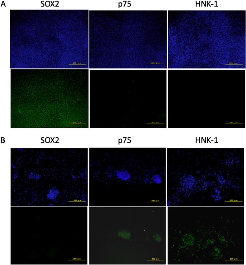
Results: Immunocytochemistry and flow cytometry were performed 12 days after induction of differentiation and revealed that the
cells generated from hiPSCs expressed the NCSC markers p75 and HNK-1, but not the hiPSC marker SOX2.
Conclusion: These findings demonstrate that hiPSCs were induced to differentiate into NCSCs in the absence of feeder cells.
Key words: induced pluripotent stem cell, neural crest stem cell, feeder-free, p75, HNK-1
(J Rural Med 2021; 16(3): 143–147)
Introduction various human diseases, they are highly relevant for clinical
applications, such as in regenerative medicine2).
Neural crest stem cells (NCSCs) represent a transient, Human NCSCs are difficult to obtain because they are
multipotent, and migratory population of cells unique to mainly harvested from fetal cells, which accounts for a few
vertebrates that emerge at the interface of the neural and relevant published studies. Therefore, our knowledge of the
non-neural ectoderm layers and migrate extensively to form role of NCSCs in development comes from studies on or-
various neural crest derivatives such as peripheral neurons, ganisms such as chickens and zebrafish3).
glia, melanocytes, endocrine cells, craniofacial tissues, Lee et al. developed protocols designed for differentiat-
bone, smooth muscle, and enteric neurons1). Because NC- ing human-induced pluripotent stem cells (hiPSCs) into NC-
SCs play major roles in development and are associated with SCs, which employ pharmacological inhibition of the bone
morphogenetic protein (BMP) and transforming growth
factor-β (TGF-β) signaling pathways. The availability of
Received: March 10, 2021
these protocols has led to numerous studies in the past de-
Accepted: March 15, 2021
Correspondence: Rei Abe, Department of Orthopaedic Surgery,
cade, dedicated to establishing robust and efficient methods
Graduate School of Medicine, Chiba University, 1-8-1 Inohana, to induce the differentiation of hiPSCs into NCSCs. Howev-
Chuo-ku, Chiba 260-8670, Japan er, most methods are cumbersome because they require nu-
E-mail: yoshihara-tuk@umin.ac.jp merous compounds and procedures and, more importantly,
This is an open-access article distributed under the terms of the rely on nonhuman materials such as stromal feeder cells and
Creative Commons Attribution Non-Commercial No Derivatives murine embryonic fibroblast-conditioned medium (MEF-
(by-nc-nd) License . the effective clinical application of NCSCs.
©2021 The Japanese Association of Rural Medicine doi: 10.2185/jrm.2021-010 | 143Journal of Rural Medicine
To solve these problems, we devised a novel method for
generating NCSCs from hiPSCs. In this study, we evalu-
ated the efficacy of this feeder-free method in establishing a
novel and useful protocol.
Materials and Methods Figure 1 Schematic of the NCSC induction methods. hiPSCs were
Cell culture maintained in mTeSR. Cell differentiation was initiated by
replacing the medium with KSR medium containing Noggin
hiPSCs were cultured in feeder-free maintenance me- and SB431542. KSR medium was then exchanged for Neu-
dium mTeSRTM1 (STEMCELL Technologies, Vancouver, robasal medium, which was added in gradually increasing
Canada) supplemented with 10 µM of the Rho-associated concentrations from day 3 to 12.
kinase inhibitor Y-27632 (Cayman Chemical Company,
Ann Arbor, MI, USA) on dishes coated with Matrigel (BD
Biosciences, San Jose, CA, USA)8, 9). bation with the primary antibody, the cells were incubated
with Alexa Fluor 488-conjugated goat anti-mouseantimouse
Generation of NCSC from hiPSCs immunoglobulin G (Abcam, Cambridge, UK). After each
The hiPSCs were plated on a Matrigel-coated dish step, the cells were rinsed three times in PBS and observed
(10,000–25,000 cells/cm 2) in mTeSR1 in the presence of under a fluorescence microscope (Olympus, Tokyo, Japan).
Y-27632. When the cells reached 50–70% confluence, DAPI (Thermo Fisher Scientific, Waltham, MA, USA) was
they were treated with different ratios of KSR:Neurobasal used to counterstain the nuclei.
medium to induce differentiation. The KSR medium con-
tained knockout DMEM, 15% KSR, 1% L-glutamine, 1% Flow cytometry
MEM-nonessential amino acids (each from Life Technolo- Cells were harvested using Accumax (Innovative Cell
gies, Carlsbad, CA, USA), and 55 µM β-mercaptoethanol Technologies, San Diego, CA, USA) for 5 min at 37 °C until
(Wako, Osaka, Japan). The Neurobasal media were made of they detached from the dishes. Cells were pelleted by cen-
NeurobasalTM supplemented with 2% B27 supplement, 1% trifugation at 160 × g for 5 min, resuspended in PBS, washed
N2 supplement, and 1% L-glutamine (each from Life Tech- with a chilled FACS buffer containing 0.5% BSA and 2-mM
nologies, Carlsbad, CA, USA). Cells were incubated daily EDTA in Dulbecco’s PBS (D-PBS), and kept on ice until
with fresh KSR:Neurobasal medium supplemented with 10 further use. Cells were resuspended in an appropriate cell
µM SB431542 (Tocris, Bristol, UK) and 500 ng/mL Nog- density in FACS buffer containing an antibody against p75
gin (R&D systems, Minneapolis, MN, USA). SB431542 is a or HNK-1 (BD Biosciences, San Jose, CA, USA). Cells were
drug candidate that inhibits activin receptor-like kinase re- filtered through a 40-µm filter, transferred to a FACS tube,
ceptors, ALK5, ALK4, and ALK7 and inhibits TGF-β. Nog- and immediately analyzed using a FACSCalibur Flow Cy-
gin is a protein that inhibits BMP signaling. The changes tometer (BD Biosciences, San Jose, CA, USA).
made to the ratio of KSR:Neurobasal medium were as fol-
lows: days 0–3 (100:0), day 5 (75:25), day 7 (50:50), day 9 Results
(25:75), and day 11 (0:100)4, 10, 11) (Figure 1).
Immunocytochemistry
Immunocytochemistry Immunocytochemistry was performed to evaluate the
The hiPSCs were seeded onto plastic chamber slides differentiation of hiPSCs into NCSCs. We used SOX2 as a
coated with Matrigel, cultured, and differentiated into NC- marker for hiPSCs, and p75 and HNK-1 were used as mark-
SCs as described above. The hiPSCs or differentiated cells ers for NCSCs. SOX2, but not p75 or HNK-1, was expressed
were fixed with 4% paraformaldehyde in a phosphate buffer by hiPSCs before they were induced to differentiate. In con-
(0.1 M, pH 7.4) for 15 min and rinsed with phosphate buff- trast, after differentiation, the cells expressed p75 and HNK-
ered saline (PBS) (Wako, Osaka, Japan). The cells were per- 1, but not SOX2 (Figure 2).
meabilized by incubation for 10 min at room temperature in
a blocking solution containing 5% BSA and PBS with 0.3% Flow cytometry
Triton X-100. Next, the cells were treated with a blocking Flow cytometry revealed that 63.4% ± 7.4% of the cells
solution for 30 min and incubated overnight at 4 °C with expressed p75 and HNK-1 after they were induced to dif-
the following mouse antibodies (diluted with blocking solu- ferentiate. In contrast, these markers were undetectable in
tion): SOX2 (R&D Systems, Minneapolis, MN, USA), p75 hiPSCs (Figure 3).
(Advanced Targeting Systems, San Diego, CA, USA), and
HNK-1 (BD Biosciences, San Jose, CA, USA). After incu-
2021; 16(3): 143–147 | doi: 10.2185/jrm.2021-010 | 144Journal of Rural Medicine
Figure 2 Immunocytochemical analysis of cells on day 12.
The upper images show DAPI-stained cells. Scale bar=200 μm. A) hiPSCs expressed SOX2 but not p75 and HNK-1.
B) After induction of differentiation, the cells expressed p75 and HNK-1 but not SOX2.
Discussion bone, and teeth. Furthermore, they differentiate into endo-
crine cells of the adrenal and thyroid glands, and melano-
In this study, we established a protocol for generating cytes of the skin. There are only a few published studies
NCSCs from hiPSCs that did not require feeder cells or xe- of human NCSCs because of the difficulty in obtaining
no-materials. The hiPSCs that were induced to differentiate 3–5-week-old human embryos and the transient nature of
expressed the NCSC markers p75 and HNK-1, in contrast this stem cell population3). However, recent developments
with untreated hiPSCs that expressed SOX2, which was as- in improving technologies for using hiPSCs have expanded
sessed by immunocytochemistry and flow cytometry. research efforts focused on human NCSCs, NC-derived tis-
NCSCs represent embryonic migratory cells with the sues, or both. The most important advantages of these ad-
potential to differentiate into various cell types that popu- vanced technologies include the high numbers (e.g., >106) of
late the peripheral nervous system, craniofacial cartilage, NCSCs that can be generated from hiPSCs, and NCSCs or
2021; 16(3): 143–147 | doi: 10.2185/jrm.2021-010 | 145Journal of Rural Medicine
Figure 3 Flow cytometric analysis of cells on day 12.
A) hiPSCs were not detected using antibodies against p75 or HNK-1. B) After hiPSC cul-
tures were induced to differentiate, 63% cells expressed p75 and HNK-1.
NC-derived tissues that can be isolated from patients with requires only eight compounds and fewer xeno-materials.
certain diseases1). Furthermore, this simple protocol achieved efficient gen-
The regulation of specific developmental pathways, such eration of NCSCs within 12 days, which is shorter than that
as the BMP/activin and Wnt signaling axes, is required for required using the protocol published by Menendez et al13).
proper development of NCs during embryogenesis12). Effi- This study has several limitations. First, the ability of
cient methods for generating NCSCs from hiPSCs employ the induced NCSCs to differentiate was not evaluated. Other
specific inhibitors such as Noggin and SB 431542 that in- studies have reported that all cells induced from hiPSCs that
hibit the BMP and activin A/nodal signaling pathways, express both p75 and HNK-1 express NCSC markers such as
respectively4, 10). Furthermore, NCSCs are generated by SOX10 and ERBB31). Therefore, it is likely that the induced
supplementing cultures of hiPSCs with an activator of Wnt NCSCs produced here that expressed p75 and HNK-1 were
signaling and an inhibitor of activin/nodal/TGF-β signal- authentic. Second, this protocol is not completely xeno-free
ing, although modulation of BMP signaling is not required 2). because mTeSR and KSR medium contains some xeno-
However, these methods require xeno-materials such as materials. However, in using this method, we were able to
feeder cells and sera, which are problematic for research reduce the xeno-materials. Third, we evaluated only one
aimed at clinical applications such as regenerative medicine. hiPSC cell line, despite the availability of several other cell
For clinical purposes, the cell induction protocol should lines with varying phenotypes14). However, all media and
not include the xeno-materials mentioned above and should compounds employed in this study, including mTeSRTM1
use a chemically defined medium. Recently, Fukuta et al. re- and KSR, are widely used to maintain hiPSCs and embry-
ported a modification of the method reported by Menendez13) onic stem cell lines, suggesting that this method is generally
that employs a feeder- and xeno-free protocol using an acti- applicable, although further studies are required to confirm
vator of Wnt signaling and inhibitor of activin/nodal/TGF-β this assumption.
signaling without BMP inhibitors. In contrast, in this study,
we developed a feeder-free protocol using a BMP inhibitor Conclusion
based on the report by Lee et al. We used a feeder-free medi-
um instead of MEF-CM, which was used in Lee’s protocol4). We developed a feeder-free protocol to induce hiPSCs
Although MEF-CM does not contain feeder cells, it contains to differentiate into NCSCs. Therefore, this novel protocol
fetal bovine serum and requires feeder cells for its produc- shows promise as a resource for generating NCSCs and NC-
tion, indicating that it is not technically feeder- or xeno-free. derived tissues that are suitable for preclinical research.
Another advantage of this protocol is its high efficiency
and use of a concise methodology. For example, 63% of hiP- Acknowledgments
SCs were induced to differentiate into NCSCs, which is con-
sistent with previous studies using xeno-materials. Previous The authors are grateful to A. Iwama and M. Ohsawa for
protocols induced 30–80% of hiPSCs to differentiate into suggesting the topics addressed in this study. The authors
NCSCs1, 3, 11, 13). Moreover, although certain protocols employ would like to thank Enago (www.enago.jp) for the English
more than 15 compounds in the medium 2, 13), our method language review.
2021; 16(3): 143–147 | doi: 10.2185/jrm.2021-010 | 146Journal of Rural Medicine
References
1. Lee G, Kim H, Elkabetz Y, et al. Isolation and directed differentiation of neural crest stem cells derived from human embryonic stem cells. Nat Biotechnol
2007; 25: 1468–1475. [Medline] [CrossRef]
2. Menendez L, Kulik MJ, Page AT, et al. Directed differentiation of human pluripotent cells to neural crest stem cells. Nat Protoc 2013; 8: 203–212. [Medline]
[CrossRef]
3. Liu Q, Spusta SC, Mi R, et al. Human neural crest stem cells derived from human ESCs and induced pluripotent stem cells: induction, maintenance, and
differentiation into functional schwann cells. Stem Cells Transl Med 2012; 1: 266–278. [Medline] [CrossRef]
4. Lee G, Chambers SM, Tomishima MJ, et al. Derivation of neural crest cells from human pluripotent stem cells. Nat Protoc 2010; 5: 688–701. [Medline]
[CrossRef]
5. Chimge NO, Bayarsaihan D. Generation of neural crest progenitors from human embryonic stem cells. J Exp Zoolog B Mol Dev Evol 2010; 314: 95–103.
[Medline] [CrossRef]
6. Milet C, Monsoro-Burq AH. Embryonic stem cell strategies to explore neural crest development in human embryos. Dev Biol 2012; 366: 96–99. [Medline]
[CrossRef]
7. Mica Y, Lee G, Chambers SM, et al. Modeling neural crest induction, melanocyte specification, and disease-related pigmentation defects in hESCs and
patient-specific iPSCs. Cell Rep 2013; 3: 1140–1152. [Medline] [CrossRef]
8. Watanabe K, Ueno M, Kamiya D, et al. A ROCK inhibitor permits survival of dissociated human embryonic stem cells. Nat Biotechnol 2007; 25: 681–686.
[Medline] [CrossRef]
9. Chan EM, Ratanasirintrawoot S, Park IH, et al. Live cell imaging distinguishes bona fide human iPS cells from partially reprogrammed cells. Nat Biotech-
nol 2009; 27: 1033–1037. [Medline] [CrossRef]
10. Chambers SM, Fasano CA, Papapetrou EP, et al. Highly efficient neural conversion of human ES and iPS cells by dual inhibition of SMAD signaling. Nat
Biotechnol 2009; 27: 275–280. [Medline] [CrossRef]
11. Kreitzer FR, Salomonis N, Sheehan A, et al. A robust method to derive functional neural crest cells from human pluripotent stem cells. Am J Stem Cells
2013; 2: 119–131. [Medline]
12. Raible DW, Ragland JW. Reiterated Wnt and BMP signals in neural crest development. Semin Cell Dev Biol 2005; 16: 673–682. [Medline] [CrossRef]
13. Fukuta M, Nakai Y, Kirino K, et al. Derivation of mesenchymal stromal cells from pluripotent stem cells through a neural crest lineage using small mol-
ecule compounds with defined media. PLoS One 2014; 9: e112291. [Medline] [CrossRef]
14. Bock C, Kiskinis E, Verstappen G, et al. Reference Maps of human ES and iPS cell variation enable high-throughput characterization of pluripotent cell
lines. Cell 2011; 144: 439–452. [Medline] [CrossRef]
2021; 16(3): 143–147 | doi: 10.2185/jrm.2021-010 | 147You can also read