The immunohistochemical expression of CD34 in human hair follicles: a comparative study with the bulge marker CK15
←
→
Page content transcription
If your browser does not render page correctly, please read the page content below
Experimental dermatology • Original article                                                                doi: 10.1111/j.1365-2230.2006.02255.x

The immunohistochemical expression of CD34 in human hair
follicles: a comparative study with the bulge marker CK15
E. Poblet, F. Jiménez,* J.M. Godı́nez,† A. Pascual-Martı́n and A. Izeta‡
Department of Pathology, and †Research Unit, Hospital General Universitario de Albacete, Universidad de Castilla La Mancha, Albacete, Spain; *Clı´nica
Dr Jiménez Acosta, Las Palmas de Gran Canaria, Spain; and ‡Fundación Inbiomed, San Sebastián, Spain

Summary                                 Background. Anti-CD34 antibodies label the bulge region of mouse hair follicles.
                                        However, in human hair follicles, CD34 immunoreactivity is found in the outer root
                                        sheath below the bulge zone. The immunohistochemical staining of CD34 in catagen
                                        and telogen follicles has not been evaluated.
                                        Aims: To characterize the expression of CD34 immunoreactivity at different stages of
                                        the hair cycle in human terminal hair follicles, and to compare the immunostaining
                                        pattern of CD34 with that of CK15, used here as a marker of the bulge region.
                                        Method. Serial vertical sections of human hair follicles in anagen, catagen and tel-
                                        ogen phases were immunostained with anti-CD34 (QBEnd 10) and anti-CK15 (LHK15
                                        and C8 ⁄ 144B) antibodies. Double-labelling immunofluorescence was also performed.
                                        Results. The catagen and telogen follicles studied did not show CD34 immuno-
                                        reactivity in the outer root sheath. The location of CD34 and CK15 immunoreactivity
                                        in anagen follicles reveals a different staining pattern: CD34-positive cells are located in
                                        the outer root sheath below the attachment zone of the arrector pili muscle, whereas
                                        CK15-positive cells are located in the outer root sheath above the attachment zone of
                                        the arrector pili muscle.
                                        Conclusions. Only anagen human hair follicles show CD34 immunoreactivity. CD34
                                        and CK15 recognize different types of cells or cells at different stages of differentiation.

                                                                                vascular and spindle cell tumours, such as Kaposi’s
Introduction
                                                                                sarcoma and dermatofibrosarcoma protuberans.
CD34 is a 110-kDa, heavily glycosylated, transmem-                                 To our knowledge, the hair follicle is the only human
brane protein encoded by a gene located on chromo-                              structure in which the expression of CD34 in epithelial
some 1q and expressed on haematopoietic stem cells,                             cells has been reported.3 CD34-positive cells were
vascular endothelial cells, embryonic fibroblasts, and                          initially described in the outermost cell layer of the
fibroblast-like dendritic cells in connective tissues.1 In                      external root sheath of anagen human hair follicles, in a
the skin, CD34 is expressed in a variety of mesenchymal-                        segment below the attachment of the arrector pili
derived cells such as vascular endothelial cells, dermal                        muscle and above the matrix cells.3 A recent study has
dendritic ⁄ spindle-shaped cells, and perifollicular cells.2                    confirmed that in human follicles, CD34 expression at
In diagnostic pathology, CD34 is used as a marker of                            the external root sheath does not include the bulge area,
                                                                                which is currently considered the niche for epithelial
                                                                                stem cells.4 In contrast, there is a significantly different
Correspondence: Dr Francisco Jiménez, Angel Guimerá, 2, 35003 Las             CD34 immunostaining pattern on mouse hair follicles,
Palmas de Gran Canaria, Spain.                                                  in which CD34-positive cells are found at a higher level
E-mail: fjimenez@clinicadelpelo.com
                                                                                of the follicle, specifically at the bulge region.5 More-
Conflict of interest: none declared.                                            over, these CD34-positive bulge cells in murine follicles
Accepted for publication 18 July 2006                                           identify keratinocytes with typical characteristics of

 2006 The Author(s)
Journal compilation  2006 Blackwell Publishing Ltd, Clinical and Experimental Dermatology, 31, 807–812                                              807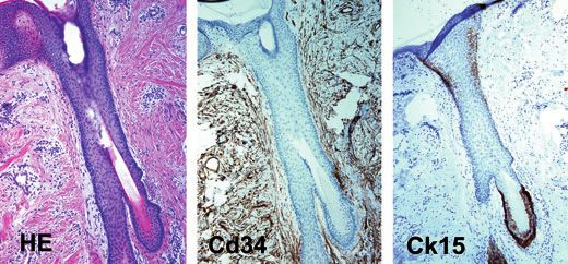
CD34 in human hair follicles. • E. Poblet et al.

stem cells: they are slow-cycling keratinocytes with the                  phases) could be examined. Serial sections were cut
capacity to form large colonies.                                          following a standard protocol to examine the same hair
   In the microscopic anatomy of the follicle, the bulge                  follicle stained with haematoxylin and eosin, CK15, and
region can be recognized as a prominent protuberance                      CD34. The protocol was as follows: sections were 3–
below the sebaceous gland in vertical sections of murine                  5 lm thick, and three consecutive single sections were
and human foetus specimens stained with haematoxy-                        retrieved and mounted on separate slides for the three
lin and eosin. In contrast, in adult human follicles, the                 different stains.
bulge region is barely prominent and is identified only                      In addition, 27 follicular units harvested from human
by its correspondence with the insertion of the arrector                  scalp were obtained from five different donors. The
pili muscle. Recent evidence suggests important struc-                    follicular units were harvested using a 1-mm circular
tural and ⁄ or biological differences between the human                   punch. The manipulation of these follicular units was
and the mouse outer root sheath, including the bulge                      very meticulous when it came to making the paraffin-
zone.4                                                                    wax blocks and the microtome cutting, as the goal was
   One of the most reliable immunohistochemical                           to obtain parallel sections that included all the length of
markers of the bulge region of human hair follicles                       the hair follicles. In order to obtain consecutive sections
in formalin-fixed, paraffin wax-embedded sections is                      of the same hair follicles, the same protocol described
cytokeratin (CK) 15. The antibodies used for the                          above was used. The only difference was that the series
detection of CK15 are derived from two different                          consisted of four slides, because two different anti-CK15
clones, C8 ⁄ 144B6 and LHK15.7 In human hair folli-                       antibodies (C8 ⁄ 144B and LHK15) were used.
cles, the C8 ⁄ 144B antibody, originally raised against                      Only the hair follicles that could be visualized along
the carboxy-terminal peptide of the T-lymphocyte                          their complete length were submitted for immunohisto-
protein, CD8, delineates the bulge region in anagen,                      chemical analysis. These included 96 hair follicles in
telogen and catagen follicles.6 Likewise, in formalin-                    anagen, 4 in catagen and 15 in telogen phase.
fixed tissues, the LHK15 antibody, raised against a
peptide derived from the last 17 amino acids of the
                                                                          Immunohistochemistry
CK15 polypeptide, delineates the isthmus area and a
small segment of the outer root sheath located above                      Two anti-CK15 antibodies (clone LHK15; Novocastra,
the hair bulb.8 However, when used in frozen sections,                    Newcastle, UK, and clone C8 ⁄ 144B; Dako, Glostrup
the antibody LHK15 appears to have a more extensive                       Denmark), and one anti-CD34 antibody (clone QBEnd
immunostaining pattern.7                                                  10; Dako) were used in our study.
   The aim of this study was to further characterize                         The immunohistochemical method used for the
the expression of CD34 immunoreactivity in human                          C8 ⁄ 144B antibody was as described by Lyle et al.8
terminal hair follicles not only in anagen human                          Briefly, the paraffin-wax sections were first dewaxed,
follicles, but also in catagen and telogen hair follicles.                and then steamed for 15 min in citrate buffer
In order to precisely define the location of the CD34                     (10 mmol ⁄ l sodium citrate pH 6.77) at 85 C prior to
immunoreactivity we used vertical serial sections of                      incubating overnight at 4 C with the anti-CD8 anti-
the entire length of the hair follicles and compared the                  body (dilution 1 : 40). The avidin–biotin-complex tech-
microscopic location of CD34 and CK15 immuno-                             nique was used for development, and diaminobenzidine
reactivity, the latter used here as a marker of the                       for visualization, followed by haematoxylin counter-
bulge region.                                                             stain.
                                                                             For the immunohistochemical staining with LHK15
                                                                          and QBEnd 10, the samples were dewaxed and
Materials and methods
                                                                          steamed for 40 min in citrate buffer pH 7 at 95 C.
                                                                          The antibody LHK15 was diluted to 1 : 80 and the
Tissue samples
                                                                          QBEnd was diluted to 1 : 50. The incubation time with
Normal human scalp samples from 15 surgical pathol-                       the primary antibodies was 30 min. The secondary
ogy specimens were selected from specimens received at                    antibodies used were those included in the kit (REAL
the Department of Pathology, Albacete University Gen-                     detection system; Dako), with an incubation time of
eral Hospital. All specimens were fixed using buffered                    15 min. Human hair follicles were immunostained
formalin and embedded in paraffin wax. About 120 hair                     using the standard avidin–streptavidin staining meth-
follicles in different stages of the hair cycle (most of them             od, and an automatic staining apparatus (Cytomation
in anagen phase, and others in catagen and telogen                        autostainer; Dako).

                                                                                                                               2006 The Author(s)
808                                        Journal compilation  2006 Blackwell Publishing Ltd, Clinical and Experimental Dermatology, 31, 807–812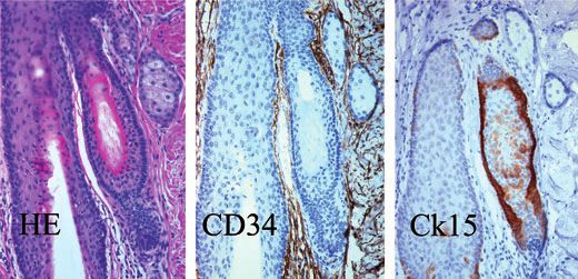
CD34 in human hair follicles. • E. Poblet et al.
Normal structures that should not be labelled with
these markers (i.e. endothelium for CK15, and epidermis
for CD34) served as internal negative controls.
Immunofluorescence double labelling
This technique was used to better delineate the location
of the expression of CD34 and CK15 by comparing in
the same section the areas stained with both antibodies.
Sections were dewaxed and then steamed for 40 min in
citrate buffer (pH 7) at 95 C for antigen retrieval. The
first antibody to be applied was the anti-CD34 QBend10
(1 : 50, 30 min at room temperature), followed by a
biotinylated secondary antibody, and by fluorescein-
labelled streptavidin (Cy2, dilution 1 : 100, goat
antirabbit; Amersham Biosciences). Slides were then
incubated with nonimmune calf serum for 30 min in
order to reduce nonspecific binding, and then with the
anti-LHK15 antibody, dilution 1 : 80, for 30 min at
room temperature. Finally, rodamine antimouse anti-
body was applied (Cy3, dilution 1 : 100; Amersham
Biosciences). The buffer used to rinse the sections
between the different incubations was a Tris-buffered
saline ⁄ Tween buffer, diluted 1 : 10 with deionized
water. The sections were imaged using a Leica DM
IRE2 confocal microscope. The laser used to observe the
fluorescence was the Leica TCS SP2.
Results
CD34 immunoreactivity
In the typical anagen human follicle, CD34 immuno-
reactivity was detected at the most peripheral layer of (a) (b)
the outer root sheath, in the transient portion of the
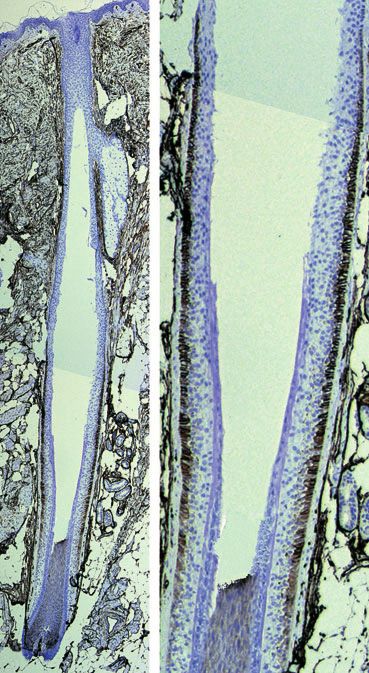
Figure 1 Longitudinal section of human scalp. Left, CD34
follicle, below the isthmus and above the matrix cells
expression in spindle-shaped dermal cells, endothelial cells and
(Figs 1 and 2). The anti-CD34 antibody stained the perifollicular spindle-shaped cells, and in epithelial cells of the
epithelial cells located at the outermost layer of the outer root sheath. Right, high-power image showing CD34-posit-
external root sheath (Fig. 1b). CD34 immunoreactivity ive epithelial cells in the most external layer of the outer root
was not detected in catagen or in telogen hair follicles sheath. CD34 immunostaining, original magnification (a) · 25;
(b) · 200.
(Figs 3 and 4). As expected, other skin structures that
stained with the CD34 antibody were the endothelial
cells, dendritic ⁄ spindle-shaped dermal cells and perifol-
licular spindle-shaped cells. muscle (Fig. 2). This CK15 staining was maintained
throughout the different phases of the hair follicle cycle
(Figs 3 and 4). The two anti-CK15 antibodies used in
Cytokeratin 15 immunoreactivity
this study, LHK15 and C8 ⁄ 144B, showed the same
In typical anagen follicles, CK15 was expressed in the immunostaining pattern. A few CK15-positive cells
outermost cell layer of the external root sheath, at the were also detected in the external root sheath just
level of the isthmus. Specifically, the CK15 immuno- above the bulb. Other skin structures that stained with
staining extended from the entrance of the sebaceous both CK15 antibodies were basal cells from the epider-
gland duct down to the insertion site of the arrector pili mis and secretory cells from eccrine glands.
2006 The Author(s)
Journal compilation 2006 Blackwell Publishing Ltd, Clinical and Experimental Dermatology, 31, 807–812 809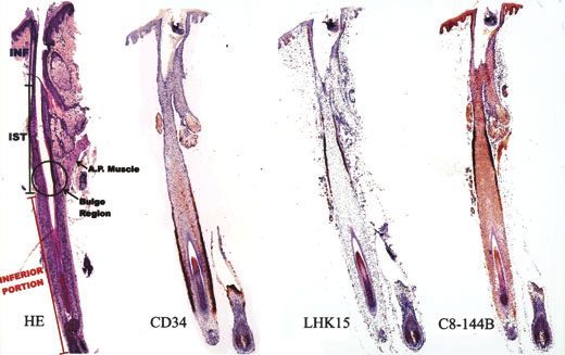
CD34 in human hair follicles. • E. Poblet et al.

Figure 2 Serial vertical sections of a human terminal anagen hair follicle, stained with (from left) haematoxylin and eosin, anti-CD34, and
two anti-CK15 antibodies (original magnification · 25). Sections are consecutive cuts of the same hair follicle. All the nonepithelial,
perifollicular, CD34-positive cells have been artificially removed in order to show the epithelial area of staining clearly. CD34 immuno-
reactivity was detected in the external root sheath, at the level of the inferior portion of the follicle, below the isthmus and above the matrix
cells. CK15 was expressed at the level of the isthmus, between the sebaceous-gland duct and down to the insertion site of the arrector pili
muscle (the bulge zone). Both anti-CK15 antibodies (LHK15 and C8 ⁄ 144B) showed similar immunoreactivity. Comparing the CD34 and
CK15 immunoreactivity reveals a different immunostaining pattern: CD34 stains below the bulge zone and CK15 stains at the isthmus
level. INF, infundibulum; IST, isthmus; AP, arrector pili. The bulge region (black circle), by definition, corresponds to the attachment site of
the AP muscle.

                                                                                                     Figure 3 Comparison of CD34 and CK15
                                                                                                     immunostaining (original magnification
                                                                                                     · 40) in human catagen hair follicles.
                                                                                                     CD34 immunoreactivity was not detected
                                                                                                     in the residual outer root sheath. CK15
                                                                                                     was strongly expressed by epithelial cells of
                                                                                                     the isthmus.

                                                                                                     Figure 4 Serial vertical sections of a telo-
                                                                                                     gen hair follicle stained with (from left)
                                                                                                     haematoxylin and eosin, CD34 and CK15
                                                                                                     (original magnification · 40). Intense
                                                                                                     positive CK15 immunostaining is clearly
                                                                                                     seen surrounding the telogen club. CD34
                                                                                                     immunoreactivity was not detected in
                                                                                                     telogen follicles.

                                                                                                                                 2006 The Author(s)
810                                          Journal compilation  2006 Blackwell Publishing Ltd, Clinical and Experimental Dermatology, 31, 807–812
CD34 in human hair follicles. • E. Poblet et al.
cycle. In anagen follicles, we demonstrated CD34
immunoreactivity in epithelial cells of the outer root
sheath below the bulge zone, an observation already
reported in our previous work.3 However, we extended
our observations to catagen and telogen follicles,
showing that CD34 immunostaining tends to disappear
in catagen and is absent in telogen follicles.
Comparing the immunostaining pattern of CD34 with
the immunostaining pattern of CK15, used in our study
as a bulge marker of human follicles, we confirmed that
CD34-positive cells are located below the bulge zone.
The results of the immunostaining pattern with two
different antibodies anti-CK15 concur with previous
reports,6,8 and show that CK15-positive cells are mainly
found at the most peripheral layer of the outer root
sheath at the level of the isthmus, specifically between
the entrance of the sebaceous duct and the insertion site
of the arrector pili muscle, although a less intense
staining can be observed in suprabulbar cells. This
CK15 immunostaining at the isthmus level was main-
tained in catagen and telogen follicles, where an intense
CK15 staining could be seen surrounding the telogen
club. The different CD34 and CK15 immunostaining
patterns indicate that these two markers recognize
different types of cells or cells at different stages of
differentiation. Lyle et al.6 showed that the CK15-
positive bulge cells possess the proliferative behaviour
and biochemical properties of epithelial stem cells.
However, the significance of the CD34 immunoreactiv-
ity in epithelial cells of the lower external root sheath in
Figure 5 Double-immunofluorescence study of a vertical section of
human hair follicles is unknown. The fact that human
an anagen hair follicle. The area of the follicle shown in this pic- catagen and telogen follicles lose their CD34 expression
ture corresponds to the lower portion of the isthmus (CK15+, could indicate that CD34 is not a lineage-specific
stained green) and the upper portion of the transient segment of antigen, but may be related to certain functions in
the hair follicle (CD34+, stained red). The areas stained with both anagen or growing cells. A possible role of CD34 in
markers appear to be clearly different and do not overlap. Original
magnification · 400.
cytoadhesion and signalling related to differentiation
and proliferation has been suggested.9 Based on this
adhesive property, we speculate that the CD34 antigen
might contribute to increasing the adhesion of the
Comparison of CD34 and CK15 immunoreactivity
external root sheath to the perifollicular stroma in
It may be deduced from consecutive sections of the same anagen follicles. However, it is also possible that the
hair follicle that the areas that stained with the anti- disappearance of the CD34 immunoreactivity in cat-
CD34 antibody and the anti-CK15 antibodies were agen and telogen follicles might be the result of the
completely different. A double immunofluorescence destruction by apoptosis that takes place in the transient
study showed that the segment of the follicle positive portion of the follicle, with the subsequent loss of CD34
for CD34 was located almost in continuity with, but expression by those cells.
below the zone that stained with CK15 (Fig. 5). Although CD34 identifies a subset of bulge keratino-
cytes with characteristics of stem cells in murine
follicles, this is not the case in human follicles. Ohyama
Discussion
et al.4 have recently characterized the gene-expression
We examined the immunohistochemical pattern of profile and cell-surface markers of the bulge region of
CD34 in human hair follicles at different phases of the human follicles, confirming that CD34 expression is low
2006 The Author(s)
Journal compilation 2006 Blackwell Publishing Ltd, Clinical and Experimental Dermatology, 31, 807–812 811CD34 in human hair follicles. • E. Poblet et al.
or absent in human bulge cells with stem cell-charac- Ms Prieto for technical assistance. JMG is supported by
teristics. These authors have shown that human bulge a fellowship from Balague Center S.A.
cells with stem cell properties have a CD200hiCD34lo
surface phenotype. Nevertheless, the possibility that the
References
CD34-positive cells of the lower outer root sheath of
human follicles still retain some clonogenic capacity 1 Krause DS, Fackler MJ, Civin CI, May WS. CD34: structure,
cannot, according to previous studies on clonal analysis, biology, and clinical utility. Blood 1996; 87: 1–13.
be excluded. The location of the CD34 immunoreactivity 2 Nickoloff BJ. The human progenitor cell antigen (CD34) is
in human follicles concurs with the area in whcih localized on endothelial cells, dermal dendritic cells, and
perifollicular cells in formalin-fixed normal skin, and on
Rochat et al.10 found the highest clonogenic capacity. In
proliferating endothelial cells and stromal spindle-shaped
this classic experiment of clonal analysis, these inves-
cells in Kaposi’s sarcoma. Arch Dermatol 1991; 127: 523–
tigators examined the in vitro proliferative behaviour of 9.
dissociated keratinocytes from microdissected portions 3 Poblet E, Jimenez-Acosta F, Rocamora A. Qbend ⁄ 10 (anti-
of human hair follicles, and found that an area of the CD34 antibody) in external root sheath cells and follicular
hair follicle below the bulge, but above the bulb, in the tumors. J Cut Pathol 1994; 21: 224–8.
lower outer root sheath, contained the highest percent- 4 Ohyama M, Terunuma A, Tock CL. Characterization and
age of clonogenicity. Other evidence indicating that isolation of stem cell-enriched human hair follicle bulge
these CD34-positive cells of the outer root sheath retain cells. J Clin Invest 2006; 116: 249–60.
significant proliferative capacity is their potential to give 5 Trempus CS, Morris RJ, Bortner CD et al. Enrichment for
rise to a neoplasm known as trichilemmoma.3 living murine keratinocytes from the hair follicle bulge
with the cell surface marker CD34. J Invest Dermatol 2003;
In summary, in this study we have clarified the
120: 501–11.
expression of CD34 in hair follicles from human scalp.
6 Lyle S, Christofidou-Solomidou M, Liu Y et al. The
We have confirmed that CD34 is expressed in epithelial C8 ⁄ 144B monoclonal antibody recognizes cytokeratin 15
cells of the external root sheath only in anagen follicles, and defines the location of human hair follicle stem cells.
and is not expressed in catagen or telogen hair follicles. J Cell Sci 1998; 111: 3179–88.
The location of CD34 and CK15 immunostaining is 7 Waseem A, Dogan B, Tidman N et al. Keratin 15 expres-
different and they do not overlap, suggesting that they sion in stratified epithelia: downregulation in activated
recognize different types of cells or cells at different keratinocytes. J Invest Dermatol 1999; 112: 362–9.
stages of differentiation. 8 Ozawa M, Aiba S, Kurosawa M, Tagani H. Ber-EP4 antigen
is a marker for a cell population related to the secondary
hair germ. Exp Dermatol 2004; 13: 401–5.
Acknowledgements 9 Majdic O, Stockl J, Pickl WF et al. Signalling and induction
of enhanced cytoadhesiveness via the hematopoietic pro-
This work has been supported by grant FIS PI03-1364.
genitor cell surface molecule CD34. Blood 1994; 83:
The authors express their appreciation to Dr de Cabo 1226–34.
and Dr Atienzar for their thoughtful review of the 10 Rochat A, Kobayashi K, Barrandon Y. Location of stem
manuscript, to Ms Sanchis for her collaboration with cells of human hair follicles by clonal analysis. Cell 1994;
the confocal laser microscopy technique, and to 76: 1063–73.
2006 The Author(s)
812 Journal compilation 2006 Blackwell Publishing Ltd, Clinical and Experimental Dermatology, 31, 807–812You can also read