The Use of Immunohistochemistry for Diagnosis of Prostate Cancer
←
→
Page content transcription
If your browser does not render page correctly, please read the page content below
Clinical Urology             Immunohistochemistry for Diagnosis of Prostate Cancer
International Braz J Urol                                                Vol. 36 (5): 583-590, September - October, 2010
                                                                                    doi: 10.1590/S1677-55382010000500008

The Use of Immunohistochemistry for Diagnosis of Prostate
Cancer
Katia R. M. Leite, Miguel Srougi, Adriana Sanudo, Marcos F. Dall’oglio, Adriano Nesrallah,
Alberto A. Antunes, Jose Cury, Luiz H. Camara-Lopes

Laboratory of Medical Investigation (KRML, MS, AS, MFDO, AN, AAA, JC), Division of Urology,
University of Sao Paulo Medical School, and Genoa Biotechnology (KRML, LHCL), Sao Paulo,
Brazil

ABSTRACT

Purpose: Atypical glands (ASAP) are diagnosed in 5.0% of prostate biopsies, and cancer identification in a rebiopsy is
higher than 40.0%. The use of antibodies to mark basal cells is currently a common practice, in order to avoid rebiopsies.
There has been no reported study that has reviewed characteristics of radical prostatectomies (RPs) when immunohisto-
chemistry (IHC) was necessary for definitive diagnosis.
Materials and Methods: Out of 4127 biopsies examined from 2004 to 2008, 144 (3.5%) were diagnosed with ASAP. IHC
was performed using antibody anti-34βE12 and p63. The results of surgical specimens of 27 patients treated by RP after
the diagnosis of prostate cancer (PC) was made using IHC (Group 1) were compared with 1040 patients where IHC was
not necessary (Group 2).
Results: IHC helped to diagnose PC in 103 patients (71.5%). Twenty-seven (26.2%) underwent RP. In Group 1, two (7.4%)
adenocarcinomas were insignificant versus 29 (2.9%) for Group 2. Patients from Group 1 were younger (p = 0.039), had lower
Gleason scores (GS) (p < 0.001), lower percentage of Gleason pattern 4 (p < 0.001), and smaller tumors (p < 0.001).
Conclusion: The use of IHC did not lead to diagnosis of insignificant tumors as illustrated by absence of differences in
pathological stage or positive surgical margins in men submitted to RP. Therefore, our results suggest that this modality
should be routinely used for a borderline biopsy and ASAP cases.

Key words: prostatic neoplasms; diagnosis; biopsy; immunohistochemistry; atypical small acinar proliferation
Int Braz J Urol. 2010; 36: 583-90

INTRODUCTION                                                     The frequency of this diagnosis is variable from 0.7 to
                                                                 23.4%, with a mean of 5.0%, as reviewed by Epstein
         Atypical glands suspicious for carcinoma,               and Herawi (1). The possibility of diagnosing cancer
also denominated atypical small acinar proliferation             in a subsequent biopsy is high, mean 40.2% (1-3).
(ASAP), is not a specific entity but represents a large           After radical prostatectomy (RP), the majority of cases
group of lesions which includes lesions that mimic               are determined to be low grade and organ-confined
cancer and, most importantly, carcinomas that lack all           (2,4,5).
the cytological and/or architectural characteristics for                  In 1984, Gown and Vogel (6) reported the use
the establishment of a definitive diagnosis of cancer.            of a monoclonal antibody anti-high molecular weight

                                                           583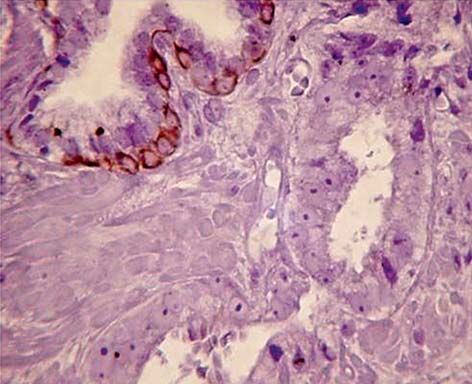
Immunohistochemistry for Diagnosis of Prostate Cancer

cytokeratin (34βE12) to mark basal cells of the pros-              median 14, ranging from 6 to 40. All the slides were
tate that was later demonstrated as a characteristic of            examined by the same uropathologist.
benign glands that retain the basal cell layer (7-9). In                    The immunohistochemistry was performed
a larger series, Wojno and Epstein (10) used 34βE12                using a mouse monoclonal antibody anti-high mo-
to diagnose adenocarcinoma in suspicious glands                    lecular weight cytokeratin (clone 34βE12, Dako,
identified in needle prostate biopsies. Shah et al. (11)            Glostrup, Denmark) at a dilution of 1/100 and p63
later proposed the combined use of p63, an homolog                 (clone 4A4, Dako, Glostrup, Denmark) at a dilution
of the p53 tumor suppressor protein, as an auxiliary               of 1/100. After paraffin removal and hydration, the
for the determination of cancer since it is also a protein         slides were immersed in 10 mM citrate buffer pH
expressed selectively by the basal cells of epithelial             6, for 15 min for antigen retrieval. The antibodies
organs, including the prostate gland (12,13).                      were incubated overnight at 4°C, and the secondary
         Recently, lower levels of prostate specific                biotin-labeled antibody was incubated for 30 min at
antigen (PSA) have been used to indicate the need                  room temperature. The streptavidin labeled strepta-
for a prostate biopsy, and there has been an increasing            vidin-biotin amplification method (Dako K0679) was
number of cores taken in each biopsy session. These                carried out for 30 minutes followed by peroxidase/di-
new practices have resulted in the representation of               aminobenzidine substrate/chromagen. The slides were
smaller tumors, often more adequately named ASAP.                  counterstained with hematoxylin.
In addition, pathologists frequently use immunohis-                         RP was carried out in 27 out of 144 patients.
tochemistry to enhance their diagnostic capabilities               The results were compared to 1040 patients who
in order to avoid rebiopsies. There have been reports              underwent RP during the same period and where the
of false-positives and false-negatives for use of the              histology was sufficient to define adenocarcinoma. All
combined 34βE12 and p63 cocktail. To date there                    patients were treated by the same group of surgeons.
has been no reported study that reviews surgical                   The surgical specimens were routinely examined
specimens of tumors from patients who underwent                    in toto by the same pathologist. To measure tumor
RP after a diagnosis of carcinoma in which the use of              volume we used the grid method as described by
immunohistochemistry was necessary for the defini-                  Humphrey and Vollmer (14).
tive diagnosis.

                                                                   RESULTS
MATERIALS AND METHODS
                                                                           Immunohistochemistry permitted the de-
                          st                st
         From January 1 2004 to July 31 2008, 4127                 finitive diagnosis of prostate cancer in 103 patients
biopsies were examined in our laboratory. ASAP was                 (71.5%) (Figure-1). The mean age of patients was
the diagnosis made for 144 (3.5%) of the biopsies. The             61.7 years old, median 61 (range 43 - 84); mean PSA
mean age of the patients was 60.8 years old, median                was 7.6 ng/mL, median 5.4 ng/mL (range 1.4 - 44
60 ranging from 40 to 84 years old. Mean PSA was                   ng/mL); and PSA free to total ratio was mean 15%,
7.11 ng/mL, with a median of 5.3 ng/mL, ranging                    median 13% (range 1 - 29%). The mean Gleason
from 1.4 to 43.5 ng/mL. The free to total relation of              score was 5.9, median 6 (range 4 to 7). The mean and
PSA was mean 15.1%, median 13.0%, ranging from                     median number of cores positive for tumor was 1.4
1 to 30%. The major reason for a biopsy among these                and 1 respectively, ranging from 1 to 4. The higher
patients was a progressive increase in PSA levels. One             percentage of a single core that made up the tumor
patient had a familiar history, and two had shown ab-              was mean 18.3%, median 10% (range 1 to 80%). The
normalities in transrectal examination. In 31 patients             mean total percentage of tumor in all cores was 1.6%,
(21.5%), ultrasound examination found abnormalities,               median 1% (range 1 to 7%). Perineural invasion was
hypoecoic and hypervascular areas. Twenty patients                 not detected in any cases.
(13.9%) had previously undergone biopsies. The mean                        We had access to the surgical specimens from
number of cores taken per biopsy session was 15.8,                 27 (26.2%) patients who underwent radical prostatec-

                                                             584
Immunohistochemistry for Diagnosis of Prostate Cancer
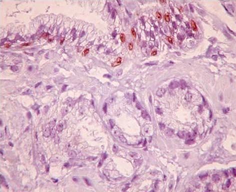
A B
Figure 1 – Immunohistochemistry study of prostate biopsy with atypical small acinar proliferation, where a diagnosis of adenocarci-
noma was made because of the absence of basal cells in the neoplastic glands. A normal gland is represented at the top of the figure
with basal cells stained by antibodies anti-high molecular weight cytokeratin 34βE12 (A) and anti-p63 (B).
tomy. For the same period, from January 1 2004 to the final diagnosis. The former will be referred to as
July 31 2008, 1040 patients also underwent radical Group 1, and the control group as Group 2. The char-
prostatectomy after a diagnosis of adenocarcinoma acteristics of both groups are shown in Table-1. There
that did not necessitate immunohistochemistry for were no pT0 in any of the two groups. Within Group
Table 1 – Demographic description and tumor characteristics of patients submitted to radical prostatectomy for treatment
of prostate cancer. Group 1 represents patients where immunohistochemistry was used for the definitive diagnosis. Group
2 represents patients where the diagnosis was made without any complementary study.
Group 1 Group 2
(27) (1040) p Value
Age years old 56 61
Median (Q1 - Q3) (53 - 65) (55 - 67) 0.039
Gleason Score 6 7
Median (Q1 - Q3) (6 - 6) (6 - 7) < 0.001
% of pattern 4 of Gleason 0 50
Median (Q1 - Q3) (0 - 17.75) (9 – 100)Immunohistochemistry for Diagnosis of Prostate Cancer
1, two (7.4%) adenocarcinomas could be considered CONCLUSION
clinically insignificant, defined as less than 2% of the
gland involved, organ-confined and with no Gleason Our data show that the usage of IHC did not
4 or 5 pattern present, versus 29 (2.9%) insignificant lead to diagnosis of insignificant tumors, as dem-
cases in Group 2 (15). onstrated by the study of the radical prostatectomy
The patients in Group 1 were younger, had specimens that had similar pathological stage and
lower Gleason scores, a lower percentage of Gleason positive surgical margins rates. Therefore, our results
pattern 4 and smaller tumors. However, the rate of show that this modality should be routinely used to
positive surgical margins was similar and there were evaluate a borderline biopsy and ASAP cases.
no differences in pathological stage.
CONFLICT OF INTEREST
COMMENTS
None declared.
The use of IHC as an auxiliary in the diagnosis
of adenocarcinoma is a common practice in uropathol-
ogy, and the use of antibodies against p63 and high REFERENCES
molecular weight cytokeratin has been recommended
as adjuncts in confirming prostatic carcinoma in 1. Epstein JI, Herawi M: Prostate needle biopsies con-
doubtful cases. taining prostatic intraepithelial neoplasia or atypical
Although basal cell markers, such as foci suspicious for carcinoma: implications for patient
34βE12 and p63 antibodies are useful for identify- care. J Urol. 2006; 175: 820-34.
ing basal cells, several benign mimickers of PC, 2. Leite KR, Mitteldorf CA, Camara-Lopes LH: Repeat
such as atrophy, atypical adenomatous hyperplasia, prostate biopsies following diagnoses of prostate
nephrogenic adenoma, and mesonephric hyper- intraepithelial neoplasia and atypical small gland
proliferation. Int Braz J Urol. 2005; 31: 131-6.
plasia, can stain negatively with these markers. In
3. Schlesinger C, Bostwick DG, Iczkowski KA: High-
addition, with patients being submitted to prostate
grade prostatic intraepithelial neoplasia and atypi-
biopsy with lower PSA levels and with larger cal small acinar proliferation: predictive value for
numbers of cores being taken in each biopsy ses- cancer in current practice. Am J Surg Pathol. 2005;
sion, concern that patients are being overtreated 29: 1201-7. Erratum in: Am J Surg Pathol. 2005;
is common. 29: 1548.
Our results show that there was no overdiag- 4. Hoedemaeker RF, Kranse R, Rietbergen JB, Kruger
nosis of PC with any pT0 after radical prostatectomy, AE, Schröder FH, van der Kwast TH: Evaluation of
with only 7.4% of cases classified as clinically insig- prostate needle biopsies in a population-based screen-
nificant. Tumor stage was similar for both groups, ing study: the impact of borderline lesions. Cancer.
but only 7.4% of patients from Group 1 had stage 1999; 85: 145-52.
pT3 tumors. On the other hand, positivity of surgical 5. Iczkowski KA, Bassler TJ, Schwob VS, Bassler IC,
Kunnel BS, Orozco RE, et al.: Diagnosis of “suspi-
margins, a very important parameter related to the
cious for malignancy” in prostate biopsies: predictive
outcome of patients submitted to radical prostatec-
value for cancer. Urology. 1998; 51: 749-57; discussion
tomy, mainly in organ-confined tumors (16,17), was 757-8.
similar for both groups; 22.2% and 24.3% in groups 6. Gown AM, Vogel AM: Monoclonal antibodies to hu-
1 and 2, respectively. man intermediate filament proteins. II. Distribution
This is the first study to our knowledge to of filament proteins in normal human tissues. Am J
show histological characteristics of radical prostatec- Pathol. 1984; 114: 309-21.
tomy specimens in men submitted to surgery to treat 7. Brawer MK, Peehl DM, Stamey TA, Bostwick DG:
adenocarcinoma where was necessary to use IHC for Keratin immunoreactivity in the benign and neoplastic
final diagnosis. human prostate. Cancer Res. 1985; 45: 3663-7.
586Immunohistochemistry for Diagnosis of Prostate Cancer
8. Hedrick L, Epstein JI: Use of keratin 903 as an adjunct 13. Signoretti S, Waltregny D, Dilks J, Isaac B, Lin D,
in the diagnosis of prostate carcinoma. Am J Surg Garraway L, et al.: p63 is a prostate basal cell marker
Pathol. 1989; 13: 389-96. and is required for prostate development. Am J Pathol.
9. O’Malley FP, Grignon DJ, Shum DT: Usefulness of im- 2000; 157: 1769-75.
munoperoxidase staining with high-molecular-weight 14. Humphrey PA, Vollmer RT: Percentage carcinoma as
cytokeratin in the differential diagnosis of small-acinar a measure of prostatic tumor size in radical prostatec-
lesions of the prostate gland. Virchows Arch A Pathol tomy tissues. Mod Pathol. 1997; 10: 326-33.
Anat Histopathol. 1990; 417: 191-6. 15. Guzzo TJ, Vira MA, Neway W, Hwang WT, Tomasze-
10. Wojno KJ, Epstein JI: The utility of basal cell-specific wski J, VanArsdalen K, et al.: Minimal tumor volume
anti-cytokeratin antibody (34 beta E12) in the diag- may provide additional prognostic information in good
nosis of prostate cancer. A review of 228 cases. Am J performance patients after radical prostatectomy. Urol-
Surg Pathol. 1995; 19: 251-60. ogy. 2007; 69: 1147-51.
11. Shah RB, Zhou M, LeBlanc M, Snyder M, Rubin 16. Psutka SD, Feldman AS, Rodin S, Wu CL, McDougal
MA: Comparison of the basal cell-specific markers, WS: Positive surgical margins do not affect recurrence
34betaE12 and p63, in the diagnosis of prostate cancer. in patients with T3a prostate cancer. J Urol. 2009; 181
Am J Surg Pathol. 2002; 26: 1161-8. (4Suppl): 776 #abstract 288.
12. Barbareschi M, Pecciarini L, Cangi MG, Macrì E, 17. Perez D, Shental J, Israel A, Salomon L, Abbou CC:
Rizzo A, Viale G, et al.: p63, a p53 homologue, is a Influence of positive surgical margins on adjuvant
selective nuclear marker of myoepithelial cells of the treatment after radical prostatectomy. J Urol. 2009;
human breast. Am J Surg Pathol. 2001; 25: 1054-60. 181 (4 Suppl): 776 #abstract 2138.
Accepted after revision:
February 18, 2010
Correspondence address:
Dr. Katia Ramos Moreira Leite
Rua Desembargador Eliseu Guilherme 69
São Paulo, SP, 04004-030, Brazil
Fax: + 55 11 3231-2249
E-mail: katiaramos@uol.com.br
EDITORIAL COMMENT
Immunohistochemistry (IHC) for 34βE12 and As the authors stressed in the present paper,
p63 is at present a diagnostic standard for determining few studies have intentionally examined surgical
the presence of prostate cancer. It is also used to dis- specimens in patients with prostate cancer, who were
criminate cancer from mimic cancer, when the definite primarily diagnosed with so-called atypical glands
diagnosis is difficult with conventional microscopic in previous biopsy specimens. Also, recent biopsy
examinations; however, second biopsy is frequently protocols such as multi-cores or saturation method
recommended in practice. Indeed, 34 - 60% patients has lead to an increase of ASAP (3). The authors
showing atypical small acinar proliferation (ASAP) elaborated patients’ demographics as well as radi-
in the primary biopsies were diagnosed with prostate cal prostatectomy specimens, of which preoperative
cancer in repeat biopsy sessions (1,2). diagnosis in biopsy cores required 34βE12 and p63
587Immunohistochemistry for Diagnosis of Prostate Cancer
IHC, to underscore the characteristics and outcome REFERENCES
of this type of prostate cancer.
It is of interest that the patients in the IHC- 1. Schlesinger C, Bostwick DG, Iczkowski KA: High-
required group were younger, had lower Gleason grade prostatic intraepithelial neoplasia and atypical
small acinar proliferation: predictive value for cancer
score and lower fraction with Gleason pattern 4, and
in current practice. Am J Surg Pathol. 2005; 29: 1201-
had smaller tumor foci, compared with those in the
7. Erratum in: Am J Surg Pathol. 2005; 29: 1548.
IHC-unnecessary group. These facts may be relevant 2. Epstein JI, Herawi M: Prostate needle biopsies
to lead-time bias in patients examined during differ- containing prostatic intraepithelial neoplasia or
ent era with a different screening protocol, or simply atypical foci suspicious for carcinoma: implications
based on earlier disease in younger patients. Although for patient care. J Urol. 2006; 175: 820-34.
pathological T-stage and positive surgical margin 3. Ploussard G, Plennevaux G, Allory Y, Salomon L,
rates were not statistically different between the two Azoulay S, Vordos D, et al.: High-grade prostatic
groups, the difference in patients’ number between intraepithelial neoplasia and atypical small acinar
them possibly explains this contradiction. Also, the proliferation on initial 21-core extended biopsy
IHC-required group was considered to include good- scheme: incidence and implications for patient care
risk cases, while the fraction of patients diagnosed and surveillance. World J Urol. 2009; 27: 587-92.
with insignificant cancer was not large (7.4%). Posi-
tive surgical margin cases were distributed uniformly Dr. Noboru Hara
between the IHC-required and IHC-unnecessary Division of Urology
groups, suggesting that 34βE12 and p63 IHC was Dep. of Regenerative and Transplant Medicine
useful as a preoperative diagnostics even for patients Graduate School of Medical and Dental Sciences
showing equivocal results in biopsy specimens with Niigata University, Niigata, Japan
routine histology. E-mail: harasho@med.niigata-u.ac.jp
EDITORIAL COMMENT
This study was carried out in order to in- include extensive sampling of the initial atypical site
vestigate the value of immunohistochemstry (IHC) as well as adjacent ipsilateral and contralateral sites
in borderline pathological prostate cancer cases. with routine extended schemes. However, extensive
Immunostaining has gained an important role in the transrectal sampling may increase the risk of infec-
evaluation of borderline biopsy cases, especially when tion (2). Herein the authors demonstrate that imuno-
high-grade prostate intraepithelial neoplasia (HGPIN) histochemical approach, using monoclonal antibody
and atypical small acinar proliferation (ASAP) are anti-high molecular weight cytokeratin and p63, may
present. The predictive values of ASAP and HPGIN assist in a more accurate diagnosis of ASAP and,
for cancer detection on repeat biopsies are 39 and therefore, may obviate the need for re-biopsies with
23%, respectively (1). Usually, the presence of ASAP the potential complications. The main benefit from
in the initial biopsy tissue requires re-biopsy within this study should be related to diagnosis and pretreat-
6-12 months considering other clinical features (age, ment judgment.
comorbidities etc.). Although this topic has not been This study shows comparable tumor stage in
sufficiently discussed in the literature, it may have a both study groups (radical prostatectomy after IHC
significant impact on patient management. It has been diagnosis and control) and recommends the inclusion
previously recommended that a second biopsy should of IHC in ASAP cases work-up.
588Immunohistochemistry for Diagnosis of Prostate Cancer
This study contributes to our daily clinical REFERENCES
practice and may have major impact on our judgment
in borderline prostate biopsy cases. Similar studies 1. Iczkowski KA: Current prostate biopsy interpretation:
evaluating novel methods to enhance biopsy-based criteria for cancer, atypical small acinar proliferation,
diagnosis accuracy should be encouraged in view high-grade prostatic intraepithelial neoplasia, and use
of the intriguing concept of active surveillance. This of immunostains. Arch Pathol Lab Med. 2006; 130:
study also leads to a yet another question - does active 835-43.
2. Epstein JI, Herawi M. Prostate needle biopsies con-
or passive surveillance miss cases of significant tu-
taining prostatic intraepithelial neoplasia or atypical
mors that should be treated more aggressively? Future
foci suspicious for carcinoma: implications for patient
studies are warranted to answer this question.
care. J Urol. 2006; 175: 820-34.
Dr. Avraham Shtricker
The Sackler School of Medicine
Tel Aviv University
Tel Aviv, Israel
E-mail: shtrickera@hotmail.com
EDITORIAL COMMENT
Authors compared the pathologic stage between REFERENCES
radical prostatectomy cases performed in patients for
which cancer was diagnosed with the help of combined 1. Molinié V, Fromont G, Sibony M, Vieillefond A, Vas-
34βE12 and p63 cocktail IHC (n = 27) or not (n = 1040). siliu V, Cochand-Priollet B, et al.: Diagnostic utility of
Their results showed that there was no overdiagnosis of a p63/alpha-methyl-CoA-racemase (p504s) cocktail
prostate cancer with any stage pT0 after radical prosta- in atypical foci in the prostate. Mod Pathol. 2004; 17:
1180-90.
tectomy, with only 7.4% of cases classified as clinically
2. Iczkowski KA, MacLennan GT, Bostwick DG: Atypical
insignificant. This shows the interest of IHC staining
small acinar proliferation suspicious for malignancy
and the quality of the pathologist reading. in prostate needle biopsies: clinical significance in 33
Pathologists frequently use immunohisto- cases. Am J Surg Pathol. 1997; 21: 1489-95.
chemistry to enhance their diagnostic capabilities in 3. Iczkowski KA: Current prostate biopsy interpretation:
order to avoid rebiopsies in cases of diagnosis diffi- criteria for cancer, atypical small acinar proliferation,
culty, which is a frequent situation in biopsy reading. high-grade prostatic intraepithelial neoplasia, and use
I would make 2 comments: of immunostains. Arch Pathol Lab Med. 2006; 130:
1) Recent studies recommend not to use the ASAP 835-43.
definition but atypical foci
2) It was also demonstrated that a different IHC of a Dr. Arnauld Villers
p63/alpha-methyl-CoA-racemase (p504s) cock- Service d’Urologie
tail in case of atypical foci in the prostate has a Hôpital HURIEZ, CHRU
diagnostic utility (1-3). Lille, France
Authors did not use this p504s in their IHC E-mail: arnauld.villers@wanadoo.fr
study. It may be discussed whether the addition of
the p504s is superior or not to the 34βE12 and p63
cocktail authors used.
589Immunohistochemistry for Diagnosis of Prostate Cancer
REPLY BY THE AUTHORS
To Dr. Arnauld Villers Comment
ASAP is a term that has been discussed in REFERENCES
uropathology meetings, and some pathologists argu-
ment that the suspicious gland is not always small, 1. Gologan A, Bastacky S, McHale T, Yu J, Cai C, Mon-
so it should be called atypical focus. ASAP was a zon-Bordonaba F, Dhir R: Age-associated changes in
very good term coined to describe doubtful lesions, alpha-methyl CoA racemase (AMACR) expression
and has been used for a long time. It means for us, in nonneoplastic prostatic tissues. Am J Surg Pathol.
pathologists, the presence of a small focus of atypi- 2005; 29: 1435-41.
2. Zhou M, Aydin H, Kanane H, Epstein JI: How often
cal glands, not necessarily small, highly suspicious
does alpha-methylacyl-CoA-racemase contribute to
for cancer and help pathologists and urologists to resolving an atypical diagnosis on prostate needle bi-
communicate. Also, used as a keyword facilitates opsy beyond that provided by basal cell markers? Am
the search in the literature, whereas atypical has a J Surg Pathol. 2004; 28: 239-43.
profuse meaning. Concerning the use of alpha-meth-
yl-CoA-racemase (p504s), it has been shown that it
stains number of benign prostate glands, periurethral The Authors
glands and mimics of prostate cancer (1), increases
the cost and help the final decision in only 50% of
cases (2).
590You can also read