Comparison of three ECG machines for electrocardiography in green iguanas (Iguana iguana)
←
→
Page content transcription
If your browser does not render page correctly, please read the page content below
Original Paper                                                    Veterinarni Medicina, 66, 2021 (02): 66–71

                                                                    https://doi.org/10.17221/39/2020-VETMED

Comparison of three ECG machines
for electrocardiography in green iguanas
(Iguana iguana)
Eva Cermakova1, Anna Piskovska2, Veronika Trhonova1,
Lionel Schilliger3, Zdenek Knotek1
1
  Avian and Exotic Animal Clinic, Faculty of Veterinary Medicine, University of Veterinary
  and Pharmaceutical Sciences Brno, Brno, Czech Republic
2
  Private Veterinarian, Nové Veselí, Czech Republic
3
  Clinique Vétérinaire du Village d’Auteuil, Paris, France
*Corresponding author: cermakovae@vfu.cz

Citation: Cermakova E, Piskovska A, Trhonova V, Schilliger L, Knotek Z (2021): Comparison of three ECG machines
for eletrocardiography in green iguanas (Iguana iguana). Vet Med-Czech 66, 66–71.

Abstract: The aim of the study was to compare the heart rate, QRS interval, and R wave amplitude across three
electrocardiogram models, and assess the ability of each of them to provide electrocardiograms (ECG) for clini-
cal interpretation. The three electrocardiogram models included ECG Seiva Praktik Veterinary, CardioStore ECG
and AliveCor Veterinary Heart Monitor. The data were collected from twelve healthy adult captive green iguanas
(Iguana iguana) monitored under a manual restraint at a room temperature of 22.6–28.0 °C. The ECGs using
the Seiva Praktik and CardioStore ECG veterinary electrocardiography were performed with standard 4 lead ECG
recordings. The AliveCor Veterinary Heart Monitor was placed (with the use of gel) directly on the lateral body
wall. The mean heart rate was 42 ± 8 beats/min (CardioStore), 50 ± 11 beats/min (Seiva Praktik Veterinary), and
51 ± 9 beats/min (AliveCor Veterinary Heart Monitor). No significant difference in the heart rate was observed.
A significant difference (P < 0.05) in the QRS duration was observed between the CardioStore and AliveCor Veterinary
Heart Monitor. Significant differences (P < 0.01) in the R wave amplitude were detected between the CardioStore
and AliveCor Veterinary Heart Monitor and between the Seiva Praktik Veterinary and AliveCor Veterinary Heart
Monitor. The ECGs produced by the Seiva Praktik VVeeterinary and CardioStore machines were interpretable
at 100%, while those produced by the AliveCor Veterinary Heart Monitor were interpretable at 66%. Seiva Prak-
tik Veterinary is most appropriately used as an anaesthesia monitoring tool. AliveCor Veterinary Heart Monitor
could be used as an additional diagnostic tool, but the results should be ideally confirmed with a standard ECG
machine. Seiva Praktik Veterinary is the most appropriate tool for monitoring the ECG within the anaesthesia, while
CardioStore might be most appropriately used as an advanced diagnostic tool by virtue of its software assistance.
The ECGs obtained with AliveCor Veterinary Heart Monitor should be confirmed using a standard ECG machine.

Keywords: reptile cardiology; reptile electrocardiogram; heart frequency; R wave; QRS complex

  Reptile cardiology is an important specialisation        (Davies et al. 1951; Mullen 1967; Valentinuzzi et al.
in veterinary exotic practice (Schillinger and Girling     1969; Valentinuzzi et al. 1970; McDonald and Heath
2019). Reptile electrocardiography (ECG) has proved        1971; Jacob and McDonald 1975; Heaton-Jones and
a promising diagnostic tool in reptile cardiology          King 1994; Holz and Holz 1995; Liu and Li 2005;

Supported by the Ministry of Education, Youth and Sports of the Czech Republic (Project No. IGA VFU 119/2016/FVL).

66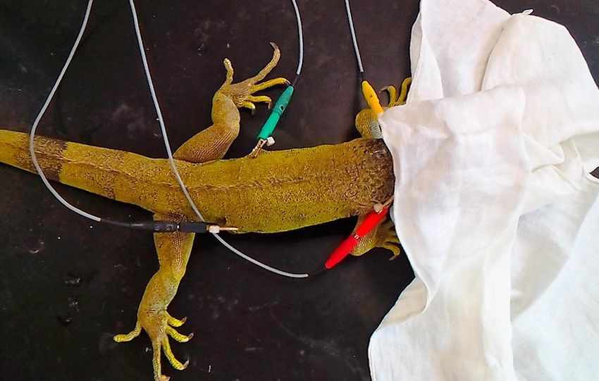
Original Paper Veterinarni Medicina, 66, 2021 (02): 66–71
https://doi.org/10.17221/39/2020-VETMED
Dahhan 2006; Hunt 2013; Germer et al. 2015; Bogan to an iPhone 5S (Apple Inc., Cupertino, CA, USA).
2017), as well as an adjunct tool for monitoring an- During the procedure, the animals were kept under
aesthesia (Mitchell 2009; Schumacher and Mans a manual restraint and the head was covered with
2014; Schillinger and Girling 2019). Because reptilian a towel to minimise the stress (Figure 1). The external
electrocardiograms differ from companion animals body temperature (range: 22.5–30.9 °C) and room
in terms of amplitude [up to 1 mV; Schillinger and temperature (range: 22.6–28.0 °C) were measured
Girling (2019), Zemanova et al. (2016)], the inter- using a contactless thermometer.
pretation of a reptile ECG requires an additional The monitoring started with the ECG Seiva
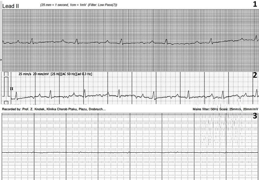
specialisation. The ECG should ideally be performed Praktik Veterinary device, which was applied us-
when the reptile is calm with a constant heart rate ing standard 4 lead ECG recordings (Figure 1).
(Dahhan 2006). The aim of this study was to compare Two electrodes with clips were attached to the skin
the heart rate, QRS interval, and R wave amplitude of the neck (yellow electrode on the left side, red
across three electrocardiogram models, and assess one on the right side) and two electrodes with clips
the ability of each model to provide ECGs for clinical were attached to the skin of the lateral body wall
interpretation in healthy adult captive green iguanas (green electrode on the left side, black one on the
(Iguana iguana). right side). An acoustic coupling gel (Topvet, Kuřim,
Czech Republic) was liberally applied to improve
the contact of the ECG leads with the scales. Two
MATERIAL AND METHODS ECG measurements were performed because the
electrocardiogram only allows recording of ECGs
Study animals in time frames of 10 s (total time was 20 s) with the
use of ECG Seiva Praktik Veterinary.
This study was performed on twelve adult captive The experiment continued directly afterwards
green iguanas (9 males, 3 females), aged 10–15 years by switching to the CardioStore ECG device – the elec-
old, with a body mass range of 1.46–3.04 kg. All the trocardiography was performed as described above.
animals were handled in accordance with the nation- The ECG values were recorded for at least 3 min-
al and European legislation (EU Council Directive utes. The ECGs were evaluated using CardioStore
86/609/EEC for the protection of animals) and with v1.33 software (Vetronic Services, Devon, UK).
the approval of the ethical committee (64-2016). The machine was then changed and we performed
Based on the clinical exams, all the iguanas were the third ECG using the AliveCor Veterinary Heart
healthy and without any clinical signs of cardio- Monitor device, which was placed directly on the
vascular pathology. The study animals were kept lateral body wall using the acoustic gel (Figure 2).
in standard husbandry conditions, in terrariums The ECG values were recorded for at least three
with a 12-hour day/12-hour night cycle provided minutes and evaluated on the iPhone (Figure 2).
by 100 W incandescent bulbs and basking areas
provided by infrared lamps. A linear UV.B (Repti-
Glo 10.0; Rolf C. Hagen, Mansfield, MA, USA) lamp Figure 1
was also in each terrarium. The temperature inside
the terrariums was 22.6 °C in the coldest part, the
temperature of the neutral zone was 27–30 °C and
the average temperature of the basking spot was
35–38 °C. Water and food were available ad libitum.
Electrocardiography
ECGs were performed using three types of electro- Figure 1. ECG electrodes attached to the skin folds on the
cardiograms: ECG Seiva Praktik Veterinary (Seiva s.r.o., neck (yellow electrode on the left, red one on the right)
Prague, Czech Republic), CardioStore ECG (Vetronic and on the lateral body wall (green electrode on the left,
Services, Devon, UK), and AliveCor Veterinary Heart black one on the right) of the green iguana. The head
Monitor (IDT Technology Ltd., P.R. China) attached is covered to reduce stress
67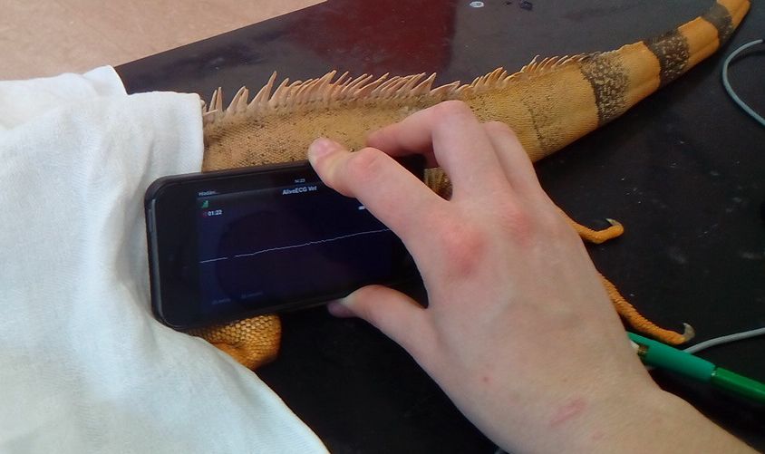
Original Paper                                                  Veterinarni Medicina, 66, 2021 (02): 66–71

                                                                  https://doi.org/10.17221/39/2020-VETMED

                                                         of the CardioStore v1.33 software), Seiva Praktik
 Figure 2                                                Veterinary ECG and AliveCor ® Veterinary Heart
                                                         Monitor devices with the use of the iPhone 5S and
                                                         AliveCor software (Figure 3). The speed of the paper
                                                         was 25 mm/sec and the amplitude was 20 mm/mV.
                                                           The indicator values (maximum, minimum,
                                                         mean, and standard deviation) were analysed
                                                         by a one-way ANOVA (analysis of variance; fac-
                                                         tor ECG machine). The significance was accepted
                                                         at P ≤ 0.05. To compare the results of the three
                                                         electrocardiograms, a post-hoc t-test was used with
Figure 2. AliveCor Veterinary Heart Monitor placed       a significance of P ≤ 0.016 7. All the analyses were
on the left body wall of green iguana                    calculated using MS Excel (Office XP; Microsoft
                                                         Co., Redmond, USA).
Recording and analysis

  The ECG recordings included the heart rate, am-        RESULTS
plitude of the R waves (millivolt, mV) and dura-
tion of the QRS complex (milliseconds, mS). The            The heart rate average and range for each electro-
heart rate was calculated from the RR intervals.         cardiogram are presented in Table 1. The one way
Each of these parameters was recorded 20 times           ANOVA showed no significant difference in the
per animal and evaluated on printed electrocar-          average heart rate [F(2.6) = 3.3; P > 0.05] between
diograms for the CardioStore ECG (with the use           the three electrocardiograms (Figure 4).

 Figure 3

Figure 3. Green iguana electrocardiograms using the CardioStore ECG (1), Seiva Praktik ECG (2), AliveCor Veteri-
nary Heart Monitor devices (3)

68
Original Paper Veterinarni Medicina, 66, 2021 (02): 66–71
https://doi.org/10.17221/39/2020-VETMED
Table 1. Heart rate, R wave amplitude and QRS duration recorded in 12 healthy adult green iguanas with the three
ECG machines
ECG machines
AliveCor Veterinary Heart
Value (units) CardioStore Seiva Praktik Veterinary
Monitor
mean ± SD range mean ± SD range mean ± SD range
Heart rate (beats/min) 42 ± 8 27–55 50 ± 11 32–63 51 ± 9 36–61
R wave amplitude (mV) 0.29 ± 0.11 0.11–0.44 0.31 ± 0.09 0.15–0.42 0.09 ± 0.04 0.04–0.16
QRS complex duration (mS) 175 ± 57 90–253 151 ± 49 88–275 110 ± 15 87–136
250
55 4
Figure Figure 5
50 200
Duration of QRS
45
complex (ms)
40 150
Heart rate/min
35
30 100
25
50
20
15 0
10 CardioStore Seiva Praktik AliveCor
5 Veterinary Veterinary
0 Heart Monitor
CardioStore Seiva Praktik AliveCor
Veterinary Veterinary Figure 5. Mean R wave amplitude of 12 healthy adult green
Heart Monitor
iguanas using the three ECG machines
Figure 4. Mean heart rate of 12 healthy adult green iguanas
0.40
using the three ECG machines Figure
0.356
0.30
R wave (mV)
Aplitude of
The mean R wave amplitude and range are dis- 0.25
0.20
played in Table 1. The one way ANOVA showed a sig- 0.15
nificant difference between mean R wave amplitude 0.10
across the three electrocardiograms [F(17.26) = 3.33; 0.05
0.00
P < 0.01]. The post-hoc t-test revealed a significant CardioStore AliveCor
Seiva Praktik
difference between the CardioStore and AliveCor® Veterinary Veterinary
Veterinary Heart Monitor devices (P < 0.01), a signif- Heart Monitor
icant difference between the Seiva Praktik Veterinary Figure 6. Mean QRS complex duration of 12 healthy adult
and AliveCor® Veterinary Heart Monitor devices green iguanas using the three ECG machines
(P < 0.01), and no significant difference between the
CardioStore and Seiva Praktik Veterinary devices on a computer monitor, whereas the ECG displayed
(P > 0.05) (Figure 5). by the CardioStore device was only visible on a small
The mean QRS duration and range are shown screen. The Seiva Praktik Veterinary machine could
in Table 1. The one way ANOVA showed a signifi- record up to 10 s ECGs, while the CardioStore de-
cant difference between the mean QRS duration vice could record up 20 minutes. The AliveCor®
for the three electrocardiograms [F(4.66) = 3.32; Veterinary Heart Monitor had an unlimited time
P < 0.05]. The post-hoc t-test revealed significant dif- recording capacity.
ferences in the mean QRS between the CardioStore
and AliveCor ® Veterinary Heart Monitor devices
(P < 0.01) while no statistically significant difference DISCUSSION
was observed between the Seiva Praktik Veterinary
and CardioStore devices (P > 0.05), or the Seiva The CardioStore ECG machine has been de-
Praktik Veterinary and AliveCor® Veterinary Heart scribed in a bearded dragon (Pogona vitticeps)
Monitor devices (P > 0.016 7) (Figure 6). study (Hunt 2013). The AliveCor Veterinary Heart
The Seiva Praktik Veterinary device enabled the Monitor has been described in many reptile spe-
high quality visualisation of the ECGs directly cies studies (Schilliger et al. 2014), while the Seiva
69Original Paper Veterinarni Medicina, 66, 2021 (02): 66–71
https://doi.org/10.17221/39/2020-VETMED
Praktik Veterinary machine has only been described machines. The AliveCor Veterinary Heart Monitor
in studies on mammals (Bado et al. 2017), and pub- could be used as an additional diagnostic tool, but
lished reports about its use in reptiles is missing. the results should be ideally confirmed with a stan-
No significant differences in the heart rate val- dard ECG machine.
ues were recorded for the three electrocardiograms
compared in this study. This finding is in accord-
ance with the results published by Haberman et al. Acknowledgement
(2015), Smith et al. (2016), Vezzosi et al. (2016) and
Vandenberk et al. (2017) who compared the AliveCor The authors would like to thank to Department
Veterinary Heart Monitor with other ECG machines. of Physiology of the University of Veterinary and
Statistical analysis showed significant differences Pharmaceutical Sciences Brno for lending them the
between the values of the R wave amplitude obtained ECG Seiva Praktik Veterinary.
with the AliveCor Veterinary Heart Monitor, Seiva
Praktik Veterinary, and CardioStore devices. This
finding is in accordance with the results of a simi- Conflict of interest
lar study in which the AliveCor Veterinary Heart
Monitor underestimated the amplitude of the R wave The authors declare no conflict of interest.
in 74.7% dogs (Vezzosi et al. 2016).
Finally, the analysis showed a significant differ-
ence in the durations of the QRS complexes, which REFERENCES
relies on the ability of the machines to measure the
durations of the waves and complexes. The post-hoc Bado O, Dlouha M, Kolmanova E, Frydrych M. Influence
t-test showed a significant difference between the of new ultra-short-acting β-blockers on selected physi-
CardioStore and AliveCor Veterinary Heart Monitor ological indicators in laboratory rats. Vet Med-Czech.
devices. This result contradicts those reported by 2017 Sep;62(9):493-507.
Chung and Guise (2015), who did not find any dif- Bogan JE. Ophidian cardiology – A review. J Herpetol Med
ferences when comparing the QT intervals in hu- Surg. 2017;27(1-2):62-77.
mans obtained with the AliveCor Veterinary Heart Boukens BJD, Kristensen DL, Filogonio R, Carreira LBT,
Monitor and standard ECG recorded intervals. Our Sartori MR, Abe AS, Currie S, Joyce W, Conner J,
result could be due to inaccuracies emerging during Opthof T, Crossley DA 2nd, Wang T, Jensen B. The elec-
the hand calculation and measurement of the ECGs, trocardiogram of vertebrates: Evolutionary changes from
compared to electronic measurement obtained with ectothermy to endothermy. Prog Biophys Mol Biol. 2019
the CardioStore software. Jul;144:16-29.
The QRS duration is significantly longer than the Dahhan M. Elektrokardiographische Untersuchungen beim
P duration in reptiles, whereas the duration of the QRS Grunen Leguan (Iguana iguana) [Electrocardiographic
complex and the P wave are similar in mammals and parameters in the green iguana (Iguana iguana)] [disser-
birds; however, in Boukens et al. (2019), the original tation]. München: Ludwig-Maximilians-Universität; 2006.
data did not find a difference between the P and QRS 101 p. German.
duration in reptiles. Davies F, Francis ETB, King TS. Electrocardiogram of the
In conclusion, the ECG measurements of the heart crocodilian heart. Nature. 1951 Jan 27;167(4239):146.
rate, R amplitude, and QRS interval did not differ Germer CM, Tomaz JM, Carvalho AF, Bassani RA, Bassani
significantly between the CardioStore and Seiva JWM. Electrocardiogram, heart movement and heart rate
Praktik Veterinary devices. in the awake gecko (Hemidactylus mabouia). J Comp
With the accompanying software interpretation, Physiol. 2015 Jan;185(1):111-8.
the CardioStore machine might be better suited as Haberman ZC, Jahn RT, Bose R, Tun H, Shinbane JS, Doshi
an advanced diagnostic tool in reptile cardiology. RN, Chang PM, Saxon LA. Wireless smartphone ECG
With the electrocardiogram clearly visible on the enables large-scale screening in diverse populations.
computer monitor, the Seiva Praktik Veterinary de- J Cardiovasc Electrophysiol. 2015 May;26(5):520-6.
vice is most appropriately used as an anaesthesia Heaton-Jones T, King R. Characterization of the electro-
monitoring tool and should be used as the gold stan- cardiogram of the American alligator (Alligator missis-
dard tool for the clinical evaluation of other ECG sippiensis). J Zoo Wildl Med. 1994;25(1):40-7.
70Original Paper Veterinarni Medicina, 66, 2021 (02): 66–71
https://doi.org/10.17221/39/2020-VETMED
Holz RM, Holz P. Electrocardiography in anesthetized red- Schumacher J, Mans CH. Anesthesia. In: Divers SJ, Mader
eared sliders (Trachemys scripta elegans). Res Vet Sci. DR, editors. Current therapy in reptile medicine and sur-
1995 Jan;58(1):67-9. gery. St. Louis: Saunders Elsevier; 2014. p. 134-53.
Hunt CJ. Electrocardiography of the normal inland bearded Smith J, Ward J, Urbano T, Mueller M. Use of AliveCor heart
dragon (Pogona vitticeps) [thesis]. [United Kingdom]: monitor for heart rate and rhythm evaluation in dairy
Royal College of Veterinary Surgeons; 2013. 43 p. water buffalo calves (Bubalis Bubalis). J Dai Vet Anim Res.
Chung EH, Guise KD. QTC intervals can be assessed with 2016;4(2):261-4.
the AliveCor heart monitor in patients on dofetilide for Valentinuzzi ME, Hoff HE, Geddes LA. Electrocardiogram
atrial fibrillation. J Electrocardiol. 2015 Jan-Feb;48(1):8-9. of the snake: Intervals and durations. J Electrocardiol.
Jacob JS, McDonald HS. Temperature preferences and elec- 1969;2(4):343-52.
trocardiography of Elaphe obsoleta (Serpentes). Comp Valentinuzzi ME, Hoff HE, Geddes LA. Electrocardiogram
Biochem Physiol Part A Physiol. 1975 Dec 1;52(4):591-4. of the snake: Effect of vagal stimulation on the Q-T dura-
Liu CH, Li R. Electrocardiogram and heart rate in response tion. J Electrocardiol. 1970;3(1):21-7.
to temperature acclimation in three representative verte- Vandenberk T, Stans J, Mortelmans C, Van Haelst R, Van
brates. Comp Biochem Physiol. 2005 Dec;142(4):416-21. Schelvergem G, Pelckmans C, Smeets CJ, Lanssens D, De
McDonald HS, Heath JE. Electrocardiographic observation Canniere H, Storms V, Thijs IM, Vaes B, Vandervoort PM.
on the tuatara, Sphenodon punctatus. Comp biochem Clinical validation of heart rate apps: Mixed-methods
Physiol. 1971 Dec;40(4):881-92. evaluation study. JMIR Mhealth Uhealth. 2017 Aug 25;
Mitchell M. Reptile cardiology. Clin North Am Exot Anim 5(8): [15].
Pract. 2009 Jan;12(1):65-79. Vezzosi T, Buralli C, Marchesotti F, Porporato F, Tognetti R,
Mullen RK. Comparative electrocardiography of the Squa- Zini E, Domenech O. Diagnostic accuracy of a smartphone
mata. Physiol Zool. 1967;40(2):114-26. electrocardiography in dogs: Comparison with standard
Schilliger L, Girling S. Cardiology. In: Divers S, Stahl SJ, 6-lead electrocardiography. Vet J. 2016 Oct;216:33-7.
editors. Mader’s reptile and amphibian medicine and Zemanova A, Sochorcova V, Knotek Z. Zakladni metody
surgery. 3rd ed. St. Louis: Elsevier; 2019. p. 669-98. kardiologickeho vysetreni plazu [Basic methods of car-
Schilliger L, Chai N, Chetboul V, Bonwitt J. Smartphone- diological examination in reptiles]. Veterinarni klinika.
based ECG monitor: Feasibility and applications in herpe- 2016;13:20-7. Czech.
tological medicine [abstract]. In: Proceedings Association
of Reptilian and Amphibian Veterinarians; 2014 Oct 18–24; Received: February 13, 2020
Orlando, Florida. Orlando, FL,USA: Arav; 2014. p. 129-30. Accepted: November 10, 2020
71You can also read