PALS Syndrome Post-LASIK - touchOPHTHALMOLOGY
←
→
Page content transcription
If your browser does not render page correctly, please read the page content below
Clinical Case Refractive Surgery
PALS Syndrome Post-LASIK
Noel Alpins1,2 and George Stamatelatos2
1. Department of Ophthalmology, Melbourne University, Melbourne, Australia; 2. NewVision Clinics, Melbourne, Australia
A
34-year-old male presented complaining of ongoing symptoms of glare, ghosting, starbursts, haloes, and reduced contrast sensitivity
commonly referred to by the acronym “GASH”. His unaided visual acuity achieved was a satisfactory 20/20 in each eye. To reduce his
symptoms and enable driving at night, he must resort to shining the torch from his cell phone into his eyes to constrict his pupils. These
symptoms are due to excess corneal astigmatism remaining, and is prevalent post refractive laser surgery when treatment is based on refractive
parameters alone without any regard for corneal measures and having ocular residual astigmatism greater than 1.00D preoperatively. In this
commonly prevalent clinical situation, corneal astigmatism postoperatively will be more than 1.00D, together with a significant risk of one or more
symptoms of glare, starbursts, or haloes which, when they occur together, is known as predictable avoidable laser-assisted in situ keratomileusis
surprise (PALS) syndrome.
Keywords A 34-year-old male presented to the clinic for a second opinion regarding ongoing symptoms of
ORA, ocular residual astigmatism, Vector glare, ghosting, starbursts, haloes and reduced contrast sensitivity three years post-laser-assisted
Planning, glare, starbursts, haloes in situ keratomileusis (LASIK) surgery. These symptoms were exacerbated under low light conditions,
particularly driving at night, and he found a reduction in their effect when shining his cell phone torch
Disclosures: Noel Alpins and George Stamatelatos report
a financial interest in ASSORT Surgical Management light in his eyes.
Systems which holds trademarks in Vector Planning™.
Review Process: Double-blind peer review. Preoperatively, manifest refraction was: right -3.25/-0.75 x 33, left -3.75/-0.50 x 152. His cycloplegic
Compliance with Ethics: All procedures were followed refraction was: right -2.50/-1.00 x 20 and left -3.00/-0.75 x 165.
in accordance with the responsible committee on human
experimentation and with the Helsinki Declaration of 1975
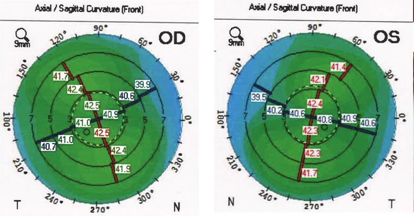
and subsequent revisions, and informed consent was Simulated keratometry from topography preoperatively was: right 41.00/42.50 @ 102 and left
received from the patient involved in this case study.
40.75/42.25 @ 75 with regular corneas in both eyes (Figure 1).
Authorship: All named authors meet the International
Committee of Medical Journal Editors (ICMJE) criteria
for authorship of this manuscript, take responsibility Pupil diameter under mesopic conditions was measured as: right 6.37 mm and left 6.94 mm.
for the integrity of the work as a whole, and have
given final approval to the version to be published.
Received: March 7, 2019 Uncomplicated bilateral LASIK surgery was performed by another clinic in July 2016 using the latest
Accepted: March 27, 2019 generation Schwind Amaris® 1050RS Hz excimer laser (Schwind eye-tech-solutions GmbH & Co. KG,
Citation: US Ophthalmic Review. 2019;12(1):37–9 Kleinostheim, Germany) with the treatment based on the manufacturer recommended practice of
Corresponding Author: Noel Alpins, NewVision manifest refraction only. The optical and the ablation zones were 6.70 mm and 7.63 mm, respectively.
Clinics, 160 Victoria Parade, East Melbourne, Victoria
3002, Australia. E: alpins@newvisionclinics.com.au
At the 7-month postoperative review, unaided visual acuity was 20/20 part in both eyes and 20/15
Support: No funding was received in with a manifest refraction of right plano/-0.75 x 9 and left plano/-0.75 x 170. Bilateral retreatment by
the publication of this article.
flap lift was then performed based again on the manifest refraction, only because of the symptoms
and remaining minor refractive error.
The patient attended our clinic 2 years post initial LASIK, complaining of significant symptoms of
glare, ghosting, starbursts, haloes, and reduced contrast sensitivity (GASH), and wanted a second
opinion on reducing these effects, which presented after the initial refractive laser surgery. On
examination, the unaided visual acuity of right 20/20 and left 20/20-2 best correcting to 20/20 in
both eyes. Manifest refraction right plano and left plano/-0.50 x 180. Cycloplegic refraction of
right +0.25DS and left +0.50/-0.25 x 180. Corneal astigmatism using simulated keratometry from
the CSO Sirius tomographer (C.S.O. Srl, Firenze, Italy), was right 1.12D @ 90 and left 1.04D @ 85. Ocular
wavefront using the ViSX WaveScan system showed higher order aberrations of right 0.27 microns
and left 0.40 microns.
It is interesting to note that there was still a significant amount (>1.00D) of corneal astigmatism
remaining in each eye after both the first treatment and the second enhancements, which were
based on the manifest refraction parameters alone. Treating the maximum amount of astigmatism
TOUC H MED ICA L MEDIA Publication Date: April 23, 2019 37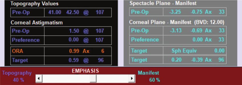
Clinical Case Refractive Surgery

     Figure 1: Preoperative axial curvature topography displaying                    Figure 2: The ocular residual astigmatism is calculated as the
     regular with-the-rule astigmatism for right and left eyes                       vectorial difference between manifest refractive cylinder at
                                                                                     the corneal plane and corneal astigmatism

                                                                                     Figure 3: Calculation of ocular residual astigmatism for the
                                                                                     right eye
     by incorporating the corneal parameters into the refractive treatment
     plan employing Vector Planning1 would have left less corneal astigmatism
     postoperatively and reduced symptoms of glare, ghosting, starbursts,
     and haloes.

     Ocular residual astigmatism
     To treat the maximum amount of astigmatism, calculation of the ocular
     residual astigmatism (ORA) is required in each case. The ORA is defined as
     the vectorial difference between the corneal astigmatism and the refractive
     cylinder at the corneal plane.1 It is expressed in dioptres together with an
                                                                                     Vector Planning places emphasis of 40% topography and 60% manifest refraction on the
     axis and has been shown to be more than 1.00D in 46% of cases2 in one           ocular residual astigmatism to target 0.59D of corneal astigmatism.
     study and 34% of cases in another study.1
                                                                                     Figure 4: Calculation of ocular residual astigmatism for the
     In this case, the ORA preoperatively was right 0.99D Ax 6 and left 1.24D Ax     left eye. Vector Planning places emphasis of 40% topography
     174 (Figure 2). Due to the pre-existing corneo-refractive differences the ORA   and 60% manifest refraction on the ocular residual
     is the minimum amount of astigmatism that can remain postoperatively.           astigmatism to target 0.74D of corneal astigmatism
     Treatment based on the refractive parameters alone, as was the case here,
     would leave all the ORA, theoretically 0.99D @ 96 for the right eye and
     1.24D @ 84 for the left eye to be directed to the cornea postoperatively.
     Note that this is 90 degrees away from the ORA axis to neutralise it.
     Even after the second treatment, which again was based on refractive
     parameters to reduce the refractive cylinder, there was still predictably a
     substantial amount of corneal astigmatism (>1.00D) remaining: right 1.12D
     @ 90 and left 1.04D @ 85 due to the significant amount of ORA.

     Incorporating the corneal parameters into the refractive treatment plan
     after calculating the ORA would treat the maximum amount of astigmatism
     and leave less corneal astigmatism postoperatively compared to                  1% refractive emphasis. Studies using the method of Vector Planning3–5
     treatment based on refractive parameters alone with the same amount             have shown 40% emphasis on corneal parameters and 60% emphasis
     of refractive cylinder remaining.3 Vector Planning is a systematic method       on refractive parameters to be suitable for most treatments with a range
     of combining both refractive and corneal parameters into the excimer            anywhere from 45–65% emphasis by refractive parameters. The important
     treatment plan. This has been shown to reduce the corneal astigmatism           consideration is the ORA, knowing that any emphasis on the ORA is treating
     remaining compared to treatments based on refractive parameters alone           the maximum amount of astigmatism.
     without compromising the refractive cylinder postoperatively.3
                                                                                     Applying Vector Planning to this case study, the ORA calculated for the right
     Vector Planning                                                                 eye is 0.99D Ax 6. By emphasising, for example, 60% of the ORA towards
     The surgeon decides how to apportion the preoperative ORA calculated by         refraction and 40% towards topography, 0.59D @ 96 would be targeted on
     placing an emphasis of corneal to refractive parameters anywhere from           the cornea and -0.39D x 96 in the refraction (Figure 3). This compares to
     1% corneal astigmatism and 99% refractive cylinder to 99% corneal and           1.12D @ 90 of corneal astigmatism that the patient has postoperatively.

38                                                                                                                                                 US OPH TH A LMIC RE VIE W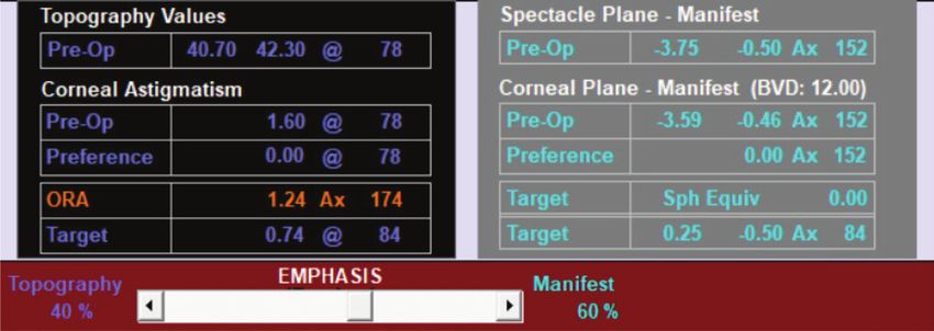
PALS Syndrome Post-LASIK

For the left eye the ORA is 1.24D @ 84. So again, emphasising the ORA by                                     The method of Vector Planning can address the excess corneal astigmatism
60% towards refractive parameters and 40% by topography, the targeted                                        causing these disturbing symptoms and improving patient vision quality and
corneal astigmatism would be 0.74D @ 84 and the refractive cylinder                                          satisfaction rates post-LASIK. The prevalence of these symptoms and signs
-0.50 x 84 (Figure 4). This compares to 1.04D @ 85 of corneal astigmatism                                    together qualifies it as a syndrome. An ORA of more than 1.00D preoperatively
that the patient now has postoperatively.                                                                    and corneal astigmatism of greater than 1.00D postoperatively, together
                                                                                                             with any one or more of the GASH symptoms constitutes preventable
Vector Planning targets a spherical equivalent of zero in the refraction                                     avoidable LASIK surprise (PALS) syndrome. This syndrome is preventable by
and because of the better corneal shape that the patient is left with                                        calculating the ORA preoperatively to ascertain how much corneo-refractive
postoperatively, compared to treatments based on refractive parameters                                       difference there exists. It is avoidable using the method of Vector Planning
alone, the targeted refractive cylinder has been shown not to be                                             to reduce excess corneal astigmatism postoperatively, it can validly apply to
accepted by the patient in the postoperative manifest refraction.                                            photorefractive keratectomy and small incision lenticule extraction as well as
                                                                                                             LASIK procedures and the surprise of GASH can be minimized or eliminated
Unfortunately, the patient did not want to undergo any further (third)                                       by maintaining postoperative corneal astigmatism at less than 1.00D.
surgery to reduce his symptoms, but was advised that Vector Planning was
a viable option.                                                                                             Laser manufacturers need to incorporate the Vector Planning method
                                                                                                             as a standard function of their laser systems, allowing all surgeons this
Excess corneal astigmatism is prevalent post refractive laser surgery with                                   option to maximally treat astigmatism and prevent PALS syndrome. This
many complaints now documented in letters to the US Food and Drug                                            is currently not available on any laser. For surgeons who want to avail
Administration and the New York Times of symptoms of glare, ghosting,                                        themselves of this facility of ORA calculation and Vector Planning with
starbursts, haloes, or decreased contrast sensitivity, now commonly                                          their refractive laser surgery, this is available as a free application at
termed GASH6 as the acronym to describe these symptoms.                                                      www.assort.com. q

1.   Alpins NA. New method of targeting vectors to treat astigmatism.          on the corneal vertex comparing vector planning with manifest    5.   Alpins NA, Stamatelatos G. Clinical outcomes of laser
     J Cataract Refract Surg. 1997;23:65–75.                                   refraction planning for the treatment of myopic astigmatism.          in situ keratomileusis using combined topography and
2.   Frings A, Katz T, Steinberg J, et al. Ocular residual astigmatism:        J Cataract Refract Surg. 2017;43:1504–14.                             refractive wavefront treatments for myopic astigmatism.
     Effect of demographic and ocular parameters in myopic laser          4.   Alpins NA, Stamatelatos G. Customized PARK treatment of myopia        J Cataract Refract Surg. 2008;34:1250–9.
     in situ keratomileusis. J Cataract Refract Surg. 2014;40:232–8.           and astigmatism in forme fruste and mild keratoconus using       6.   LASIK Complaints Filed With the FDA. Available at:
3.   Arbelaez MC, Alpins N, Verma S et al. Clinical outcomes of laser          combined topographic and refractive data. J Cataract Refract          www.lasikcomplications.com/lasik-complaints.htm
     in-situ keratomileusis with an aberration -neutral profile centred        Surg. 2007;33:591–602.                                                (accessed February 20, 2019).

US OPH TH A L MIC REVIEW                                                                                                                                                                                       39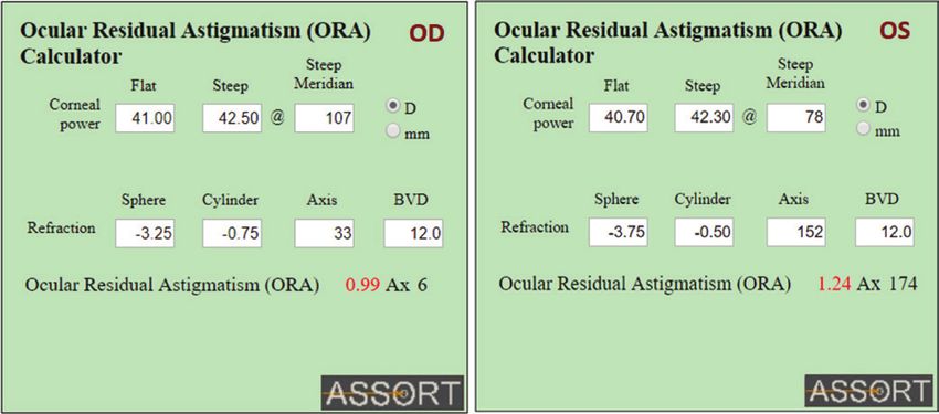
You can also read