NEUROTROPHIC KERATITIS: A "Rare Disease" Yet Common Problem - Cataract & Refractive Surgery ...
←
→
Page content transcription
If your browser does not render page correctly, please read the page content below

Supplement to                                                                                                         October 2021

                                                                                           FACULTY:

       NEUROTROPHIC                                                                        Francis S. Mah, MD
                                                                                           Marjan Farid, MD

       KERATITIS:
                                                                                           Sumitra Khandelwal, MD
                                                                                           Elizabeth Yeu, MD

       A “Rare Disease” Yet
       Common Problem

                                                                                                         Provided by

       A continuing medical education activity provided by Evolve Medical Education LLC.
       This activity is supported by an unrestricted educational grant from Dompé.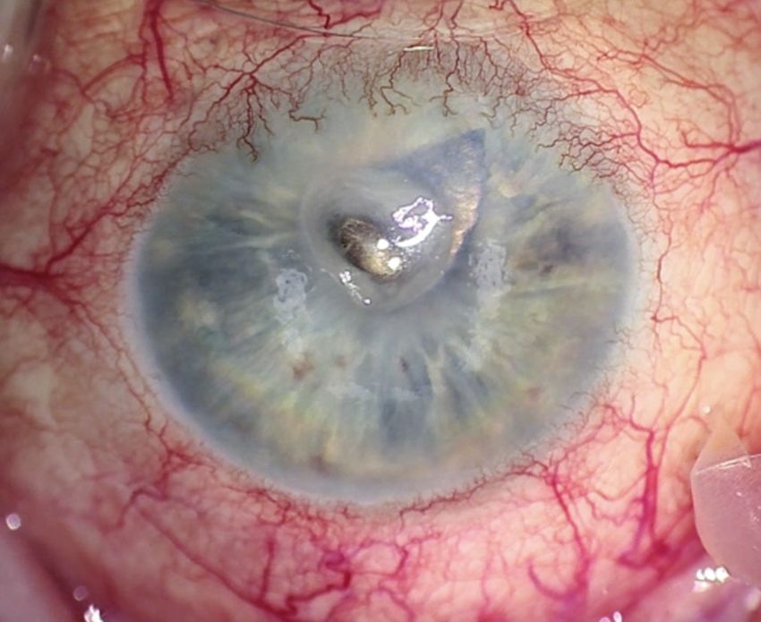
Neurotrophic Keratitis:
                                                                                                                                                               Release Date: October 2021
                                                                                                                                                             Expiration Date: October 2022

   A “Rare Disease” Yet Common Problem
   FACULTY

         FRANCIS S. MAH, MD                          MARJAN FARID, MD                                 SUMITRA                                ELIZABETH YEU, MD
                 Program Chair                        Professor of Ophthalmology                   KHANDELWAL, MD                               Virginia Eye Consultants
      Director, Cornea and External Disease                School of Medicine                          Associate Professor                 Medical Director, CVP Mid-Atlantic
          Co-Director, Refractive Surgery           Vice Chair, Ophthalmic Faculty           Medical Director, Lions Eye Bank of Texas   Cornea, Cataract, External Disease, and
                   Scripps Clinic             Director, Cornea/External Disease, Cataract,   Cornea, Cataract, and Refractive Surgery              Refractive Surgery
                    La Jolla, CA                         and Refractive Surgery                    Baylor College of Medicine                      Assistant Professor
                                                       Gavin Herbert Eye Institute                         Houston, TX                       Department of Ophthalmology
                                                     University of California, Irvine                                                       Eastern Virginia Medical School
                                                               Irvine, CA                                                                              Norfolk, VA

   CONTENT SOURCE                                                                            ACCREDITATION STATEMENT
     This continuing medical education (CME) activity captures                                 Evolve Medical Education LLC (Evolve) is accredited by the
   content from a live symposium.                                                            Accreditation Council for Continuing Medical Education (ACCME)
                                                                                             to provide continuing medical education for physicians.
   ACTIVITY DESCRIPTION
     Based on a live satellite symposium held during the American                            CREDIT DESIGNATION STATEMENT
   Society for Cataract and Refractive Surgeons Annual Meeting,                                Evolve designates this enduring material for a maximum of
   this supplement summarizes the presentations on the stages of                             1 AMA PRA Category 1 Credit™. Physicians should claim only the credit
   neurotrophic keratitis and the available treatments.                                      commensurate with the extent of their participation in the activity.

   TARGET AUDIENCE                                                                           TO OBTAIN CREDIT
      This certified CME activity is designed for ophthalmologists.                             To obtain credit for this activity, you must read the activity
                                                                                             in its entirety and complete the Pretest/Posttest/Activity
   LEARNING OBJECTIVES                                                                       Evaluation/Satisfaction Measures Form, which consists of a series
      Upon completion of this activity, the participant should be able to:                   of multiple-choice questions. To answer these questions online
      • Identify the stages of neurotrophic keratitis                                        and receive real-time results, go to https://evolvemeded.com/
      • Recognize how to differentiate neurotrophic keratitis from                           course/2132-supp. Upon completing the activity and self-assessment
        similar diseases                                                                     test, you may print a credit letter awarding 1 AMA PRA Category
      • Describe the mechanisms of action of newer treatments and                            1 Credit™. Alternatively, please complete the Posttest/Activity
        when they should be introduced into treatment regimens for                           Evaluation/Satisfaction Form and mail or fax to Evolve Medical
        neurotrophic keratitis, including the stepwise approach                              Education LLC, 353 West Lancaster Avenue, Second Floor, Wayne,
      • Explain the relationships between disease characteristics,                           PA 19087; Fax: (215) 933-3950.
        drug therapies, treatment frequency, visual, and anatomic
        outcomes                                                                             DISCLOSURE POLICY
                                                                                               It is the policy of Evolve that faculty and other individuals who
   GRANTOR STATEMENT                                                                         are in the position to control the content of this activity disclose
      This activity is supported by an unrestricted educational grant                        any real or apparent conflicts of interest relating to the topics of
   from Dompé.                                                                               this educational activity. Evolve has full policies in place that will

2 SUPPLEMENT TO CATARACT & REFRACTIVE SURGERY TODAY / MILLENNIALEYE | OCTOBER 2021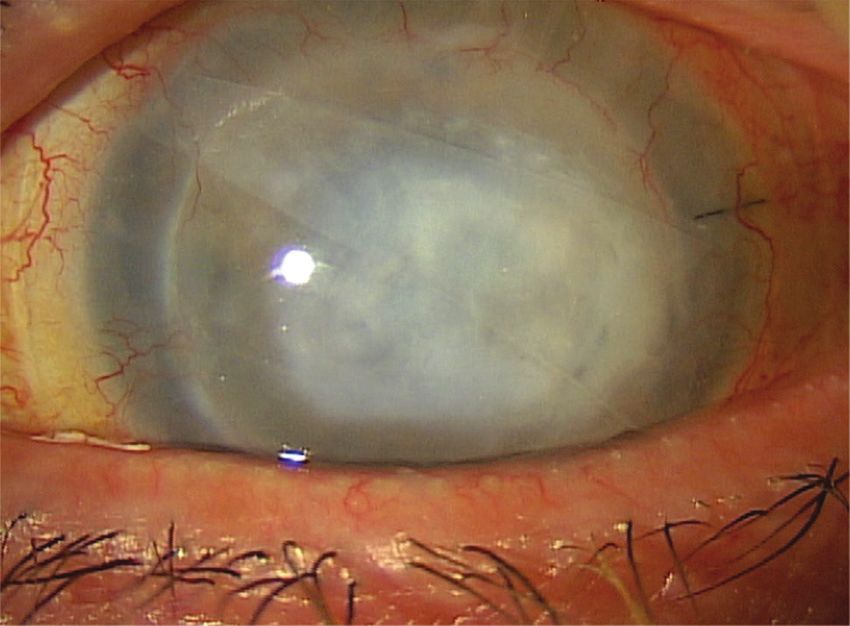
identify and resolve all conflicts of interest prior to this educa-       Merck, Mynosys, Novartis, Ocular Science, Ocular Therapeutix,
tional activity.                                                          Ocusoft, Omeros, Oyster Point Pharma, Science Based Health, Shire,
                                                                          Sight Sciences, Sun Pharmaceutical Industries, Surface, Thea, Tarsus,
   The following faculty/staff members have the following financial       TopCon, TearLab, Visus Therapeutics. Grant/Research Support:
relationships with commercial interests:                                  Alcon Vision, BioTissue, Ocular Science, TopCon, and TearLab.
                                                                          Stock/Shareholder: BlephEx, CorneaGen, Melt, Ocular Science, Oyster
  Francis S. Mah, MD, has had a financial agreement or affiliation        Point Pharma, and Tarsus.
during the past year with the following commercial interests in the
form of Consultant: Alcon Vision, Allergan, Bausch + Lomb, Eyenovia,      EDITORIAL SUPPORT DISCLOSURES
Glaukos, iView, Johnson & Johnson Vision, Nevakar, Novartis, Ocular         The Evolve staff and planners have no financial relationships
Science, and RxSight.                                                     with commercial interests. Michelle Dalton, writer, and Nisha
                                                                          Mukherjee, MD, peer reviewer, have no financial relationships with
   Sumitra Khandelwal, MD, has had a financial agreement or               commercial interests.
affiliation during the past year with the following commercial
interests in the form of Consultant: Alcon Vision, Bausch & Lomb,         OFF-LABEL STATEMENT
Carl Zeiss Meditec, Dompé, Novartis, and Ocular Therapeutix.                 This educational activity may contain discussion of published
                                                                          and/or investigational uses of agents that are not indicated by
   Marjan Farid, MD, has had a financial agreement or affiliation         the FDA. The opinions expressed in the educational activity
during the past year with the following commercial interests              are those of the faculty. Please refer to the official prescribing
in the form of Consultant: Allergan, Bausch + Lomb, BioTissue,            information for each product for discussion of approved indications,
Carl Zeiss Meditec, CorneaGen, Dompé, Tarsus, Orasis, Johnson             contraindications, and warnings.
& Johnson Vision, Kala Pharmaceuticals, Novartis, and Sun
Pharmaceutical Industries.                                                DISCLAIMER
                                                                            The views and opinions expressed in this educational activity are
  Elizabeth Yeu, MD, has had a financial agreement or affiliation         those of the faculty and do not necessarily represent the views of
during the past year with the following commercial interests in           Evolve, Cataract & Refractive Surgery Today, MillennialEYE, or Dompé.
the form of Consultant: Alcon Vision, Allergan, Avedro, Bausch +
Lomb, BioTissue, Beaver Visitec, BlephEx, Bruder, Carl Zeiss Meditec,     DIGITAL EDITION
CorneaGen, Dompé, Expert Opinion, EyePoint Pharmaceuticals,                 To view the online version of the material, go to
Guidepoint, Johnson & Johnson Vision, Kala Pharmaceuticals, Lensar,       https://evolvemeded.com/course/2132-supp.

                                                                      OCTOBER 2021 | SUPPLEMENT TO CATARACT & REFRACTIVE SURGERY TODAY / MILLENNIALEYE 3
PRETEST QUESTIONS
    PLEASE COMPLETE PRIOR TO ACCESSING THE MATERIAL AND SUBMIT WITH POSTTEST/ACTIVITY EVALUATION/
                                SATISFACTION MEASURES FOR CME CREDIT.

1.	Please rate your confidence in your ability to describe the                   5.	Endogenous nerve growth factor helps preserve and restore the ocular
    mechanisms of action of newer treatments and when they should be                  surface by which of the following mechanisms?
    introduced into treatment regimens for neurotrophic keratitis (NK)                a. 	Strengthening tight junctions between epithelial cells to enhance
    (based on a scale of 1 to 5, with 1 being not at all confident and 5                   corneal epithelial barrier functions.
    being extremely confident).                                                       b. 	Providing nutrition to conjunctival goblet cells and eyelid tear glands
    a. 1                                                                                   in order to increase tear production and improve tear quality.
    b. 2                                                                              c. Stimulating limbal stem cells to generate new epithelial cells.
    c. 3                                                                              d. 	Increasing tear production at the lacrimal gland, stimulating nerve
    d. 4                                                                                   regeneration, and supporting epithelial cell proliferation and
    e. 5                                                                                   differentiation.

2.	A 52-year-old female with history of LASIK OU, pars plana vitrectomy          6.	According to the Mackie Severity Classification for NK, all but which
    OS, and herpes keratitis OS is referred by her regular optometrist for            clinical feature is classified as Stage 1?
    decreased vision during the past few months in the left eye despite               a. Punctate epitheliopathy
    intensive therapy. Vision is 20/20 OD and 20/200 OS. IOP is 15 mm                 b. Stromal opacity
    Hg in both eyes. Slit lamp examination of the right eye reveals a well            c. Decreased tear break-up time
    healed LASIK flap but is otherwise unremarkable. The left eye has                 d. Stromal haze
    a 3 x 4 mm epithelial defect with mild stromal haze, diffuse corneal
    staining outside the defect, and a mild anterior segment reaction.
    Qualitative testing of corneal sensation shows decreased sensation            7.	Ms. Smith is referred into your clinic for a dry eye evaluation. She has
    in the left eye. Fundus examination is normal in the right eye and a              a history of type 2 diabetes, proliferative diabetic retinopathy, and
    B-scan is unremarkable in the left eye. Based on the patient’s history            bacterial corneal ulcers with poor healing that required penetrating
    and clinical evaluation, this presentation appears to be consistent with          keratoplasty; 2 months later the epithelium has not yet healed.
    neurotrophic keratitis. According to the Neurotrophic Keratitis Study             Previous treatments included antibiotics, steroids, bandage contact
    Group, this patient has what stage of disease?                                    lenses, and self-retaining amniotic membrane. Corneal sensitivity
    a. Stage 1                                                                        testing reveals centrally absent sensation. What would be considered
    b. Stage 3                                                                        an appropriate next step?
    c. Stage 4                                                                        a. Continue with Prokera
    d. Stage 5                                                                        b. Increase steroid-free artificial tears to 5 times daily
                                                                                      c. Initiate a 6-week course of cenegermin 8 times daily
3.	Corneal innervation is essential for good epithelial health. How do
                                                                                      d. Initiate an 8-week course of cenegermin 6 times daily
    corneal nerves maintain a healthy corneal surface?
    a. Provide stromal, epithelial, and Bowman’s structural support               8.    _____________ is the only way to diagnose NK.
    b.	Maintain sensory functions that are essential to tear film                     a. Visual acuity testing
         maintenance                                                                   b. In vivo confocal microscopy
    c. 	Facilitate protective functions of blinking and tear production as            c. Corneal sensation testing
         well as trophic support                                                       d. Imaging with a slit lamp
    d. 	Provide key nutrients to the epithelium while also serving as a
         physical barrier to microbes                                             9.	Which of the following systemic conditions has been shown in studies
                                                                                      to exacerbate NK?
4.	The Mackie Neurotrophic Keratitis Classification System breaks                    a. Diabetes
    neurotrophic keratitis into three stages. Recently, the Neurotrophic              b. Multiple sclerosis
    Keratitis Study Group has developed a new 7-step staging system. The              c. Leprosy
    purpose for this new system is:                                                   d. Congenital syndromes
    a. To replace an outdated system                                                  e. All of the above
    b. 	To allow for more accurate monitoring of progression of the disease          f. None of the above
         as well as delineate which patients may respond well to particular
         therapies and evaluate response to treatment                             10.	In the REPARO clinical study, ________ of patients who achieved
    c. 	To better educate patients about their disease and help them                  corneal healing remained healed 48 weeks after completing one full
         understand the prognosis and possible consequences of the condition           course of treatment with cenegermin.
    d. Determine which patients need amniotic membrane grafting                        a. 40%
                                                                                       b. 60%
                                                                                       c. 80%
                                                                                       d. 100%

 4 SUPPLEMENT TO CATARACT & REFRACTIVE SURGERY TODAY / MILLENNIALEYE | OCTOBER 2021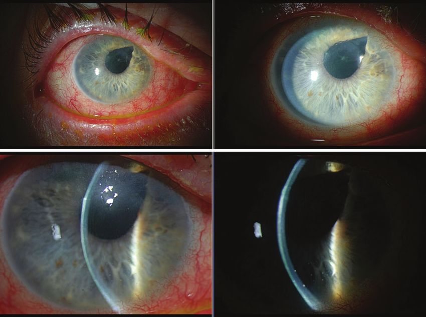
NEUROTROPHIC KERATITIS:
                                                                                                                    A “Rare Disease” Yet Common Problem

Neurotrophic Keratitis:
A “Rare Disease” Yet Common Problem
Neurotrophic keratitis (NK) is characterized by decreased or absent corneal sensation, which can lead to epithelial breakdown, impairment of
healing, and ultimately, to the development of corneal ulceration, melting, and perforation.1 This rare, degenerative disease (affecting fewer than
65,000 people in the United States2) is ultimately caused by trigeminal nerve damage, which, in turn, causes the loss of corneal sensitivity.3 NK
remains a challenge to successfully diagnose and treat—systemic conditions including diabetes, multiple sclerosis, congenital syndromes, and
leprosy have also been associated with the development of corneal anesthesia,4 which can make diagnosis more difficult. Treating NK should be
prompt and based on the stage and severity of the disease. Despite various medical and surgical therapies having been proposed, NK remains
difficult and challenging to treat, and the lack of positive response is commonly observed in the clinical practice.5 We’ve brought together
corneal experts to discuss this disease and offer case-based examples.
                                                                                                            — Francis S. Mah, MD, Program Chair

INTRODUCTION                                                                anyone with ocular surface issues and finding that decreased
Q         DR. MAH: NK is a real problem we face as cornea
          specialists. Why is it so important to identify it?
                                                                            sensation is more common than we think.

   Marjan Farid, MD: NK is classified as a rare/orphan disease,             DIAGNOSING NEUROTROPHIC KERATITIS
but it is actually more prevalent than I thought. The current                  Dr. Farid: Testing or checking corneal sensation is the only way
prevalence estimate is five or less individuals in 10,000.6 Its true        to diagnose NK because it's an issue with the trigeminal nerves
prevalence is difficult to determine because many mild cases are            in the cornea. Patients with NK don't receive that biofeedback to
missed. Even in that mild phase, and certainly in the later stages,         the brain to have good healthy tears, to have the epithelial growth
it can be visually significant. NK is a degenerative corneal disease        factors released by the nerve endings, and so on.5,7 If you don't
that includes damage to the trigeminal nerve (cranial nerve V),             test that corneal sensation, you won’t clinch the diagnosis.
loss of corneal sensation, breakdown of the corneal epithelium,                I always checked corneal sensation as a resident and fellow, but
impaired corneal healing, and persistent epithelial defect, which           that fell to the wayside in a busy clinical practice. It’s important to
leads to corneal ulceration, stromal melting, and perforation.3 If          bring corneal sensation testing back to the forefront. We need to
you don't identify it and treat it quickly, it can cause subepithelial      educate our technicians and ancillary staff and remind them not
scarring, fibrosis, and long-term visual sequelae.                          to use topical anesthetic solution in patients who are suspicious
                                                                            for NK. If they do, you must bring the patient back and obtain
  Dr. Mah: That vision-threatening issue is obviously critical.             that sensation testing. Remember, patients won’t complain of
Because of the neurotrophic component, many patients don't                  pain; it’s the visual disturbance they notice.
realize how serious the issue is. Have you started noticing an
increase in the number of patients with NK?
                                                                            Q         DR. MAH: Let's say you do notice there's some
                                                                                       decreased corneal sensation, how do you educate the
   Dr. Farid: Yes, I have noticed an increase. Diabetes plays a                        patients on the condition?
large role in exacerbating any underlying etiologies of NK. I have             Dr. Farid: I relate it to a diabetic foot ulcer. I tell patients
patients with severe diabetes and renal disease and their corneas           that, like patients with diabetic foot ulcers, they won’t feel pain
look terrible. I'm definitely seeing it more as I look for it, and I’m      when they have an eye ulceration. It’s quite dangerous because
able to address in earlier with our newer therapies.                        it can become infected. If we don't treat it, it can scar and affect
                                                                            long-term vision. With that little bit of education, patients are
   Dr. Mah: I’m also looking for it more than I did previously. I’m         much more willing to adhere to treatment.
testing corneal sensitivity in anyone who comes in for ocular surface
disease or consult for dry eyes. Many of these patients have NK.               Dr. Khandelwal: I educate patients about the importance
                                                                            of follow-up for any change in symptoms. I tell them
  Sumitra Khandelwal, MD: It has been a surprise to me that                 “the body doesn’t heal what it doesn’t feel” and this is why it
once you start having NK in your differential diagnosis, you start          is important to address this challenge even though it doesn’t
seeing it more in your clinic. I agree with Dr. Mah; I’m testing            cause them discomfort.

                                                                         OCTOBER 2021 | SUPPLEMENT TO CATARACT & REFRACTIVE SURGERY TODAY / MILLENNIALEYE 5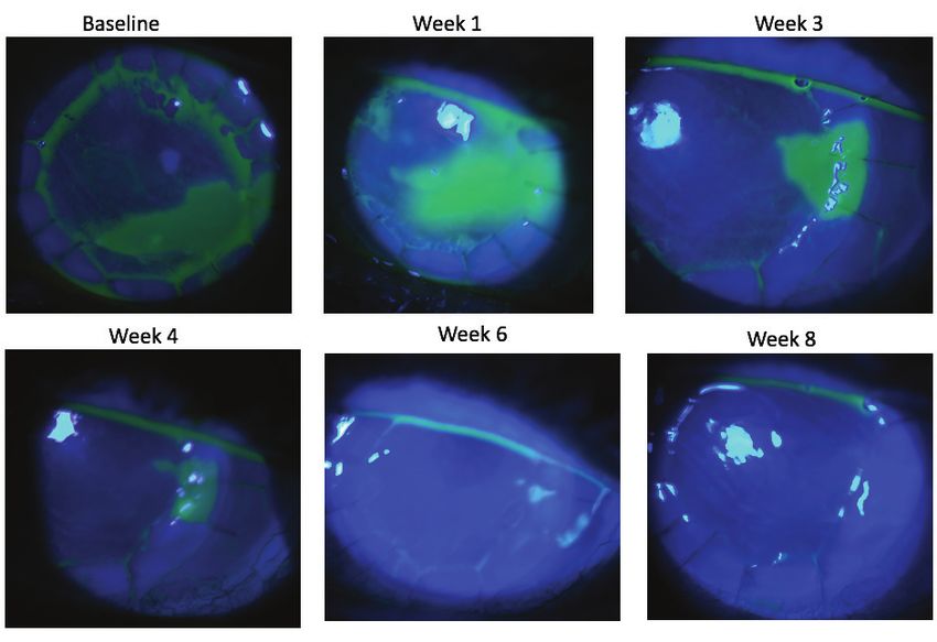
NEUROTROPHIC KERATITIS:
  A “Rare Disease” Yet Common Problem

     Dr. Mah: The classic NK schema, the Mackie Severity                             Many patients will present to you for a routine follow-up exam,
   Classification, was described decades ago.8 The Mackie                         and their eyes don’t look that red. They may come in for evaluation
   Classification includes three stages. A new definition and schema              of “blurrier vision” that is progressively worsening in the last 3 to
   have been proposed that increase the number of stages to six and               4 days. On clinical presentation, they have decreased sensation with
   includes education on how to differentiate it from similar diseases.           or without pain; corneal epithelium irregularities with or without
                                                                                  epithelial defect; stromal involvement that is usually oval in shape with
      Elizabeth Yeu, MD: Do you think it really affects fewer than                smooth and rolled edges; and corneal ulcer, melting, and perforation.2
   five people per 10,000?                                                           A large part of diagnosing NK is taking a thorough clinical history
                                                                                  followed by an examination checking for corneal sensitivities.
     Dr. Farid: I think perhaps in the stage 2 and 3, the numbers are low.        Corneal staining, corneal cultures, in vivo confocal microscopy,
   But I think there’s a great deal of stage 1 or early disease that is missed.   and Schirmer, which can be impaired as a result of reduction in
                                                                                  corneal sensitivity, are also helpful.2 Corneal sensitivity tests can be
      Dr. Yeu: I agree. Moderate to severe classes of NK should be                qualitative or quantitative. Qualitative examples include a cotton
   in the rare category. There are two types of patients on opposite              swab, cotton wisp, dental floss, or tissue tip. Quantitative is often
   ends of the spectrum: patients with a ton of pain without corneal              used in basic research and clinical trial settings with, for example,
   stain (keratoneuralgia, corneal neuropathic pain) and patients with            the Cochet-Bonnet esthesiometer.2 I use a cotton wisp, checking
   no pain and a lot of significant keratopathy. Some diseases, such              their blink response in both eyes and ask how they feel.
   as severe chronic dry eye disease, contact lens-related disorders,
   blepharitis, stem cell deficiency, topical drug toxicity, and exposure            Dr. Khandelwal: It is interesting because once I come close to
   keratopathy, have overlapping features of NK and corneal sensation             the patient and check their sensation in both eyes, they suddenly
   can be affected.2,6 By definition, there's a degeneration of the nerves,       understand there is something wrong with their corneas, especially
   whether it's iatrogenic or not, that leads to a vicious cycle, including       in unilateral cases. Then we go back in time and discuss how their
   a breakdown of the corneal epithelium (Figure 1). That breakdown               signs and symptoms of blurry vision or recurrent infections do not
   without the neural triggers prevents the actual growth factors from            correlate with their pain. I agree with all the testing options, although
   coming in naturally to reepithelialize naturally.                              in a busy clinic a qualitative test like cotton swab is quick and easy.
      The cornea is the most sensitive and densely innervated tissue
   in the human body.5,7 Corneal innervation is essential. Corneal                       Dr. Mah: I also use qualitative methods with a cotton swab.
   epithelial cells act in a mutually supportive relationship with corneal
   nerves, which maintain corneal integrity; it provides the reason for             Dr. Farid: I use cotton swabs as well. You can also use plain
   us to actually blink and tear.6 Corneal nerve damage leads to loss of          dental floss or a sterile Gore-Tex suture.
   corneal sensation, epithelial breakdown, and poor healing.5,7 When
   we lack corneal sensation, we’re not performing those motor skills               Dr. Yeu: Regarding staging, the Mackie Severity Classification of
   of blinking and refreshing the tear and preventing the desiccation.            NK has been well documented, but honestly, not many of us use it
      Risk factors for NK include infections such as herpes, systemic             (Table 1).6,8 With stage 1, there is no defect. The patient may have
   diseases such as diabetes, contact lens use, and fifth-nerve palsy             epithelial erosions and decreased tear break-up time. There may
   due to trigeminal neuralgia and surgery. Chronic comorbidities
   such as dry eye, blepharitis, topical drug toxicity, and contact
   lens-related disorder may worsen the prognosis of NK.2
                                                                                                TABLE 1. MACKIE SEVERITY CLASSIFICATION FOR
                                                                                                         NEUROTROPHIC KERATITIS. 6,8
                                                                                     Stage        Clinical Features
                                                                                     1            • P unctate epitheliopathy (punctate corneal
                                                                                                     fluorescein/ lissamine green staining)
                                                                                                  • Decreased tear break-up time
                                                                                                  • Stromal haze
                                                                                     2            • P ersistent epithelial defect with smooth
                                                                                                     rolled edges
                                                                                                  • Stromal opacity

                                                                                     3            • Stromal thinning/ulceration
                                                                                                  • Corneal perforation

   Figure 1. The cycle of neurotrophic keratitis.5,7

6 SUPPLEMENT TO CATARACT & REFRACTIVE SURGERY TODAY / MILLENNIALEYE | OCTOBER 2021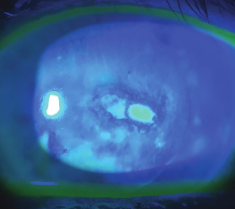
NEUROTROPHIC KERATITIS:
                                                                                                                   A “Rare Disease” Yet Common Problem

be stromal haze from either old scarring or devitalized epithelium               TABLE 2. NEUROTROPHIC KERATITIS STUDY GROUP 7-STEP
that is giving off the perception of haze, but there's not an overt                           CLINICAL STAGING SYSTEM.
epithelial defect. In stage 2, the patient has a persistent epithelial
                                                                           Stage                    Clinical Features                Imaging
defect with heaped-up edges; stromal opacity may or may not
exist. When a patient gets to stage 3, there is true stromal thinning      0                        Altered sensation without
with potential perforation.                                                                         keratopathy
                                                                           1 (mild)                 Epitheliopathy without
Q         DR. YEU: In your experiences, what are some of the
          loopholes in the Mackie Severity Classification?
                                                                                                    stromal haze

  Dr. Farid: It’s still very limited; we have patients who
are in between these stages. We have patients who have a
                                                                           2 (moderate)             Epitheliopathy with stromal
new persistent epithelial defect without any long-standing
                                                                                                    haze
subepithelial haze, versus the ones where its edges are more
chronic and where there's subepithelial haze and scarring. So even
within stage 2, we have degrees of severity that are not caught by
this classification.

   Dr. Mah: The biggest issue is the Mackie categories are too             Stage 3 (severe)         Persistent or recurrent
broad and nonspecific. The recent advent of more effective                                          epithelial defects
treatment options has necessitated a more highly defined staging
system that better reflects the evolution of the disease and alerts
clinicians to the earlier stages of NK. The Neurotrophic Keratitis
Study Group (NKSG) has worked to separate the differences so
treatment can begin earlier before there’s vision loss.

   Dr. Yeu: Adding stages makes sense. Stage 2, in particular, is          Stage 4 (severe)         Persistent or recurrent
too broad. Just like we can have marginally compensated corneas,                                    epithelial defect and stromal
you can have a marginally compensated neurotrophic disease                                          scarring without corneal
that breaks down because of some other comorbid state. You                                          ulceration
can also have someone who has zero sensation after trigeminal
neuralgia surgery. Because of the zero sensation, they're rapidly          Stage 5 (severe)         Persistent or recurrent
and more aggressively declining. Stage 2 can include a spectrum,                                    epithelial defect with corneal
which is why the Mackie Classification may not be overly helpful                                    ulceration
to clinicians. To address this, the NKSG has proposed a new 7-step
clinical staging system (Table 2) to more precisely classify the           Stage 6 (severe)         Corneal perforation
signs and symptoms of NK, allowing for earlier diagnosis, earlier
recognition of where they are in their process, and identifying the
underlying etiology or etiologies. The underlying etiology may
impact their response to treatment.
   The NKSG staging system, which is currently unpublished, starts
with stage 0, which is altered sensation without corneal findings.
Stage 1 is mild NK, including epitheliopathy without stromal haze.
Stage 2 includes epitheliopathy with stromal haze. That stromal             This new classification system gives you a better sense of
haze may be from chronic edema, which can lead to that difficulty         where they are in that disease process. Patient history is going
with deturgescence of the cornea leading to the haze. It can also be      to be key in understanding the iatrogenic causes of NK and how
haze secondary to infiltrate or early white blood cell recruitment,       not to further exasperate it. These patients can turn into that
which could indicate that there is a kind of superinfection that is       superimposed or secondary infection rather quickly.
going on. Stage 3, severe disease, includes persistent or recurrent
epithelial defects. Stage 4 is much more severe and also includes           Dr. Khandelwal: It is so important to recognize these stages can
persistent or recurrent epithelial defect, but with stromal scarring      progress quickly. I have seen patients with stage 1 disease that are
without corneal ulceration. In stage 5 disease, the patient has           completely neurotrophic progress to stage 3 within weeks on the
persistent or recurrent epithelial defect with corneal ulceration.        Mackie classification, so the NKSG staging will hopefully provide
Finally, the last stage, stage 6, is corneal perforation.                 us with more data on each patient.

                                                                      OCTOBER 2021 | SUPPLEMENT TO CATARACT & REFRACTIVE SURGERY TODAY / MILLENNIALEYE 7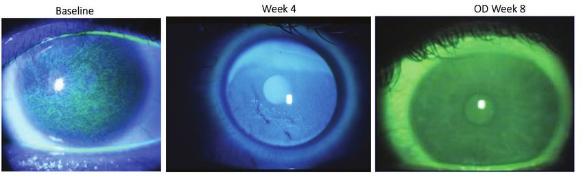
NEUROTROPHIC KERATITIS:
  A “Rare Disease” Yet Common Problem

   TREATMENTS FOR NEUROTROPHIC KERATITIS                                         cornea is the best course. For others, I may do a more stepwise
      Dr. Mah: NK requires severity-based therapy (Table 3).2,7 Going            approach with cenegermin and medical management with drops
   back to the Mackie Classification, which includes three stages,               or amniotic membrane. Each case is unique and often based on
   treatment for stage 1 disease is similar to dry eye management with           what the patient can tolerate.
   the use of preservative-free artificial tear formulations. Patients with
   stage 1 disease present with blurred vision and vision fluctuation.           Serum and plasma as primary or adjunct therapy
   They have some punctate epithelial keratopathy, a little bit of                  Dr. Mah: Serum and plasma have reported efficacy as primary
   corneal edema, and there may be a little stromal scarring. But for all        or adjunct therapy in these patients. The reported success of
   intents and purposes, they look like they've got an epitheliopathy            serum alone at 20 to 50% concentration ranges from 71 to 100%
   that could be consistent with dry eyes. That's why it's critical to           within 90 days.9-11 It's very successful for stage 1 NK. Umbilical
   test patients for corneal sensitivity.2,7 In addition to artificial tears,    cord serum may be more effective, as it has a higher concentration
   you can use punctal occlusion, hydrogel contact lenses, human                 of substance P as well as NGF and peripheral blood serum.12,13
   recombinant nerve growth factor (rhNGF) or cenegermin, and                    In one case series, epithelial defects healed 97.4% in stage 2 and
   serum drops, plasma, or platelet-rich plasma (PRP).                           stage 3 NK after 11 weeks of plasma rich in growth factors. Serum
                                                                                 can be used safely in combination with a silicone hydrogel contact
            TABLE 3. SEVERITY-BASED THERAPY FOR NEUROTROPHIC                     lens; no inflammation or contact lens deposits were observed in a
               KERATITIS BASED ON MACKIE CLASSIFICATION. 2,7                     study by Choi et al.14
    Stage       Therapy
    1           • Preservative-free artificial tears formulations                Q         What are your experiences with serum and the
                                                                                           plasma-rich platelets?
                • Punctal occlusion
                                                                                    Dr. Farid: I use serum generously in my patients. I can get serum
                • Hydrogel contact lens (consider large diameter)
                                                                                 into the hands of my NK patients quickly while waiting for rhNGF.
                • Recombinant human NGF (rhNGF, cenegermin)
                                                                                 I use a company called Vital Tears, of which I have no financial
                • Serum/plasma/platelet-rich plasma
                                                                                 interest. They’ve systematized the whole process and come to the
    2           Supportive therapies plus:                                       patient’s home to draw blood. Their turnaround time is usually
                • rhNGF                                                          2 to 3 days. Serum can be formulated at 100%, 75%, 50%, or 20%.
                • Scleral lens (± serum/plasma)                                  I usually start around 50 to 75%, and then lower the percentage
                • Amniotic membrane                                              once the patient improves.
                • Botulinum-induced ptosis, tarsorrhaphy
    3           • rhNGF                                                            Dr. Yeu: I don't have much experience with plasma-rich protein
                • Keratoplasty + scleral lens, tarsorrhaphy, neurotization       therapy. I do think serum tears are excellent in the management
                                                                                 of neurotrophic disease and epithelial defects. I don't see serum
      Patients with Mackie Classification stage 2 disease have an                tears as helpful for dry eye, necessarily, but they are extremely
   epithelial defect. They may have a bit of edema and a little                  useful in combination with other medical therapies and scleral
   scarring, but they do not have melting or excavation of the                   lenses. I typically start with a serum concentration between
   stroma. Potential therapies include rhNGF and scleral lenses,                 20 and 50%, and increase the concentration, if needed.
   plus or minus serum or PRP. Amniotic membrane, self-retained
   or sutured or glued, and botulinum-induced ptosis or                          Amniotic membrane transplantation
   tarsorrhaphy, are other therapeutic choices.2,7                                  Dr. Mah: Amniotic membrane transplantation (AMT) has been
      Stage 3 disease comes with stromal melting and the potential               a godsend for ocular surface disease specialists. In randomized
   for corneal perforation. If the cornea is already perforated,                 clinical trials, there have been reports of refractory neurotrophic
   then we would think about cyanoacrylate glue for smaller                      ulcers healing with conventional therapies such as lubrication plus
   (
NEUROTROPHIC KERATITIS:
                                                                                                                  A “Rare Disease” Yet Common Problem

begin with the Prokera Slim (Bio-Tissue), because that helps with            Dr. Mah: Corneal neurotization is a newer technique where
comfort.18 I always ensure a protective ptosis by performing a             corneal sensitivity is resorted after sural nerve grafts.21 It's a
tape tarsorrhaphy across the upper lid using sterile strips. The           procedure where there's a free sural nerve graft that is coapted
cryopreserved AMT usually lasts about a week unless the patient’s          end-to-side with the supratrochlear nerve. The distal portion
eye is really hot and angry. If it looks like they need a second, I        of the nerve is separated into fascicles and distributed around
will repeat it with a conventional one with the thicker amniotic           the limbus. The thought is, if the patient doesn't have sensation
membrane. If that strategy doesn’t work, then I’ll consider                because of the sensory nerves being damaged or deadened,
something more permanent, like a pillar or lateral tarsorrhaphy.           then why not produce or supply new nerves to the cornea? A
                                                                           study by Elbaz found that corneal sensitivity measured pre- and
   Dr. Farid: I agree. Cryopreserved AMT tends to have more of             postoperatively with the Cochet-Bonnet esthesiometer returned
the antiinflammatories and the regenerative properties. Although           to normal after 5 months.21 The procedure is very long and
it can be uncomfortable, NK patients don’t feel it in their eyes, so       arduous, requiring multiple specialties.
you can repeat it. I use the Prokera Plus (Bio-Tissue), which has a
double layer and longer use.
                                                                           Q       Has anyone used corneal neurotization?
                                                                                      Dr. Farid: It's long, arduous, and requires a
  Dr. Khandelwal: There are some patients with NK that I                   multidisciplinary team approach to the surgery. But anecdotally, it
have found are very sensitive to the symblepharon ring that                has been done with success in extremely severe cases.
cryopreserved amniotic membrane graft (AMG) is on. For these
patients, you can cut out the AMG and glue it using Tisseal or               Dr. Yeu: I also have no personal experience with it, but I have
even place under a bandage lens or scleral lens.                           heard results can be very good.

Scleral lenses                                                               Dr. Mah: It appears to be a nice possibility for some patients with
   Dr. Mah: Scleral lenses are another excellent technique for these       very severe disease who are recalcitrant to every other intervention.
patients. It takes a little more time because they must be fit properly
so it’s not really an option for a rapidly evolving or urgent/emergent        Dr. Khandelwal: We have performed this procedure at our
situation. To optimize success, corneal specialists need a contact         center–it is an invasive procedure, so using alternate treatment
lens fitting specialist nearby who can perform the scleral lens fitting.   first is ideal. However, for cases that do not improve with
But it is a fantastic long-term solution, especially for patients who      cenegermin or patients who are unable to use scleral lenses,
have recovered from stage 2 or stage 3 NK with some corneal                this can be an excellent treatment. In the beginning, the surgery
scarring. The use of fluid-filled scleral contact lens for the treatment   was performed with neurosurgery. However, our oculoplastics
of NK was initially reported decades ago.19 Ling et al reported that       specialists now use cadaver donor nerve tissue that allows it to be
nonhealing corneal epithelial defects healed without recurrence in         performed just by them. It is important the patient understand
all nine eyes treated with the PROSE scleral lens.20 Overnight wear        the sensation takes months to return, so they will still need a
with close monitoring may accelerate healing.                              scleral lens or tarsorrhaphy for a while. In addition, for cases in
                                                                           which there is also lack of sensation on the lids or forehead, a
Q        What are your experiences with scleral lenses?
           Dr. Farid: Scleral lenses are a great option. In the
                                                                           contralateral nerve dissection, which is more invasive, may be
                                                                           needed as well.
acute phases, it's sometimes challenging to get a scleral lens
for these patients. But it can work very well long term for                Cenegermin: A new treatment for neurotrophic keratitis
visual rehabilitation.                                                        Dr. Mah: Cenegermin is an ophthalmic solution containing
                                                                           20 µg/mL of rhNGF. It is the first US FDA-approved therapy for NK
  Dr. Khandelwal: At our practice, we have an extra set of PROSE           and should be considered a first-line option for NK treatment.22
lenses which we can use for a patient to get them through a                   As a recumbent form of NGF, cenegermin acts through specific
nonhealing epithelial defect, similar to the previous mentioned            high-affinity (ie, TrkA) and low-affinity (ie, p75NTR) NGF in
studies. But not all practices will have this option.                      the anterior segment of the eye to support corneal innervation
                                                                           and integrity through three mechanisms of action.3 First, NGF
   Dr. Yeu: I’ve found that it’s extremely helpful to add something        stimulates the regeneration and survival of sensory nerves.2,23
inside the bed of the scleral lens, such as one drop of antibiotic,        Second, NGF helps with receptors on the lacrimal glands and
either serum tears or amniotic cytokine extract. It helps the              promotes sensory-mediated reflex tearing secretions.24 Third,
patient reepithelialize relatively quickly, so long as they don't          NGF maintains corneal integrity through cell proliferation and
have endothelial dysfunction. I wouldn’t use it in patients with           differentiation and survival of the corneal epithelial cell.3 The
endothelial disease because they can't pump out the fluid that's           active ingredient of cenegermin is structurally identical to hNGF
within the scleral lens.                                                   and naturally occurring.

                                                                       OCTOBER 2021 | SUPPLEMENT TO CATARACT & REFRACTIVE SURGERY TODAY / MILLENNIALEYE 9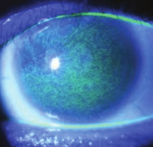
NEUROTROPHIC KERATITIS:
  A “Rare Disease” Yet Common Problem

      Neurotrophin is responsible for differentiation, growth, and
   maintenance of neurons. It is structurally identical and discovered
   by Nobel Prize-winning scientists in the early 1950s.25 Efficacy was
   established in two different studies, one from the United States
   (NGF0214) and one from Europe (REPARO).26-28 Both trials led
   to the FDA approval of cenegermin. NGF0214 showed complete
   corneal healing in up to 65.2% of patients receiving cenegermin six
   times a day or every 2 hours for a total of 8 weeks. The endpoint
   was complete epithelial healing with no epithelial defect. Every
   patient in both studies had at least stage 2 or stage 3 NK, meaning
   they had at least an epithelial defect. REPARO had a slightly                 Figure 2. Baseline presentation.         Figure 3. After 4 weeks on cenegermin.
   different endpoint, which was a reduction in the size of the
   epithelial defect to less than 0.5 mm. In REPARO, 80% of patients
   who achieved complete corneal healing remained healed 48 weeks
   after completing one 8-week cenegermin treatment cycle.26 This is
   very encouraging and remarkable.
      As far as the pooled safety reports, there were no serious
   treatment-related adverse reactions to the treatment. I do like
   to prepare my patients for the treatment regimen since it’s so
   intense. A small proportion of patients, about 16%, experience
   some eye pain following installation, typically 2 to 3 weeks after
   beginning treatment. How do you educate your patients on
   cenegermin treatment?
                                                                                 Figure 4. After 8 weeks on cenegermin.   Figure 5. Nonhealing epithelial defect post-PK at baseline.
     Dr. Farid: When I put my patients on cenegermin, I spend a lot
   of time reviewing the instructions. I inform them that they will
   experience some pain around week 2 or 3. I let them know it’s                 treatment. The corneal sensitivity testing showed absent sensation
   a good thing; it means the nerves are regenerating. I have found              on that cornea. By the Mackie Classification, this is stage 2 NK.
   that explaining that helps with compliance.                                   We prescribed cenegermin, and by 4 weeks on treatment, the
                                                                                 epithelial defect had significantly shrunk to about 1 to 2 mm of
     Dr. Mah: The studies concluded that after 8 weeks of                        central opening (Figure 3). By week 8, what I found remarkable
   6-times-daily treatment, 65.2% of patients completely healed                  was the epithelial defect had completely healed, and there was no
   and up to 80% remained healed for up to a year.26-28 These are                other punctate staining on the cornea (Figure 4). He’s now 2 years
   extremely encouraging data.                                                   out, and his cornea still looks like it did in Figure 4 with smooth
                                                                                 epithelium and a healthy tear film.
   CASE 1: NONHEALING EPITHELIAL DEFECT
      Dr. Farid: Our first case is a patient referred to me by a                 CASE 2: NONHEALING EPITHELIAL DEFECT
   colleague in the community. He is a 75-year-old man who had                   POSTCORNEAL TRANSPLANT
   a 3-month history of nonhealing epithelial defect. The referring                Dr. Farid: Our second case is also one of a nonhealing epithelial
   doctor had gone through all the appropriate treatment regimens                defect. This patient, a 78-year-old woman, had undergone a
   with this patient, but the eye would not heal. The patient had                full-thickness penetrating keratoplasty (PK), and developed a
   bilateral LASIK several years earlier, and when asked about                   nonhealing corneal epithelial defect, with a duration of 8 weeks
   shingles, reported “blistering on one side of the face” about                 at presentation (Figure 5). This is certainly atypical, as we expect
   5 to 6 years ago on the same side as his current epithelial defect.           an epithelial defect posttransplant to heal within the first 1 to
   He also mentioned an abrasion about a year earlier that had                   2 weeks. Part of her medical history included diabetes, which as
   healed after 2 weeks of aggressive lubrication and antibiotic                 we discussed earlier can exacerbate NK.
   treatments. These are all red flags for NK.                                     We tried all the typical treatments: antibiotic drops, steroids
      See Figure 2 for his baseline at presentation to our clinic. At this       drops for antirejection, bandage contact lenses, self-retaining
   point, the patient had been treated with two rounds of bandage                amniotic membrane, and took her off of all preserved eye drops.
   contact lens therapy as well as a self-retaining cryo-preserved               Her corneal sensation was decreased as we would expect in a
   amniotic membrane and then autologous serum. We kept him                      post-PK eye.
   on antibiotics to ensure there were no preservatives in his system;             We decided to prescribe cenegermin, and you can see in Figure 6
   his medical history included valaciclovir for systemic antiviral              the improvement progression from week 1 through week 8.

10 SUPPLEMENT TO CATARACT & REFRACTIVE SURGERY TODAY / MILLENNIALEYE | OCTOBER 2021
NEUROTROPHIC KERATITIS:
A “Rare Disease” Yet Common Problem
Dr. Mah: I've used cenegermin after PKs. I wasn't able to treat
the NK before the corneal transplant.
CASE 3: STAGE 1 NEUROTROPHIC KERATITIS
Dr. Mah: Our next case is a 45-year-old woman who was
referred by a retina colleague. This patient had type 2 diabetes,
diabetic neuropathy, and proliferative diabetic retinopathy, which
then led to a vitreous hemorrhage, and my colleague performed
a pars plana vitrectomy for the vitreous hemorrhage. She was
bilaterally pseudophakic because of her diabetes. She wears a
contact lens and was being treated for dry eye; my retina colleague
tried cyclosporine, lifitegrast, loteprednol, erythromycin ointment,
serum tears, and even self-retained amniotic membrane punctal
plugs. Believe it or not, her only real complaint before referral
into our clinic was that she wanted to see so she could drive.
Figure 7 shows her cornea at baseline presentation to us. The
Figure 6. Baseline Through Week 8 whirling pattern almost looks like limbal stem cell failure. Corneal
sensitivity was reduced in all quadrants as well as centrally. As I
said earlier, I now incorporate corneal sensitivity into all my dry
Dr. Yeu: Did you use a bandage contact lens alongside eye referrals/evaluations.
treatment, or it was just the cenegermin? Because this was a bilateral case, I put a Prokera lens in one eye
and a bandage contact lens in the other while we were waiting
Dr. Farid: That's a great question. So, it’s off label to use a for cenegermin (this was during the clinical trials for cenegermin,
contact lens with cenegermin. It’s recommended to take off the so we did not have it readily on hand). We removed the bandage
contact lens to allow the drug to really penetrate into the ocular contact lens because that is an off-label use, and prescribed
surface. I'll have these patients in a contact lens early on, but I'll tell cenegermin for 8 weeks, every 2 hours six times daily. Figure 8
them once on cenegermin we need to remove the contact lens shows the beautiful improvement progression at weeks 4 and 8.
during the 8-week course. At week 4, the central corneal visual axis is clear, which resulted
in her improved vision. She still has some punctate erosions, but
Dr. Yeu: This was a beautiful case; it’s not often we see that kind at 8 weeks there was no corneal staining, which was remarkable.
of progress for post-PK eyes that have those persistent defects. Again, she didn't really complain about pain. Her only complaints
were related to her vision. How do you address patients who want
Dr. Mah: Have you ever used cenegermin before you were going to know if they have to take the cenegermin for the full 8 weeks?
to do a PK?
Dr. Farid: If you dig into the data from the cenegermin studies,
Dr. Farid: I haven't yet, unless they have NK. I think it's at 4 weeks more than 50% of patients had fully healed, but they
reasonable to try. You do need to document NK as a diagnosis remained on the study medication through 8 weeks. So we don’t
code in your chart in order for the insurance to kick in and cover know yet if we’ll get the same long-term results with a 4- or
the cenegermin. 6-week treatment instead.
Figure 7. Baseline imaging with a stage 1
neurotrophic keratitis diagnosis. Figure 8. Improvement over time.
OCTOBER 2021 | SUPPLEMENT TO CATARACT & REFRACTIVE SURGERY TODAY / MILLENNIALEYE 11NEUROTROPHIC KERATITIS:
A “Rare Disease” Yet Common Problem
Q DR. MAH: What about the reverse? After 8 weeks,
cenegermin proved effective for 72% of the patients, but
that leaves 28% who have not healed. Has anyone used
more than one round of 8-week treatment?
Dr. Yeu: I’m very bullish about the 8 weeks because I want to
regenerate as many nerves as possible. I had a patient who had
repeated herpes simplex virus keratitis, epithelial keratitis, and
then had the breakdown because of the neurotrophic disease.
They were treated with topical trifluridine, which is so toxic it
creates its own neurotrophic disease and cycle. If there’s still
staining after 8 weeks or even a defect, I will try another course. If
patients break down again, I’ll do another round in the future.
Dr. Mah: I’ll also do a second or even third round to get them
fully healed.
CASE 4: NONHEALING CORNEAL ULCER
Figure 10. Possible epithelial basement membrane dystrophy.
Dr. Khandelwal: This is a 56-year-old man who presents with
a nonhealing corneal ulcer that developed after a Descemet
stripping endothelial keratoplasty (DSEK) procedure with removal the scarring, we performed a PK with AMG and tarsorrhaphy.
of epithelium. He was treated with fortified antibiotics for several Finally, we removed the tarsorrhaphy and fit the patient with a
weeks. He was referred into our clinic because of a nonhealing scleral lens.
ulcer. His cultures were negative and patient was found to be
neurotrophic as well. Our diagnosis was sterile neurotrophic ulcer Dr. Farid: Complex and slow responding cases like this often
from weeks of fortified antibiotics. See Figure 9. require multimodal treatment approaches. The key is to support
the ocular surface from further breaking down with protective
Q What are your thoughts?
Dr. Mah: Once the persistent epithelial defect forms,
mechanisms such as scleral lens or tarsorrhaphy while at the same
time providing growth factors to regenerate the nerves and the
then the ulcer, obviously it’s an urgent matter to get things self-healing properties of the cornea.
healed as quickly as possible. It would be interesting to hear if
there was anything in the history that could have been a tip off of Dr. Mah: This is a difficult case because the patient
preexisting NK. Otherwise, obviously, surgery could have been the underwent a DSEK procedure to improve vision but he had a
cause of NK. complication postoperatively that could compromise vision.
Most likely, he had some NK prior to the DSEK surgery. If the
Dr. Farid: A neurotrophic ulcer can often be misdiagnosed as patient had diabetes mellitus or contact lens wear, or other
infectious and the attempted topical treatment for the infection, possible reasons for NK, there is a possibility it could have been
whether antibiotics for bacteria or antivirals for herpetic disease, identified prior to DSEK, and the surgeon could have pretreated
can often worsen the NK ulcer. If the epithelial defect is not with cenegermin.
improving or responding with the antimicrobials after a week,
then the diagnosis of NK has to be considered. CASE 5: LONG-TERM DISEASE
Dr. Khandelwal: Our next case is a 77-year-old man, who
Dr. Khandelwal: presented in May 2020 after 6 months of redness, irritation, and
There was a blurry vision in his right eye. He was referred to us for evaluation
suboptimal response, of possible basement membrane dystrophy. His ocular history
so we next tried includes a retinal detachment in his right eye after pars plana
amniotic membrane, vitrectomy with scleral buckle in 2003. He has bilateral primary
but that fell off. open-angle glaucoma. He’s pseudophakic, reporting no
He’s had multiple postoperative complications from cataract surgery. Based on
bandage lenses. We what you see, how would you proceed? (Figure 10.)
treated the patient
with cenegermin Dr. Mah: As we’ve discussed, I’ve incorporated corneal
six times daily for sensitivity into all my ocular surface consults. The patient has
Figure 9. Nonhealing corneal ulcer. 8 weeks. To treat several reasons to possibly have at least some mild NK.
12 SUPPLEMENT TO CATARACT & REFRACTIVE SURGERY TODAY / MILLENNIALEYE | OCTOBER 2021NEUROTROPHIC KERATITIS:
A “Rare Disease” Yet Common Problem
underwent PK without complications. We’re still following him,
and have plans for repeating cenegermin in the future.
Dr. Mah: Nice job escalating quickly to stabilize the
situation. I want to thank the faculty for this incredibly
informative discussion. n
1. Feroze KB, Patel BC. Neurotrophic keratitis. Last updated December 6, 2018. Treasure Island, FL: StatPearls Publishing, 2018.
2. Sacchetti M, Lambiase A. Diagnosis and management of neurotrophic keratitis. Clin Ophthalmol. 2014;8:571-579.
3. Mastropasqua L, Massaro-Giordano G, Nubile M, Sacchetti M. Understanding the Pathogenesis of Neurotrophic Keratitis: The Role of
Corneal Nerves. J Cell Physiol. 2017;232(4):717-724.
4. Bonini S, Rama P, Olzi D, Lambiase A. Neurotrophic keratitis. Eye (Lond). 2003;17(8):989-995.
5. Versura P, Giannaccare G, Pellegrini M, et al. Neurotrophic keratitis: current challenges and future prospects. Eye Brain. 2018;10:37-45.
6. Dua HS, Said DG, Messmer EM, et al. Neurotrophic keratopathy. Prog Retin Eye Res. 2018;66:107-31.
7. Sheha H, Tighe S, Hashem O, Hayashida Y. Update On Cenegermin Eye Drops In The Treatment Of Neurotrophic Keratitis. Clin Ophthalmol.
2019;13:1973-1980.
8. Mackie IA. Current Ocular Therapy: WB Saunders, 1995.
9. Guadilla AM, Balado P, Baeza A, Merino M. [Effectiveness of topical autologous serum treatment in neurotrophic keratopathy]. Arch Soc
Esp Oftalmol. 2013;88(8):302-306.
10. Jeng BH, Dupps WJ, Jr. Autologous serum 50% eyedrops in the treatment of persistent corneal epithelial defects. Cornea.
2009;28(10):1104-1108.
11. Pflugfelder SC. Is autologous serum a tonic for the ailing corneal epithelium? Am J Ophthalmol. 2006;142(2):316-317.
12. Yoon KC, You IC, Im SK, et al. Application of umbilical cord serum eyedrops for the treatment of neurotrophic keratitis. Ophthalmology.
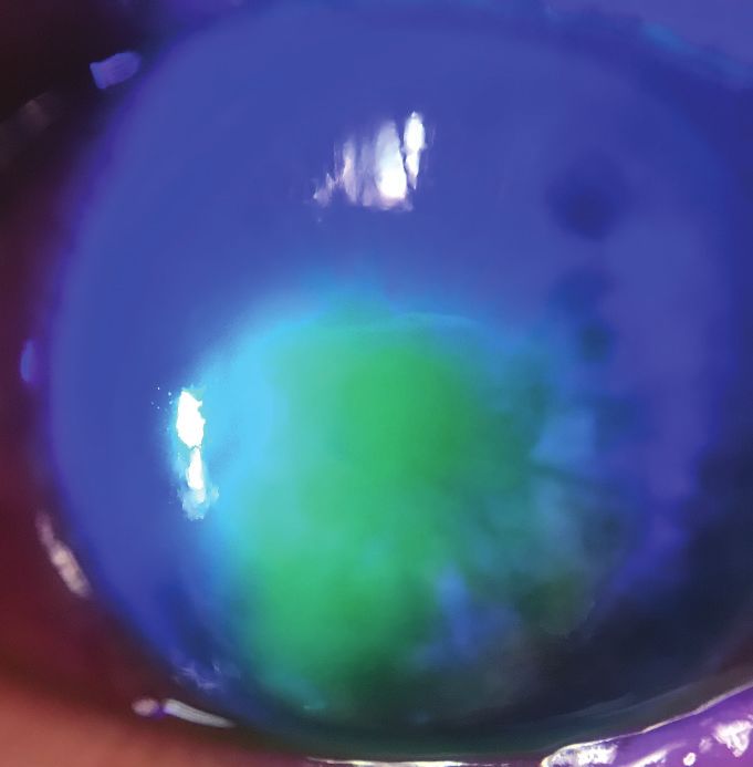
Figure 11. Follow-up exam in June 2020. 2007;114(9):1637-1642.
13. Sanchez-Avila RM, Merayo-Lloves J, Riestra AC, et al. Treatment of patients with neurotrophic keratitis stages 2 and 3 with plasma rich
in growth factors (PRGF-Endoret) eye-drops. Int Ophthalmol. 2018;38(3):1193-1204.
14. Choi JA, Chung SH. Combined application of autologous serum eye drops and silicone hydrogel lenses for the treatment of persistent
Dr. Khandelwal: You’re exactly right. This was a patient epithelial defects. Eye Contact Lens. 2011;37(6):370-373.
with stage 1 NK that we started planning to treat. The patient 15. Khokhar S, Natung T, Sony P, et al. Amniotic membrane transplantation in refractory neurotrophic corneal ulcers: a randomized,
returned about a month later, with worsening symptoms, and controlled clinical trial. Cornea. 2005;24(6):654-660.
16. Turkoglu E, Celik E, Alagoz G. A comparison of the efficacy of autologous serum eye drops with amniotic membrane transplantation in
we determined he now had stage 3 NK with microperforation. neurotrophic keratitis. Semin Ophthalmol. 2014;29(3):119-126.
(Figure 11). 17. Kruse FE, Rohrschneider K, Völcker HE. Multilayer amniotic membrane transplantation for reconstruction of deep corneal ulcers.
Ophthalmology. 1999;106(8):1504-1510; discussion 11.
18. Brocks D, Mead OG, Tighe S, Tseng SCG. Self-Retained Cryopreserved Amniotic Membrane for the Management of Corneal Ulcers. Clin
Dr. Mah: It’s amazing how quickly things can progress, and Ophthalmol. 2020;14:1437-1443.
why it’s so important to check and identify possible NK. 19. Romero-Rangel T, Stavrou P, Cotter J, et al. Gas-permeable scleral contact lens therapy in ocular surface disease. Am J Ophthalmol.
2000;130(1):25-32.
20. Ling JD, Gire A, Pflugfelder SC. PROSE therapy used to minimize corneal trauma in patients with corneal epithelial defects. Am J
Dr. Khandelwal: I agree. In August 2020, we proceeded with Ophthalmol. 2013;155(4):615-9, 9.e1-2.
layered amniotic membrane with tarsorrhaphy, which solved the 21. Elbaz U, Bains R, Zuker RM, et al. Restoration of corneal sensation with regional nerve transfers and nerve grafts: a new approach to a
difficult problem. JAMA Ophthalmol. 2014;132(11):1289-1295.
acute issues with the descmetocele with microperforation. 22. Deeks ED, Lamb YN. Cenegermin: A Review in Neurotrophic Keratitis. Drugs. 2020;80(5):489-494.
23. Müller LJ, Marfurt CF, Kruse F, Tervo TM. Corneal nerves: structure, contents and function. Exp Eye Res. 2003;76(5):521-542.
24. Muzi S, Colafrancesco V, Sornelli F, et al. Nerve growth factor in the developing and adult lacrimal glands of rat with and without
Dr. Farid: I have had several patients that have required PK inherited retinitis pigmentosa. Cornea. 2010;29(10):1163-1168.
eventually and I believe that the use of cenegermin for the full 25. Lambiase A, Rama P, Bonini S, et al. Topical treatment with nerve growth factor for corneal neurotrophic ulcers. N Engl J Med.
8-week course prior to the keratoplasty significantly improved 1998;338(17):1174-1180.
26. Bonini S, Lambiase A, Rama P, et al. Phase II Randomized, Double-Masked, Vehicle-Controlled Trial of Recombinant Human Nerve
their postoperative course and healing time. The stem cells and Growth Factor for Neurotrophic Keratitis. Ophthalmology. 2018;125(9):1332-1343.
the nerve plexus at the limbus do respond and regenerate so that 27. Dompe Inc. Dompé receives FDA approval of Oxervate eye drops (cenegermin-bkbj ophthalmic solution), first-in-class recombinant
epithelialization subsequent to a transplant is improved. human nerve growth factor with potential to completely heal rare neurotrophic keratitis. www.biospace.com/article/releases/dompe-
receives-fda-approval-of-oxervate-eye-drops-cenegermin-bkbj-ophthalmic-solution-first-in-class-recombinant-human-nerve-growth-
factor-with-potential-to-completely-heal-rare-neurotrophic-keratitis/2018.
Dr. Khandelwal: Once the amniotic graft dissolved, he was 28. Chao W. Healing of persistent epithelial defects or corneal ulcers by recombinant human nerve growth factor eye drops in patients
started on cenegermin six times daily for 8 weeks. He then with stage 2 or 3 neurotrophic keratitis. Congress of the European Society of Ophthalmology. Barcelona, Spain 2017.
OCTOBER 2021 | SUPPLEMENT TO CATARACT & REFRACTIVE SURGERY TODAY / MILLENNIALEYE 13NEUROTROPHIC KERATITIS: Release Date: October 2021
A “Rare Disease” Yet Common Problem Expiration Date: October 2022
INSTRUCTIONS FOR CME CREDIT
To receive credit, you must complete the attached Pretest/Posttest/Activity Evaluation/Satisfaction Measures Form and mail or fax to Evolve
Medical Education LLC, 353 West Lancaster Avenue, Second Floor, Wayne, PA 19087; Fax: (215) 933-3950. To answer these questions online and
receive real-time results, please go to https://evolvemeded.com/course/2132-supp. If you experience problems with the online test, email us at
info@evolvemeded.com. NOTE: Certificates are issued electronically.
Please type or print clearly, or we will be unable to issue your certificate.
Full Name______________________________________________________________________________________________________________________
Phone (required) ________________________________ o Email (required*) __________________________________________________________________
Address/P.O. Box_________________________________________________________________________________________________________________
City ________________________________________________________________State/Country_____ Zip/Postal Code______________________________
*Evolve does not share email addresses with third parties.
License Number __________________________________________________ OE Tracker Number ________________
DEMOGRAPHIC INFORMATION
Profession Years in Practice Patients Seen Per Week (with the Region
___ MD/DO ___ >20 disease targeted in this activity) ___ Northeast
___ OD ___ 11-20 ___ 0 ___ Northwest
___ NP ___ 6-10 ___ 1-15 ___ Midwest
___ Nurse/APN ___ 1-5 ___ 16-30 ___ Southeast
___ PA ___ 50
LEARNING OBJECTIVES
Did the program meet the following educational objectives? Agree Neutral Disagree
Identify the stages of neurotrophic keratitis _____ _____ _____
Recognize how to differentiate neurotrophic keratitis from similar diseases _____ _____ _____
Describe the mechanisms of action of newer treatments and when they should _____ _____ _____
be introduced into treatment regimens for neurotrophic keratitis, including
the stepwise approach
Explain the relationships between disease characteristics, drug therapies, _____ _____ _____
treatment frequency, visual, and anatomic outcomes
14 SUPPLEMENT TO CATARACT & REFRACTIVE SURGERY TODAY / MILLENNIALEYE | OCTOBER 2021PLEASE COMPLETE AT THE CONCLUSION OF THE PROGRAM.
1. Based on this activity, please rate your confidence in your ability to 5. Endogenous nerve growth factor helps preserve and restore the ocular
describe the mechanisms of action of newer treatments and when surface by which of the following mechanisms?
they should be introduced into treatment regimens for neurotrophic a. Strengthening tight junctions between epithelial cells to enhance
keratitis (based on a scale of 1 to 5, with 1 being not at all confident and corneal epithelial barrier functions.
5 being extremely confident). b. Providing nutrition to conjunctival goblet cells and eyelid tear
a. 1 glands in order to increase tear production and improve tear
b. 2 quality.
c. 3 c. Stimulating limbal stem cells to generate new epithelial cells.
d. 4 d. Increasing tear production at the lacrimal gland, stimulating nerve
e. 5 regeneration, and supporting epithelial cell proliferation and
differentiation.
2. A 52-year-old female with history of LASIK OU, pars plana vitrectomy
OS, and herpes keratitis OS is referred by her regular optometrist for 6. According to the Mackie Severity Classification for NK, all but which
decreased vision during the past few months in the left eye despite clinical feature is classified as Stage 1?
intensive therapy. Vision is 20/20 OD and 20/200 OS. IOP is 15 mm a. Punctate epitheliopathy
Hg in both eyes. Slit lamp examination of the right eye reveals a well b. Stromal opacity
healed LASIK flap but is otherwise unremarkable. The left eye has c. Decreased tear break-up time
a 3 x 4 mm epithelial defect with mild stromal haze, diffuse corneal d. Stromal haze
staining outside the defect, and a mild anterior segment reaction.
Qualitative testing of corneal sensation shows decreased sensation
in the left eye. Fundus examination is normal in the right eye and a 7. Ms. Smith is referred into your clinic for a dry eye evaluation. She has
B-scan is unremarkable in the left eye. Based on the patient’s history a history of type 2 diabetes, proliferative diabetic retinopathy, and
and clinical evaluation, this presentation appears to be consistent with bacterial corneal ulcers with poor healing that required penetrating
neurotrophic keratitis. According to the Neurotrophic Keratitis Study keratoplasty; 2 months later the epithelium has not yet healed.
Group, this patient has what stage of disease? Previous treatments included antibiotics, steroids, bandage contact
a. Stage 1 lenses, and self-retaining amniotic membrane. Corneal sensitivity
b. Stage 3 testing reveals centrally absent sensation. What would be considered
c. Stage 4 an appropriate next step?
d. Stage 5 a. Continue with Prokera
b. Increase steroid-free artificial tears to 5 times daily
3. Corneal innervation is essential for good epithelial health. How do c. Initiate a 6-week course of cenegermin 8 times daily
corneal nerves maintain a healthy corneal surface? d. Initiate an 8-week course of cenegermin 6 times daily
a. Provide stromal, epithelial, and Bowman’s structural support
b. Maintain sensory functions that are essential to tear film 8. _____________ is the only way to diagnose NK.
maintenance a. Visual acuity testing
c. Facilitate protective functions of blinking and tear production as b. In vivo confocal microscopy
well as trophic support c. Corneal sensation testing
d. Provide key nutrients to the epithelium while also serving as a d. Imaging with a slit lamp
physical barrier to microbes
9. Which of the following systemic conditions has been shown in studies
4. The Mackie Neurotrophic Keratitis Classification System breaks NK to exacerbate NK?
into three stages. Recently, the Neurotrophic Keratitis Study Group a. Diabetes
has developed a new 7-step staging system. The purpose for this new b. Multiple sclerosis
system is: c. Leprosy
a. To replace an outdated system d. Congenital syndromes
b. To allow for more accurate monitoring of progression of the disease e. All of the above
as well as delineate which patients may respond well to particular f. None of the above
therapies and evaluate response to treatment
c. To better educate patients about their disease and help them 10. In the REPARO clinical study, ________ of patients who achieved
understand the prognosis and possible consequences of the corneal healing remained healed 48 weeks after completing one full
condition course of treatment with cenegermin.
d. Determine which patients need amniotic membrane grafting a. 40%
b. 60%
c. 80%
d. 100%
OCTOBER 2021 | SUPPLEMENT TO CATARACT & REFRACTIVE SURGERY TODAY / MILLENNIALEYE 15You can also read