Corneal confocal microscopy identifies small fibre damage and progression of diabetic neuropathy
←
→
Page content transcription
If your browser does not render page correctly, please read the page content below
www.nature.com/scientificreports
OPEN Corneal confocal microscopy
identifies small fibre damage
and progression of diabetic
neuropathy
Shaishav Dhage1,2,3, Maryam Ferdousi2, Safwaan Adam1,2,3, Jan Hoong Ho1,2,3,
Alise Kalteniece2, Shazli Azmi1,2, Uazman Alam4, Georgios Ponirakis5, Ioannis Petropoulos5,
Andrew J. Atkinson2, Andrew Marshall6, Maria Jeziorska2, Handrean Soran1,2 &
Rayaz A. Malik1,2,5*
Accurately quantifying the progression of diabetic peripheral neuropathy is key to identify individuals
who will progress to foot ulceration and to power clinical intervention trials. We have undertaken
detailed neuropathy phenotyping to assess the longitudinal utility of different measures of
neuropathy in patients with diabetes. Nineteen patients with diabetes (age 52.5 ± 14.7 years, duration
of diabetes 26.0 ± 13.8 years) and 19 healthy controls underwent assessment of symptoms and signs
of neuropathy, quantitative sensory testing, autonomic nerve function, neurophysiology, intra-
epidermal nerve fibre density (IENFD) and corneal confocal microscopy (CCM) to quantify corneal
nerve fibre density (CNFD), branch density (CNBD) and fibre length (CNFL). Mean follow-up was
6.5 years. Glycated haemoglobin (p = 0.04), low-density lipoprotein-cholesterol (LDL-C) (p = 0.0009)
and urinary albumin creatinine ratio (p < 0.0001) improved. Neuropathy symptom profile (p = 0.03),
neuropathy disability score (p = 0.04), vibration perception threshold (p = 0.02), cold perception
threshold (p = 0.006), CNFD (p = 0.03), CNBD (p < 0.0001), CNFL (p < 0.0001), IENFD (p = 0.04), sural
(p = 0.02) and peroneal motor nerve conduction velocity (p = 0.03) deteriorated significantly. Change
(∆) in CNFL correlated with ∆CPT (p = 0.006) and ∆Expiration/Inspiration ratio (p = 0.002) and
∆IENFD correlated with ∆CNFD (p = 0.005), ∆CNBD (p = 0.02) and ∆CNFL (p = 0.01). This study shows
worsening of diabetic neuropathy across a range of neuropathy measures, especially CCM, despite
an improvement in HbA1c and LDL-C. It further supports the utility of CCM as a rapid, non-invasive
surrogate measure of diabetic neuropathy.
The natural history of diabetic peripheral neuropathy (DPN) is poorly defined with limited studies assessing pro-
gression of neuropathy1. As a consequence, clinical trials of disease modifying therapies in patients with diabetic
neuropathy have not been able to identify the optimal neuropathy end points to adequately assess progression
or improvement in D PN2. Indeed, whilst the DCCT in patients with T1DM showed that intensive glycaemic
control reduced the incidence of clinical DPN and nerve conduction abnormalities by 60%3; in patients with
T2DM, the U KPDS4 and VA-CSDM t rial5 reported no effect on DPN and cardiac autonomic neuropathy and
whilst the Kumamoto s tudy6 showed a prevention of nerve conduction slowing, the ACCORD t rial7 showed no
effect on VPT over 6-years.
Quantitative sensory testing (QST) is relatively easy to perform but has limited reproducibility and a high
degree of s ubjectivity8. Nerve conduction studies (NCS) are the established ‘gold standard’ for evaluating DPN
but require standardization in a clinical trial and cannot evaluate small fibres9. Whilst small nerve fibre damage
and repair can be identified by performing a skin biopsy and quantifying intra-epidermal nerve fibre density
1
Department of Medicine, Manchester University NHS Foundation Trust, Manchester, UK. 2Cardiovascular
Research Group, University of Manchester, Manchester, UK. 3The Christie NHS Foundation Trust, Manchester,
UK. 4Institute of Cardiovascular and Metabolic Medicine and The Pain Research Institute, University of Liverpool
& Liverpool University NHS Hospital Trust, Liverpool, UK. 5Department of Medicine, Weill Cornell Medicine-Qatar,
Qatar Foundation, Education City, Doha, Qatar. 6Institute of Life Course and Medical Sciences and The Pain
Research Institute, University of Liverpool & Liverpool University NHS Hospital Trust, Liverpool, UK. *email:
ram2045@qatar‑med.cornell.edu
Scientific Reports | (2021) 11:1859 | https://doi.org/10.1038/s41598-021-81302-8 1
Vol.:(0123456789)
www.nature.com/scientificreports/
(IENFD), it is invasive and requires e xpertise10–12. Other techniques for the assessment of small nerve fibres
include microneurography, Laser doppler image flare (LDIflare), nociceptive-evoked potentials and electro-
chemical skin conductance, but have considerable variability and are not routinely available13,14. Corneal confo-
cal microscopy (CCM) is a rapid non-invasive imaging technique for the quantitative assessment of small fibre
damage. Several studies have shown that it has good diagnostic utility for sub-clinical DPN, predicts incident
DPN15,16 and correlates with other measures of neuropathy16. Furthermore, automated quantification of corneal
nerve parameters allows rapid, unbiased and objective assessment of small fibre damage17 with comparable
diagnostic capability to I ENFD18,19.
Longitudinal studies of patients with diabetic neuropathy have been of relatively short duration and lacked
detailed neuropathy phenotyping20–23. In this study we compare the change in CCM and IENFD with symptoms,
signs, QST, autonomic function and neurophysiology over 6.5 years in a cohort of patients with diabetes.
Results
Clinical and metabolic assessment (Tables 1, 2). Age (p = 0.2), weight (p = 0.9) and body mass index
(BMI) (p = 0.5) did not differ significantly between patients and controls and also between patients at baseline
and follow up. Systolic (p = 0.9, p = 0.37) and diastolic (p = 0.5, p = 0.08) blood pressure did not differ between
controls and patients at baseline and between patients at baseline and follow up, respectively. HbA1c was signifi-
cantly higher in patients with diabetes compared to controls at baseline (p = 0.0002) and decreased significantly
in patients at follow up (p = 0.04). Low density lipoprotein cholesterol (LDL-C) was significantly lower in dia-
betic patients compared to controls at baseline (p = 0.05) and decreased further at follow up (p = 0.0009), whilst
triglycerides did not differ between patients and controls at baseline (p = 0.9) and did not change at follow up
(p = 0.9). eGFR did not differ significantly between diabetic patients and controls at baseline and decreased at
follow up (p = 0.004). Albumin creatinine ratio (ACR) was significantly higher in diabetic patients compared to
controls at baseline (p < 0.0001) and increased further at follow up (p < 0.0001).
Neuropathy assessments. Neuropathic symptoms and deficits (Tables 1, 2, Fig. 3). Neuropathy symp-
tom profile (NSP) (p = 0.0005) and neuropathy disability score (NDS) (p < 0.0001) were significantly higher in
patients at baseline compared to controls and increased significantly (p = 0.03, p = 0.04, respectively) in patients
at follow up.
Quantitative sensory testing (QST) (Tables 1, 2, Fig. 3). Vibration perception threshold (VPT), cold perception
threshold (CPT), warm perception threshold (WPT), cold induced pain (CIP), warm induced pain (WIP) and
percentage colour change in Neuropad did not differ significantly (p > 0.05) in patients at baseline compared to
controls. Whilst VPT increased (p = 0.02) and CPT (p = 0.006) decreased significantly there was no change in
WPT, CIP, WIP and Neuropad.
Electrophysiology (Tables 1, 2, Fig. 3). Sural (p = 0.01) and peroneal (p = 0.007) nerve conduction velocity and
peroneal nerve amplitude (p = 0.004) were significantly lower in patients at baseline compared to controls. Sural
(p = 0.02) and peroneal (p = 0.03) nerve conduction velocity decreased significantly, with no change in sural
(p = 0.75) or peroneal (p = 0.29) nerve amplitudes in patients at follow up.
Autonomic neuropathy (Tables 1, 2, Fig. 3). Deep breathing heart rate variability (DB-HRV) was significantly
lower in patients at baseline compared to controls (p = 0.005). Expiration/inspiration (E/I) ratio (p = 0.004), Val-
salva ratio (p = 0.001), and 30:15 ratio (p = 0.003) increased significantly with no change in DB-HRV (p = 0.67)
and sympathetic low frequency area (LFa)/parasympathetic respiratory frequency area (RFa) ratio (p = 0.42) at
follow up.
IENFD (Tables 1, 2, Figs. 1 and 3). Intraepidermal nerve fibre density (IENFD) was significantly lower in
patients at baseline (p = 0.04) compared to controls and decreased (p = 0.04) in patients at follow up.
CCM (Tables 1, 2, Figs. 2 and 3). Corneal nerve fibre density (CNFD) (p < 0.0001), Corneal nerve branch den-
sity (CNBD) (p = 0.009) and Corneal nerve fibre length (CNFL) (p = 0.0007) were significantly lower in patients
at baseline compared to controls and CNFD (p = 0.03), CNBD (p < 0.0001) and CNFL (p < 0.0001) decreased at
follow up.
Associations between the change in clinical and neuropathy measures (Table 3, Fig. 3). ΔIENFD correlated with
age (r = − 0.56, p = 0.01), BMI (r = − 0.47, p = 0.04), waist to hip ratio (r = − 0.66, p = 0.001), ΔE/I ratio (r = 0.595,
p = 0.0071) and ΔValsalva ratio (r = 0.59, p = 0.0078). ΔCNFD correlated with ΔVPT (r = − 0.54, p = 0.03),
ΔDBHRV (r = 0.55, p = 0.02) and ΔIENFD (r = 0.62, p = 0.005). ΔCNFL correlated with ΔCPT (r = 0.66, p = 0.006),
ΔE/I ratio (r = 0.68, p = 0.002) and ΔIENFD (r = 0.56, p = 0.014). ΔCNBD correlated with ΔVPT (r = − 0.55,
p = 0.02) and ΔIENFD (r = 0.53, p = 0.02). There was no correlation between change in HbA1c, lipids and neuro-
physiological parameters with change in CCM or IENFD (Supplementary Table 1).
Discussion
In this study we show a progressive worsening of diabetic neuropathy in diabetic patients despite an improvement
in HbA1c and LDL cholesterol, although there was no correlation between change in HbA1c, and LDL choles-
terol with change in any measure of neuropathy. In T1DM the DCCT showed that intensive glycaemic control
Scientific Reports | (2021) 11:1859 | https://doi.org/10.1038/s41598-021-81302-8 2
Vol:.(1234567890)www.nature.com/scientificreports/
Variable Controls (n = 19) Patients (Baseline) (n = 19) p value
Clinical and laboratory parameters
Age (years) 47.4 ± 14.2 52.5 ± 14.7 0.20
Duration of diabetes (years) NA 26.0 ± 13.8 NA
Weight (kg) 80.7 ± 18.0 82.0 ± 19.8 0.9
BMI (kg/m2) 27.5 ± 4.0 29.0 ± 5.7 0.50
BP (mmHg) 131 ± 23/74.0 ± 11.0 132 ± 21/ 71 ± 8 0.90/0.50
HbA1c (mmol/mol) 37.5 ± 3 63.5 ± 18.7 0.0002
Triglycerides (mmol/l) 1.4 ± 0.7 1.8 ± 1.7 0.9
LDL—C (mmol/l) 2.7 ± 0.9 2.23 ± 0.9 0.05
eGFR (ml min/ [1.73 m]2) 83 ± 7 82 ± 20 0.70
ACR (mg/mmol) 0.3 ± 0.1 7.5 ± 15.7 < 0.0001
Clinical neuropathy and QST measures
NSP (/38) 0.15 ± 0.5 3.5 ± 4.5 0.0005
NDS (/10) 0.57 ± 1.0 3.7 ± 2.4 < 0.0001
VPT (V) 7.5 ± 6.9 13.0 ± 8.0 0.06
CPT (°C) 28.4 ± 2.3 26.5 ± 3.5 0.51
WPT (°C) 36.9 ± 2.2 40.0 ± 3.7 0.20
CIP (°C) 9.0 ± 8.3 8.0 ± 8.5 0.90
WIP (°C) 45.0 ± 2.8 47.0 ± 2.5 0.15
Autonomic neuropathy measures
DB-HRV (beats/min) 30 ± 12 21 ± 15 0.005
Neuropad (%) 91.0 ± 21 62.4 ± 34 0.13
Nerve conduction studies
SNAP (µV) 17.9 ± 9.7 11.41 ± 10.9 0.06
SNCV (m/s) 49.8 ± 4.5 43.5 ± 9.0 0.01
PNAP (mV) 6.0 ± 2.2 3.8 ± 1.9 0.004
PMNCV (m/s) 48.7 ± 4.1 43.5 ± 3.6 0.0007
Corneal confocal microscopy
CNFD (no./mm2) 37.7 ± 6.5 28.8 ± 6.5 < 0.0001
CNBD (no./mm2) 96.5 ± 38.6 67.6 ± 30.2 0.009
CNFL (mm/mm2) 27.2 ± 3.4 22.2 ± 4.9 0.0007
Skin biopsy
IENFD (no./mm) 9.8 ± 3.8 6.6 ± 4.3 0.04
Table 1. Clinical and neuropathy parameters in control subjects and patients at baseline. BMI—body mass
index, BP—blood pressure, HbA1c—Glycosylated haemoglobin, eGFR—estimated glomerular filtration rate,
ACR—albumin creatinine ratio, LDL-C—low density lipoprotein cholesterol, NSP—neuropathy symptom
profile, NDS—neuropathy disability score, VPT—vibration perception threshold, DBHRV—deep breathing
heart rate variability, sural nerve action potential (SNAP), Sural nerve conduction velocity (SNCV), Peroneal
nerve amplitude (PNAP), Peroneal motor nerve conduction velocity (PMNCV), CNFD—corneal nerve fibre
density, CNBD—corneal nerve branch density, CNFL—corneal nerve fibre length, IENFD—intraepidermal
nerve fibre density. Data is presented as mean ± standard deviation (SD). Bold values show statistically
significant results. Continuous variables were compared between controls and baseline patient visits using the
paired t-test for normally distributed data and Wilcoxon matched-pairs signed rank test for non-normally
distributed data.
reduced the incidence of D PN3. However, in patients with T2DM, the U KPDS4, VA-CSDM t rial5 and A CCORD7
trials showed no effect of improved glycaemic control on DPN. A major problem in these clinical trials was the
end points utilised to assess neuropathy including symptoms and signs of neuropathy and quantitative sensory
testing, which were unable to accurately measure change in n europathy2.
Neurophysiology is considered to be the gold standard for the diagnosis of DPN and has been adopted as
an endpoint in multiple clinical trials9, but has failed to show a significant change in these trials24. Indeed, our
longitudinal data now shows a relatively small magnitude of reduction in peroneal and sural nerve conduction
with no change in amplitudes over 6.5 years. It is therefore not surprising that most trials lasting 12–24 months
show no change in neurophysiology.
Small fibre damage usually precedes large fibre damage and contributes to clinically meaningful end-points
like painful diabetic neuropathy and foot ulceration due to altered skin blood flow and delayed wound healing2.
Skin biopsy with IENFD quantification is the current gold standard for the evaluation of small fibre damage9
and whilst it is reliable and reproducible it is invasive and resource-intensive11. CCM is a rapid, non-invasive and
reproducible ophthalmic imaging technique which can be used to objectively quantify small fibre damage in a
Scientific Reports | (2021) 11:1859 | https://doi.org/10.1038/s41598-021-81302-8 3
Vol.:(0123456789)www.nature.com/scientificreports/
Variable Patients (Baseline) (n = 19) Patients (Follow up) (n = 19) p value
Clinical and laboratory parameters
Age (years) 52.5 ± 14.7 59.5 ± 15.6 NA
Duration of diabetes (years) 26.0 ± 13.8 32.5 ± 13.8 NA
Weight (kg) 82.0 ± 19.8 81.75 ± 18 0.49
BMI (kg/m2) 29.0 ± 5.7 28.7 ± 5.2 0.53
BP (mmHg) 132 ± 21/ 71 ± 8 127 ± 20 / 67 ± 9 0.37/0.08
HbA1c (mmol/mol) 63.5 ± 18.7 55.9 ± 12 0.04
Triglycerides (mmol/l) 1.8 ± 1.7 1.5 ± 1.1 0.9
LDL-C (mmol/l) 2.2 ± 0.9 1.9 ± 1.2 0.0009
eGFR (ml min−1 [1.73 m]−2) 82 ± 20 69 ± 21 0.004
ACR (mg/mmol) 7.5 ± 15.7 41.3 ± 123.6 < 0.0001
Clinical neuropathy measures and QST
NSP (/38) 3.5 ± 4.5 5.5 ± 5.7 0.03
NDS (/10) 3.7 ± 2.4 4.7 ± 2.5 0.04
VPT (V) 13.0 ± 8.0 18.0 ± 9.0 0.02
CPT (°C) 26.5 ± 3.5 21.8 ± 9.2 0.006
WPT (°C) 40.0 ± 3.7 41. 2 ± 4.8 0.38
CIP (°C) 8.0 ± 8.5 8.0 ± 7.7 0.81
WIP (°C) 47.0 ± 2.5 47.1 ± 2.8 0.622
Autonomic neuropathy measures
DB-HRV (beats/min) 21.0 ± 15.0 19.0 ± 7.0 0.67
LFa/RFa 2.8 ± 2.5 2.7 ± 2.7 0.42
E/I ratio 1.3 ± 0.2 1.2 ± 0.2 0.004
Valsalva ratio 1.6 ± 0.7 1.4 ± 0.5 0.001
30:15 ratio 1.3 ± 0.1 1.1± 0.1 0.0003
Neuropad (%) 62.4 ± 34.0 75.0 ± 31.0 0.47
Nerve conduction studies
SNAP (µV) 11.4 ± 10.9 10.5 ± 11.3 0.75
SNCV (m/s) 43.5 ± 9.0 40.4 ± 7.4 0.02
PNAP (mV) 3.8 ± 1.9 3.5 ± 1.9 0.299
PMNCV (m/s) 43.5 ± 3.6 42.4 ± 4.3 0.03
Corneal confocal microscopy
CNFD (no./mm2) 28.8 ± 6.5 25.6 ± 5.2 0.03
CNBD (no./mm2) 67.6 ± 30.2 43.7 ± 19.0 < 0.0001
CNFL (mm/mm2) 22.2 ± 4.9 16.1 ± 3.6 < 0.0001
Skin biopsy
IENFD (no./mm) 6.6 ± 4.3 5.2 ± 3.7 0.04
Table 2. Clinical and neuropathy parameters in patients at baseline and follow up. BMI—body mass index,
BP—blood pressure, HbA1c—glycosylated haemoglobin, e GFR—estimated glomerular filtration rate, ACR—
albumin creatinine ratio, LDL-C—low density lipoprotein cholesterol, NSP—neuropathy symptom profile,
NDS—neuropathy disability score, VPT—vibration perception threshold, CPT—cold perception threshold,
WPT—warm perception threshold, CIP—cold induced pain, WIP—warmth induced pain, DBHRV—deep
breathing heart rate variability, LFa/RFa ratio—low frequency area (sympathetic) and respiratory frequency
area (parasympathetic) ratio, E/I—expiration/inspiration ratio, Sural nerve action potential (SNAP), Sural
nerve conduction velocity (SNCV), Peroneal nerve amplitude (PNAP), Peroneal motor nerve conduction
velocity (PMNCV), CNFD—corneal nerve fibre density, CNBD—corneal nerve branch density, CNFL—
corneal nerve fibre length, IENFD—intraepidermal nerve fibre density. Data is presented as mean ± standard
deviation (SD). Bold values show statistically significant results. Continuous variables were compared between
baseline and follow up visits using the paired t-test for normally distributed data and Wilcoxon matched-pairs
signed rank test for non-normally distributed data.
range of peripheral neuropathies15,25–29. We have previously shown comparable diagnostic utility of CCM and
IENFD in diabetic n europathy19. Furthermore, in longitudinal studies reduced corneal nerve fibre length predicts
incident DPN30,31 and those at risk of developing D
PN32. Indeed, CCM has shown corneal nerve regeneration
6 months after pancreas and kidney transplantation in T1DM with no change in quantitative sensory testing and
an improvement in neuropathic symptoms and nerve conduction only after 24 and 36 months, r espectively21,33.
A recent study from Japan showed that an improvement in glycaemic control, body weight and blood pressure in
Scientific Reports | (2021) 11:1859 | https://doi.org/10.1038/s41598-021-81302-8 4
Vol:.(1234567890)www.nature.com/scientificreports/
Figure 1. Representative images from skin biopsies from healthy control (A) and diabetes patient of similar age
at baseline (B) and a follow-up visit after 6.5 years (C). Note numerous branching nerves reaching top layers of
epidermis (A; red arrows) and sparse short single nerve and two dividing nerves (red arrows) in epidermis of
the baseline biopsy (B) and more difficult to discern shorter nerves in the follow-up biopsy (red arrows). Scale
bar for A–C = 100 µm.
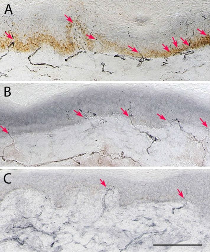
Figure 2. Corneal confocal microscopy image from a healthy control (A) and patient with diabetes at baseline
(B) and follow-up (C) showing a progressive loss of nerve fibres (red arrows main nerves, yellow arrows
branches) in patients with diabetes.
patients with T2DM was associated with an improvement in corneal nerve fibres, neurophysiology and vibration
perception over 4 years and correlated with a reduction in H bA1c34.
Studies have also shown an association between CCM and LDIflare in healthy control s ubjects35 and with
LDIflare, cooling detection thresholds and HRV in patients with diabetes16. In the present study CCM measures
worsened with greater magnitude than IENFD and large fibre (VPT, CPT, sural and peroneal nerve conduction
velocities) and autonomic (E/I ratio, Valsalva ratio and 30:15 ratio) measures of neuropathy. The worsening of
corneal nerve fibre measures was associated with worsening of other small fibre measures including cold percep-
tion threshold, IENFD and autonomic neuropathy, but not neurophysiology. Indeed, a number of studies have
shown corneal nerve loss in patients with diabetic autonomic neuropathy36–38 and a correlation between CCM
and a wide range of other measures of neuropathy including peroneal and sural nerve conduction36 and both
cold and warm perception thresholds16,39.
Scientific Reports | (2021) 11:1859 | https://doi.org/10.1038/s41598-021-81302-8 5
Vol.:(0123456789)www.nature.com/scientificreports/
A B C
Change in CCM Parameters over study period Change in neuropathy symptoms over the study period Change in NCV and IENFD over the study period
85 26 60 15
80 12
75 24
10
70 40
SNAP(µV)/PMNCV(m/s)
22
CNBD/CNFD (no./mm2)
65 8 10
CNFL (mm/mm2)
IENFD(no./mm)
60
NSP/DNS/NDS
20
55 6
20
50
18 4
45
5
40 2
16
0
35
30 0
14
25
-2
20 12 -20 0
Time from start of study(years)
Time from start of study(years) Time from start of study(years)
CNBD CNFD CNFL NSP DNS NDS SNAP PMNCV IENFD
D E F
Change in quantitative sensory testing over the study period Changes in autonomic neuropathy parameters Changes in autonomic neuropathy parameters
60 20 6 1.0
120 120
50
0.8 100 100
18
40
CPT/WPT/CIP/WIP(°C)
Valsalva/30:50 ratio
4
E/I, LFA:RFA ratio
80 80
0.6
Neuropad (%)
30
VPT(V)
DBHRV
16 60 60
20 0.4
2
40 40
10
14 0.2
0 20 20
0 0.0
-10 12 0 0
Time from start of study(years)
Time from start of study Time from start of study(years)
Valsalva ratio 30 : 50 ratio E/I Ratio LFA/RFA Ratio
VPT CIP CPT WIP WPT Patients baseline DBHRV Neuoropad
Figure 3. Percentage change from baseline values in CCM parameters (A), neuropathy symptoms (B), NCV
and IENFD (C), quantitative sensory testing (D) and autonomic neuropathy (E,F).
Variable CNBD CNFD CNFL IENFD
r = 0.53 r = 0.62 r = 0.56
IENFD
p = 0.02 p = 0.005 p = 0.01
r = − 0.26 r = − 0.43 r = − 0.045 r = − 0.07
NSP
p = 0.29 p = 0.08 p = 0.86 p = 0.76
r = − 0.13 r = − 0.43 r = − 0.11 r = − 0.05
NDS
p = 0.58 p = 0.08 p = 0.66 p = 0.82
r = 0.076 r = 0.29 r = 0.66 r = 0.27
CPT
p = 0.77 p = 0.26 p = 0.006 p = 0.26
r = − 0.55 r = − 0.54 r = − 0.08 r = − 0.12
VPT
p = 0.02 p = 0.03 p = 0.76 p = 0.37
r = − 0.19 r = − 0.55 r = − 0.14 r = − 0.03
DB-HRV
p = 0.42 p = 0.02 p = 0.57 p = 0.87
r = 0.26 r = 0.09 r = 0.017 r = 0.13
LFA/RFA ratio
p = 0.27 p = 0.70 p = 0.95 p = 0.58
r = 0.24 r = 0.31 r = 0.68 r = 0.595
E/I ratio
p = 0.32 p = 0.21 p= 0.002 p= 0.007
r = 0.41 r = 0.14 r = 0.25 r = 0.59
Valsalva ratio
p = 0.08 p = 0.56 p = 0. 32 p= 0.008
Table 3. Correlations between percentage change in small fibre pathology and other measures of diabetic
neuropathy from baseline to follow up. NSP—neuropathy symptom profile, NDS—neuropathy disability score,
DNS—diabetic neuropathy symptom score, VPT—vibration perception threshold, CPT—cold perception
threshold, DB-HRV—deep breathing heart rate variability, LFA/RFA ratio—low frequency area (sympathetic)
and high frequency area (parasympathetic) ratio, E/I—expiration/inspiration ratio, CNFD—corneal nerve fibre
density, CNBD—corneal nerve branch density, CNFL—corneal nerve fibre length, IENFD—intraepidermal
nerve fibre density. Bold values show statistically significant results.
Scientific Reports | (2021) 11:1859 | https://doi.org/10.1038/s41598-021-81302-8 6
Vol:.(1234567890)www.nature.com/scientificreports/
A limitation of this study is the relatively small number of patients assessed at follow up. However, the main
strength of this study is the comprehensive phenotyping of diabetic neuropathy over 6.5 years, enabling a detailed
comparison of the change in small and large fibre measures of diabetic neuropathy.
In conclusion, CCM identifies progressive nerve damage despite an improvement in glycaemic control and
LDL cholesterol. Furthermore, corneal nerve loss was associated with a loss of IENFD and worsening of other
measures of small fibre neuropathy. CCM is a rapid, non-invasive test to identify progression of neuropathy and
may have greater utility than symptoms, signs, QST and nerve conduction studies in longitudinal follow-up
studies and clinical trials of DPN.
Methods
Participant selection. Nineteen patients with diabetes [type 1 DM (n = 15) and type 2 DM (n = 4)], from
the Manchester University Hospital Diabetes Centre and 19 age-matched healthy control participants were
recruited and assessed between 2009 and 2011 and at follow up in 2017. The control group comprised of healthy
volunteers without DM and were not on any regular medications for any co-morbidities. Patients with a history
of neuropathy from any other cause, ocular disease, corneal trauma or surgery, systemic disorders affecting the
skin or cornea were excluded. All the tests performed at baseline were repeated in the follow up study using
the same protocol and equipment. This study has approval from the Health Research Authority (HRA), North
West—Greater Manchester South Research Ethics Committee. Written informed consent was obtained from all
individuals prior to participation. This research adhered to the tenets of the declaration of Helsinki.
Anthropometric and laboratory measurements. All participants underwent assessment of height,
weight and body mass index (BMI). Glycated haemoglobin (HbA1c), total cholesterol, low-density lipopro-
tein (LDL)-cholesterol, triglycerides (TG), serum creatinine and urinary albumin creatinine ratio (ACR) were
measured using routine laboratory methods in the Department of Biochemistry, Manchester University NHS
Foundation Trust. Estimated glomerular filtration rate (eGFR) was calculated using the abbreviated Modifi-
cation of Diet in Renal Disease (MDRD) equation: 186 × (creatinine/88.4) − 1.154 × (age) − 0.203 × (0.742 in
females) × (1.210 if Afro-Caribbean race).
Assessment of neuropathy. The neuropathy symptom profile (NSP) was used to assess the symptoms
of neuropathy. The modified neuropathy disability score (NDS) which is comprised of an assessment of vibra-
tion perception, pinprick, temperature sensation and presence or absence of ankle reflexes was used to evaluate
neurological deficits. A Horwell Neurothesiometer (Scientific Laboratory Supplies, Wilford, Nottingham, UK)
was used to establish the Vibration Perception Threshold (VPT). Cold (CT) and warm (WT) perception thresh-
olds and cold (CIP) and warm induced pain (WIP) thresholds were tested on the dorsolateral aspect of left foot
using the TSA-II NeuroSensory Analyser (Medoc, Ramat-Yishai, Israel). Electrodiagnostic nerve conduction
studies (NCS) were undertaken using a Dantec Keypoint System (Dantec Dynamics, Bristol, UK), equipped
with a DISA temperature regulator to keep the limb temperature constant at 32–35 °C. The ANX 3.0 autonomic
nervous system monitoring device (ANSAR Medical Technologies, Philadelphia, PA, USA) was used to assess
deep breathing heart rate variability (DB-HRV), sympathovagal balance via the sympathetic low frequency area
(LFa)/parasympathetic respiratory frequency area (RFa) ratio, expiratory/inspiratory (E/I ratio), Valsalva ratio
and 30:15 ratio. Sudomotor dysfunction was assessed by quantifying the percentage colour change after applying
the Neuropad to the area over the base of the first metatarsal head using our previously established protocol and
automated quantification40.
Skin biopsy. Local anaesthetic (1% lignocaine) was applied to the dorsum of the foot, 2 cm above the second
metatarsal head and two 3 mm punch biopsies were performed. Sections of 50 µm were stained using anti-human
PGP 9.5 antibody (Abcam, Cambridge, UK). SG chromogen (Vector Laboratories, Peterborough, UK) was used
to demonstrate nerve fibres and IENFD was quantified using previously established criteria and expressed as the
number per millimetre length of epidermis41. The follow-up skin biopsy was taken from the same foot, in close
proximity to the first biopsy. IENFD was quantified by the same investigator in a masked fashion.
Corneal confocal microscopy (CCM). CCM examination (Heidelberg Retinal Tomography III Rostock
Cornea Module; Heidelberg Engineering, Heidelberg, Germany) was performed using our previously estab-
lished protocol42. Six non-overlapping images, three per eye, were selected from the centre of the cornea. Three
corneal nerve parameters were quantified: Corneal nerve fibre density (CNFD): the total number of major
nerve fibres per square millimetre of corneal tissue, corneal nerve fibre branch density (CNBD): the number of
branches emanating from the major nerve trunks per square millimetre of corneal tissue and corneal nerve fibre
length (CNFL): the total length of all nerve fibres and branches (millimetre per square millimetre) using manual
quantification software [CCMetrics (Manchester, UK)]43.
Statistical analyses. Statistical analyses were performed using GraphPad Prism for Mac OS X (version
8.3.0, GraphPad Software, San Diego, California USA, www.graphpad.com). Data were tested for normality
using the Shapiro–Wilk normality test. All data are expressed as mean ± standard deviation (SD). Continuous
variables were compared between baseline and follow up visits using the paired t-test for normally distrib-
uted data and Wilcoxon matched-pairs signed rank test for non-normally distributed data. Ordinary one-way
ANOVA was performed (Kruskal–Wallis test was used for non-normally distributed data) to compare between
group differences of controls and baseline patient values. Post-hoc corrections for multiple comparison testing
Scientific Reports | (2021) 11:1859 | https://doi.org/10.1038/s41598-021-81302-8 7
Vol.:(0123456789)www.nature.com/scientificreports/
was done using Tukey’s test. Correlations were performed between the percentage change in IENFD and CCM
parameters and other variables using Pearson’s or Spearman’s Rank Test according to the distribution of the data.
A two-way p-value of less than 0.05 was considered to be statistically significant.
Data availability
The datasets generated and analysed during the current study are available from the corresponding author on
reasonable request.
Received: 7 November 2020; Accepted: 5 January 2021
References
1. Malik, R. A. et al. Small fibre neuropathy: Role in the diagnosis of diabetic sensorimotor polyneuropathy. Diabetes Metab. Res.
Rev. 27(7), 678–684 (2011).
2. Tavee, J. & Zhou, L. Small fiber neuropathy: A burning problem. Cleve Clin. J. Med. 76(5), 297–305 (2009).
3. Diabetes, C. et al. The effect of intensive treatment of diabetes on the development and progression of long-term complications in
insulin-dependent diabetes mellitus. N. Engl. J. Med. 329(14), 977–986 (1993).
4. Intensive blood-glucose control with sulphonylureas or insulin compared with conventional treatment and risk of complications
in patients with type 2 diabetes (UKPDS 33). UK Prospective Diabetes Study (UKPDS) Group. Lancet. 352(9131), 837–853 (1998).
5. Azad, N. et al. The effects of intensive glycemic control on neuropathy in the VA cooperative study on type II diabetes mellitus
(VA CSDM). J. Diabetes Complicat. 13(5–6), 307–313 (1999).
6. Ohkubo, Y. et al. Intensive insulin therapy prevents the progression of diabetic microvascular complications in Japanese patients
with non-insulin-dependent diabetes mellitus: A randomized prospective 6-year study. Diabetes Res. Clin. Pract. 28(2), 103–117
(1995).
7. Ismail-Beigi, F. et al. Effect of intensive treatment of hyperglycaemia on microvascular outcomes in type 2 diabetes: An analysis
of the ACCORD randomised trial. Lancet 376(9739), 419–430 (2010).
8. Dyck, P. J. et al. Vibratory and cooling detection thresholds compared with other tests in diagnosing and staging diabetic neuropa-
thy. Diabetes Care 10(4), 432–440 (1987).
9. Dyck, P. J. et al. Signs and symptoms versus nerve conduction studies to diagnose diabetic sensorimotor polyneuropathy: Cl vs
NPhys trial. Muscle Nerve 42(2), 157–164 (2010).
10. Nebuchennykh, M., Loseth, S., Lindal, S. & Mellgren, S. I. The value of skin biopsy with recording of intraepidermal nerve fiber
density and quantitative sensory testing in the assessment of small fiber involvement in patients with different causes of polyneu-
ropathy. J. Neurol. 256(7), 1067–1075 (2009).
11. Lauria, G. & Devigili, G. Skin biopsy as a diagnostic tool in peripheral neuropathy. Nat. Clin. Pract. Neurol. 3(10), 546–557 (2007).
12. Lauria, G. et al. European federation of neurological societies/peripheral nerve society guideline on the use of skin biopsy in the
diagnosis of small fiber neuropathy. Report of a joint task force of the European Federation of Neurological Societies and the
Peripheral Nerve Society. Eur. J. Neurol. 17(7), 903–12, e44-9 (2010).
13. Kramer, H. H., Schmelz, M., Birklein, F. & Bickel, A. Electrically stimulated axon reflexes are diminished in diabetic small fiber
neuropathies. Diabetes 53(3), 769–774 (2004).
14. Serra, J. Re-emerging microneurography. J. Physiol. 587(2), 295–296 (2009).
15. Petropoulos, I. N. et al. Corneal nerve loss detected with corneal confocal microscopy is symmetrical and related to the severity
of diabetic polyneuropathy. Diabetes Care 36(11), 3646–3651 (2013).
16. Sivaskandarajah, G. A. et al. Structure-function relationship between corneal nerves and conventional small-fiber tests in type 1
diabetes. Diabetes Care 36(9), 2748–2755 (2013).
17. Petropoulos, I. N. et al. Rapid automated diagnosis of diabetic peripheral neuropathy with in vivo corneal confocal microscopy.
Invest. Ophthalmol. Vis. Sci. 55(4), 2071–2078 (2014).
18. Alam, U. et al. Diagnostic utility of corneal confocal microscopy and intra-epidermal nerve fibre density in diabetic neuropathy.
PLoS ONE 12(7), e0180175 (2017).
19. Chen, X. et al. Small nerve fiber quantification in the diagnosis of diabetic sensorimotor polyneuropathy: comparing corneal
confocal microscopy with intraepidermal nerve fiber density. Diabetes Care 38(6), 1138–1144 (2015).
20. Dehghani, C. et al. Risk factors associated with corneal nerve alteration in type 1 diabetes in the absence of neuropathy: A longi-
tudinal in vivo corneal confocal microscopy study. Cornea 35(6), 847–852 (2016).
21. Azmi, S. et al. Early nerve fibre regeneration in individuals with type 1 diabetes after simultaneous pancreas and kidney transplan-
tation. Diabetologia 62(8), 1478–1487 (2019).
22. Deak, E. A. et al. Longitudinal changes in corneal cell and nerve fiber morphology in young patients with type 1 diabetes with and
without diabetic retinopathy: A 2-year follow-up study. Invest. Ophthalmol. Vis. Sci. 60(2), 830–837 (2019).
23. Jia, X. et al. In vivo corneal confocal microscopy detects improvement of corneal nerve parameters following glycemic control in
patients with type 2 diabetes. J Diabetes Res. 2018, 8516276 (2018).
24. Malik, R. A. Wherefore art thou, O treatment for diabetic neuropathy?. Int. Rev. Neurobiol. 127, 287–317 (2016).
25. Petropoulos, I. N. et al. Repeatability of in vivo corneal confocal microscopy to quantify corneal nerve morphology. Cornea 32(5),
e83–e89 (2013).
26. Kemp, H. I. et al. Use of corneal confocal microscopy to evaluate small nerve fibers in patients with human immunodeficiency
virus. JAMA Ophthalmol. 135(7), 795–800 (2017).
27. Stettner, M. et al. Corneal confocal microscopy in chronic inflammatory demyelinating polyneuropathy. Ann. Clin. Transl. Neurol.
3(2), 88–100 (2016).
28. Pagovich, O. E. et al. Corneal confocal microscopy: Neurologic disease biomarker in Friedreich ataxia. Ann. Neurol. 84(6), 893–904
(2018).
29. Tavakoli, M. et al. Corneal confocal microscopy detects small-fiber neuropathy in Charcot-Marie-Tooth disease type 1A patients.
Muscle Nerve 46(5), 698–704 (2012).
30. Lovblom, L. E. et al. In vivo corneal confocal microscopy and prediction of future-incident neuropathy in type 1 diabetes: A
preliminary longitudinal analysis. Can. J. Diabetes 39(5), 390–397 (2015).
31. Pritchard, N. et al. Corneal confocal microscopy predicts 4-year incident peripheral neuropathy in type 1 diabetes. Diabetes Care
38(4), 671–675 (2015).
32. Lewis, E. J. et al. Using in vivo corneal confocal microscopy to identify diabetic sensorimotor polyneuropathy risk profiles in
patients with type 1 diabetes. BMJ Open Diabetes Res. Care 5(1), e000251 (2017).
33. Tavakoli, M. et al. Corneal confocal microscopy detects early nerve regeneration in diabetic neuropathy after simultaneous pancreas
and kidney transplantation. Diabetes 62(1), 254–260 (2013).
Scientific Reports | (2021) 11:1859 | https://doi.org/10.1038/s41598-021-81302-8 8
Vol:.(1234567890)www.nature.com/scientificreports/
34. Ishibashi, F., Taniguchi, M., Kosaka, A., Uetake, H. & Tavakoli, M. Improvement in neuropathy outcomes with normalizing HbA1c
in patients with type 2 diabetes. Diabetes Care 42(1), 110–118 (2019).
35. Sharma, S., Tobin, V., Vas, P. R. J., Malik, R. A. & Rayman, G. The influence of age, anthropometric and metabolic variables on
LDIFLARE and corneal confocal microscopy in healthy individuals. PLoS ONE 13(3), e0193452 (2018).
36. Ziegler, D. et al. Early detection of nerve fiber loss by corneal confocal microscopy and skin biopsy in recently diagnosed type 2
diabetes. Diabetes 63(7), 2454–2463 (2014).
37. Wang, H., Fan, D., Wang, W., Zhang, S. & Wang, X. Early diagnosis of diabetic autonomic neuropathy by corneal confocal micros-
copy. Zhonghua yi xue za zhi 95(35), 2851–2856 (2015).
38. Misra, S. L. et al. In vivo confocal microscopy of corneal nerves: An ocular biomarker for peripheral and cardiac autonomic
neuropathy in type 1 diabetes mellitus. Invest. Ophthalmol. Vis. Sci. 56(9), 5060–5065 (2015).
39. Quattrini, C. et al. Surrogate markers of small fiber damage in human diabetic neuropathy. Diabetes 56(8), 2148–2154 (2007).
40. Ponirakis, G. et al. Automated quantification of neuropad improves its diagnostic ability in patients with diabetic neuropathy. J.
Diabetes Res. 2015, 847854 (2015).
41. Lauria, G. et al. Intraepidermal nerve fiber density at the distal leg: a worldwide normative reference study. J. Peripher. Nerv. Syst.
15(3), 202–207 (2020).
42. Malik, R. A. et al. Corneal confocal microscopy: A non-invasive surrogate of nerve fibre damage and repair in diabetic patients.
Diabetologia 46(5), 683–688 (2003).
43. Dabbah, M. A., Graham, J., Petropoulos, I. N., Tavakoli, M. & Malik, R. A. Automatic analysis of diabetic peripheral neuropathy
using multi-scale quantitative morphology of nerve fibres in corneal confocal microscopy imaging. Med Image Anal. 15(5), 738–747
(2011).
Acknowledgements
We acknowledge Mitra Tavakoli for undertaking some of the corneal confocal scans and Hassan Fadavi for
undertaking some of the neurological evaluation, QST and AFT testing at baseline. We acknowledge support
from Manchester Comprehensive Local Research Network and The National Institute for Health Research/
Wellcome Trust Clinical Research Facility in Manchester.
Author contributions
All authors were involved in revising the manuscript critically for important intellectual content and for final
approval of the version to be published. S.D. and M.F. were involved in acquisition of data, analysis and interpreta-
tion of data and wrote the manuscript. S.D., S.A., J.H.H., M.F. and A.K. recruited patients for follow up. S.D., S.A.
and J.H.H. contributed to acquisition and analysis of the data. S.Az., U.A., G.P., I.P. and M.F. recruited patients
at baseline. S.Az., U.A. performed skin biopsies for patients and controls at baseline and SD performed skin
biopsies for all patients at follow up. G.P., I.P. and M.F. performed C.C.M. for patients and controls at baseline.
M.F. and A.K. performed C.C.M. for patients at follow up. A.A. processed skin biopsies on follow up patients.
M.J. analysed and reported skin biopsies for all patients and controls at baseline and follow up. A.M. performed
and analysed nerve conduction studies for all patients and controls at baseline and at follow up. H.S. contributed
to conception, interpretation of the data, wrote and revised the manuscript. R.A.M. contributed to conception
and design of the study, wrote and revised the manuscript and is principal investigator of the study. R.A.M. is
the guarantor of this work and, as such, had full access to all the data in the study and takes responsibility for the
integrity of the data and the accuracy of the data analysis.
Funding
This research was funded from a National Institutes of Health Grant (R105991).
Competing interests
The authors declare no competing interests.
Additional information
Supplementary Information The online version contains supplementary material available at https://doi.
org/10.1038/s41598-021-81302-8.
Correspondence and requests for materials should be addressed to R.A.M.
Reprints and permissions information is available at www.nature.com/reprints.
Publisher’s note Springer Nature remains neutral with regard to jurisdictional claims in published maps and
institutional affiliations.
Open Access This article is licensed under a Creative Commons Attribution 4.0 International
License, which permits use, sharing, adaptation, distribution and reproduction in any medium or
format, as long as you give appropriate credit to the original author(s) and the source, provide a link to the
Creative Commons licence, and indicate if changes were made. The images or other third party material in this
article are included in the article’s Creative Commons licence, unless indicated otherwise in a credit line to the
material. If material is not included in the article’s Creative Commons licence and your intended use is not
permitted by statutory regulation or exceeds the permitted use, you will need to obtain permission directly from
the copyright holder. To view a copy of this licence, visit http://creativecommons.org/licenses/by/4.0/.
© The Author(s) 2021
Scientific Reports | (2021) 11:1859 | https://doi.org/10.1038/s41598-021-81302-8 9
Vol.:(0123456789)You can also read