INTERLEUKIN 25, THYMIC STROMAL LYMPHOPOIETIN AND HOUSE DUST MITES IN PATHOGENESIS OF ATOPIC DERMATITIS
←
→
Page content transcription
If your browser does not render page correctly, please read the page content below
JOURNAL OF PHYSIOLOGY AND PHARMACOLOGY 2020, 71, 2, 291-297
                                                 www.jpp.krakow.pl | DOI: 10.26402/jpp.2020.2.14

                       A.K. JAWOREK1, K. SZAFRANIEC2, Z. ZUBER3, A. WOJAS-PELC1, J. JAWOREK4

    INTERLEUKIN 25, THYMIC STROMAL LYMPHOPOIETIN AND HOUSE DUST MITES
                     IN PATHOGENESIS OF ATOPIC DERMATITIS

   Department of Dermatology, Faculty of Medicine, Jagiellonian University Medical College, Cracow, Poland; 2Department of
   1

   Epidemiology and Population Studies, Institute of Public Health, Faculty of Health Sciences, Jagiellonian University Medical
  College, Cracow, Poland; 3Andrzej Frycz Modrzewski Cracow University, Cracow, Poland; 4Department of Medical Physiology,
                      Faculty of Health Sciences, Jagiellonian University Medical College, Cracow, Poland

              Atopic dermatitis (AD) is the most common chronic inflammatory skin disease in the world. It is characterized by
              recurrent eczematous skin lesions, fluctuating course and chronic pruritus. Increasing evidence suggest that AD is more
              common in adults than previously thought. The disease is characterized by an impaired skin barrier, aberrant Th2-type
              cytokine production and intensive pruritus. Epithelial keratinocytes constitute the first physical, chemical and
              immunological barrier, classified as a part of the innate defense system. These keratinocytes secrete various factors, e.g.
              alarmins such as thymic stromal lymphopoietin (TSLP) and interleukin 25 (IL-25). Serum levels of substance P (SP)
              have been reported to be increased in patients with AD and correlated with itch intensity. Several previous studies
              reported a positive association between AD severity and house dust mites (HDM) sensitization. The aim of the study
              was to analyze IL-25, TSLP and SP concentrations in blood serum of adult patients with severe AD, depending on the
              degree of allergy to HDM. There were 31 adult AD patients enrolled into the study and a control group that consisted
              of 20 healthy subjects. AD was diagnosed on the basis of Hanifin and Rajka criteria. SCORing Atopic Dermatitis
              (SCORAD) and visual analogue (VAS) scores were used to assess the intensity of pruritus and blood content of specific
              IgE to HDM, as well as TSLP, IL-25 cytokines and SP was measured. Our study presents the evidence that IL-25 serum
              concentration is increased in patients with atopic dermatitis and this cytokine plays an important role in pathogenesis of
              this disease. HDM could stimulate the release of IL-25 which aggravates the disease severity. Our results corroborate
              previous findings on the role of TSLP in atopic dermatitis.

    K e y w o r d s : atopic dermatitis, epithelial cell-derived cytokines, interleukin-25, substance P, house dust mites, thymic stromal
                      lymphopoietin

                       INTRODUCTION                                    Despite intensive research, a precise etiopathogenesis of AD
                                                                       remains unclear. Among the numerous AD-related
     Atopic dermatitis (AD; atopic eczema, AE) is a chronic and        morphological and functional factors, dysfunction of skin
recurrent skin disease manifested by eczematous lesions                barrier and immunological disorders were singled out as the
(acute, subacute or chronic) with a characteristic, age-               main factors which, by interaction, contribute to the disease
dependent localization and intensive itching. AD is currently          development. It is noteworthy that pruritus, which is a
the most common inflammatory dermatosis in the world, with             fundamental symptom of AD, provokes the skin scratching,
a lifetime prevalence of up to 20% in developed countries (1).         leading to the epidermal damage and aggravation of skin
The clinical manifestations of the disease vary widely: from           inflammation (6-8).
mild lesions located in the hollows of the limbs to                        Substance P (SP) is a neuropeptide associated with
erythroderma - a generalized inflammation involving more               neurogenic skin inflammation and pruritus. The role of SP in the
than 90% of the skin surface, which is reflected in the                pathogenesis of AD is the subject of many reports. Elevated SP
differentiation of disease phenotypes (2). The presence of AD          levels can be used as an indicator of disease activity (9, 10).
significantly impairs patients’ quality of life not only due to the        In recent years, a growing body of evidence has indicated
irritating symptoms of the disease itself but also because of          that the development of skin inflammation in AD patients is
several potential comorbidities (e.g. bronchial asthma, mental         triggered mainly by epidermal damage resulting in epidermal
disorders, autoimmune diseases) (3). Although for years AD             keratinocytes releasing alarmins, such as thymic stromal
was considered a childhood disease that patients could ‘grow           lymphopoietin (TSLP), and interleukin IL-25 (11, 12). Also
out’ of, an increasing number of clinical reports indicate that        HDM, domestic and storage mites can aggravate skin damage
the disease is also prevalent in the adult population (4, 5).          and exacerbate lesions in AD patients, by affecting the skin both
292

directly (due to the activity of the mite cysteine proteases) and      mellitus), taking immunosuppressive medications (up to 3
indirectly (due to their allergenic properties) (13).                  months prior to the study) and undergoing phototherapy up to 6
    The aim of the study was to analyze IL-25, TSLP and SP             months prior to the study.
serum concentrations in adult patients with severe AD in
comparison to the severity of HDM allergy.                             Sample collection and laboratory analysis

                                                                           Blood samples were taken from ulnar vein between 7:00 am
                MATERIALS AND METHODS                                  and 8:00 am in a volume of 10 mL by the same nurse under the
                                                                       same circumstances. Standard blood sampling protocols were
Compliance with ethical standards
                                                                       followed; patients were fasting, after 30 minutes of rest. The
                                                                       samples were kept for 2 hours at room temperature for a clot to
    The study protocol was approved by the Bioethics                   form, then centrifuged at 3500 rpm for 10 minutes, frozen and
Committee of Jagiellonian University Medical College (consent          stored at –80ºC until use. The serum content of TSLP, IL-25 and
no. 122.6120.117.2016). The design and procedures of the study         SP was assessed using an ELISA assay (R&D System,
were carried out in accordance with the principles of the              Minneapolis, MN) according to the manufacturer’s protocol.
Declaration of Helsinki. All subjects provided a written               Total IgE concentration was evaluated during the routine
informed consent to participate in this study after a full             diagnostic tests using ELISA method with the fluorimeter
explanation of the study had been provided.                            UniCAP (Pharmacia, Sweden). The values above 100 IU/mL
                                                                       were considered as increased. Dust mite specific IgE (sIgE; d1:
Study sample                                                           Dermatophagoides pteronyssinus, d2: Dermatophagoides
                                                                       farinae) antibodies were assessed using ELISA method with the
    The group of 31 adult Caucasian patients (F/M: 16/15;              fluorimeter UniCAP (Pharmacia, Sweden) according to
median age: 34 years old; range 20 – 72 years) suffering from          manufacturer’s instruction. The level of IgE antibodies was
childhood-onset of extrinsic AD were qualified to the study. The       classified as follows: Class I: 0.35 – 0.68 IU/mL, Class II: 0.70
control group consisted of 20 healthy Caucasian volunteers             – 3.49 Iu/mL, Class III: 3.5 – 17.49 IU/mL, Class IV: 17.5 – 49.9
(F/M: 10/10; median age: 27 years old; range 23 – 72 years). A         IU/mL, Class V: 50.0 – 100.0 Iu/mL, Class VI: > 100 Iu/mL.
study group participants were recruited at the Dermatology
Department of Jagiellonian University Medical College from             Statistical analysis
January 2017 to May 2018. The diagnosis of AD was based on
the diagnostic criteria of Hanifin and Rajka (14). The severity of         To describe the sample, standard measures of mean (SD) and
AD was assessed based on the SCORing Atopic Dermatitis                 median (min-max) were used, depending on the distribution of
(SCORAD) index, where score over 50 points indicates a severe          the parameter. To compare the mean values of IL-25 and TLSP
form of the disease (15). All of the patients were classified as       between the analyzed groups, the Student’s t-test and the
having severe AD (median SCORAD index: 61.4 points; min–               ANOVA test were used; similar non-parametric methods were
max: 50.3 – 80.4 points). The worst itch during last 24 hours was      applied to SP analysis. The calculations were made using the
evaluated with Visual-Analogue-Scale (WI-VAS; where 0                  StatSoft STATISTICA package version 13.0. Tests of
points: no pruritus, 1 – 3: mild pruritus, 4 – 6: moderate pruritus,   significance were 2-tailed with α = 0.05.
7 – 8: severe pruritus, 9 – 10: very severe pruritus) (16). The
characteristics of the study group are presented in Table 1.
    The exclusion criteria of the selected subjects (patients and                                 RESULTS
controls) included: lack of consent, age below 18 years, pregnant
and lactating females, systemic inflammatory disorders,                   Nearly all patients with severe AD (96.8%) were allergic to
presence of other cutaneous inflammatory disorders,                    HDM. Only one patient from the study group revealed O class for
cardiovascular diseases, metabolic diseases (e.g. diabetes             house dust mite specific IgE (d1, d2). As much as 84% patients

Table 1. Characteristics of the atopic dermatitis (AD) patients and control group.
                    Parameter                                               Patients                  Control
                                                                             (n=31)                   (n=20)
                    Gender                                    Male          15 (48.5)                 10 (50)
                    n (%)                                    Female         16 (51.5)                 10 (50)
                    Age                                                         34                      27
                    median; min-max: (years)                                (20 – 72)                (23 – 72)
                    SCORAD                                                     61.4                     0.0
                    median; min-max: (points)                             (50.3 – 80.4)
                    VAS                                        10           27 (87.1)                  0.0
                    n (%) points                                9            4 (12.9)                  0.0
SCORAD, SCORing atopic dermatitis index; VAS, visual analogue scale.

Table 2. Distribution of atopic dermatitis patients by house dust mite specific IgE classes.

                    Classes            0           I           II        III          IV         V            VI
                    d1                 2           0           1          2            1         2            23
                    d2                 1           1           1          2            1         2            23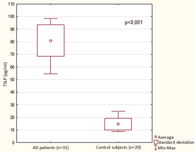
293

                                                                                                    Fig. 1. Serum IL-25 in adult
                                                                                                    patients suffering from atopic
                                                                                                    dermatitis (AD) and in control
                                                                                                    subjects.

                                                                                                    Fig. 2. Serum TSLP in adult
                                                                                                    patients suffering from atopic
                                                                                                    dermatitis (AD) and in control
                                                                                                    subjects.

from the study group was categorized into higher sIgE classes           The serum concentration of IL-25 was significantly higher in
(Class IV – VI), the majority of them was classified as Class VI    the severe AD patients (average: 17.2 ± 3.0 pg/mL) than in the
(23 (74%) subjects d1, d2) (Table 2). All patients from the study   control subjects (average: 15.2 ± 3 pg/mL ) (P < 0.033) (Fig. 1).
group (31/31; 100%) suffered from severe AD (SCORAD                 Moreover, there was a significant statistical difference (P <
median: 61.4 points; min-max: 50.3 – 80.4 points). According to     0.001) in TSLP serum level which was markedly higher in AD
the visual assessment scale of pruritus (VAS) all patients          subjects (average: 80.8 ± 12.5 pg/mL ), as compared to the
experienced very intensive itching (VAS ≥ 9, 100%). The levels      healthy control group (14.6 ± 4.5 pg/mL) (Fig. 2).
of total IgE antibodies were extraordinarily high in AD patients,       However, there was no statistical difference in serum SP
ranging between 1030 to 58 600 IU/mL; median 10 600 IU/mL.          concentration between patients with AD and controls (P =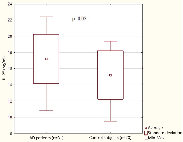
294

Table 3. Blood level of substance P in patients with atopic dermatitis (AD) and healthy control group.

                                              AD patients                        Control group
                                               (n = 31)                            (n = 20)
                    Substance P        Median    Min           Max         Median     Min      Max           P
                    (pg/ml)            170.6    107.6          749.0       163.5     107.9     191.6       0.148

                                                                                                      Fig. 3. Serum IL-25 (average
                                                                                                      and 95% CI) in classes of HDM
                                                                                                      specific IgE (d1 d2) and in
                                                                                                      control subjects.

                                                                                                      Fig. 4. Serum TLSP (average
                                                                                                      and 95% CI) in classes of HDM
                                                                                                      specific IgE (d1 d2) and in
                                                                                                      control subjects.

0.148), although 29% of patients had SP levels above the               in the mean values of IL-25 concentration with the increase of
maximum SP levels revealed in the control group (Table 3).             HDM d1, d2 classes (P = 0.015) (Fig. 3). Similar TSLP analysis
    Analysis of IL-25 concentration in relation to HDM classes         showed no differentiation of mean concentrations in HDM
showed the existence of a significant linear trend of the increase     classes (Fig. 4).
295
DISCUSSION suggested that mRNA and protein levels of IL-25 and its receptor
are elevated in the skin of individuals with AD, and their
AD is most frequently demonstrated among babies and expressions are higher in lesional skin compared to unchanged
children. The number of AD patients in the population of adults skin. IL-25 significantly reduced the mRNA signal of filaggrin in
is reduced, but still considerably high, amounting to about 7% a dose-dependent manner, supporting that IL-25 has a direct
(5, 17). International survey by Harrop et al. (18) revealed that effect on the skin structure. Secondary impetiginisation is a
AD was diagnosed in 2 – 17.6% of population in individual common phenomenon in AD. The effect of lipopolysaccharide
countries. The results of an extensive study by Barabarot et al. (LS) stimulation on IL-25 is well known (32).
(19) including the population of people from the USA, Japan, To the best of our knowledge, the IL-25 blood level in AD
Canada and Europe indicated a significant underestimation of patients has not been investigated yet. Herein it is demonstrated
the disease prevalence in adult population (the prevalence of for the first time that serum concentration of IL-25 in subjects
previously diagnosed and active AD ranged from 2.1% to 4.9%). with severe form of AD was significantly higher, compared to
Dysfunction of skin barrier, both in lesional and non-lesional healthy controls. Furthermore, an interesting relationship has
skin, plays a fundamental role in pathogenesis of allergic skin been found between IL-25 concentration and HDM. Notably, IL-
disease (20). The most important elements related to epidermal 25 serum concentration shows a significant positive linear tend
defect in patients with AD are: deficiencies of filaggrin and with increased HDM d1, d2 classes. It seems that this is the first
claudin-1, which are proteins vitally important for normal skin result showing that HDM could stimulate IL-25 production and
structure, reduction of acylceramides, and impairment of genes aggravate the symptoms of AD in patients with severe form of
encoding Epidermal Differentiation Complex (7). Experiments this disease.
on filaggrin-deficient mice revealed that skin permeability for Neuropeptides are important in the development of skin
allergic protein, including HDM protein, was strongly increased inflammation in AD. The skin of AD patients presents the
in these animals (21, 22). excessive density of cutaneous sensory nerve fibers in skin
The key role in allergic reactions is played by the Th2- lesions with increased SP - positive nerve fibers and amplified
mediated immune response and the processes it triggers: IgE mast-cell-nerve fibers in skin lesions (33, 34). Substance P, an
overproduction by plasma cells, activation of innate lymphomatoid endogenous neuropeptide of tachykinine family, mediates
cells 2 (ILC2), eosinophils, mast cells and basophils. These intracellular signaling, mainly through G-protein coupled
processes are accompanied by the release of Th-2 mediated receptors (35). This receptor was found on mast cells,
cytokines, such as IL-4, IL-5, IL-9 and IL-13 from the immune keratinocytes, and cutaneous nerve ending. Binding SP to its
system cells. The keratinocyte activation is a hallmark of the receptor resulted in the release of additional itch mediators (36).
development of AD in acute and chronic phases. Keratinocytes, as SP activates mast cell (MC) to release inflammatory mediators
part of the innate immune defense, contribute to the inflammatory such as leukotriene B4, prostaglandin D2, and TNF-α (37).
reactions and immune responses in AD by regulating the release of Serum levels of SP have been reported to be increased in patients
cytokines, chemokines, proteases, and bioactive lipids. Epidermal with AD and correlated with itch intensity. It is noteworthy that
keratinocytes secrete three cytokines, which are considered crucial serum SP remained elevated even after AD remission (9, 10). In
in mediating the Th2-dependent response: TSLP, IL-25 and IL-33 our study, SP serum concentrations in AD patients and healthy
(11, 23). TSLP belongs to the group of IL-7-like cytokines. It was controls were not statistically different, which could probably be
initially discovered in a mouse thymic stromal cell line supernatant related to the limited number of individuals in the study group
as a T and B cell growth factor (24) and subsequently identified in and considerable differences in SP concentration among
human tissue (25). The epidermis releases TSLP as a result of individual patients. Nevertheless, in about 30% of AD patients
mechanical trauma, e.g. scratching, infection or the activity of SP serum concentration exceeded the maximum concentration
inflammatory cytokines and proteases, e.g. papain and trypsin. The measured in healthy controls.
role of TLSP in direct stimulation of itch-sensory neurons in vivo Furthermore, chemokine C-X-C motif ligand 1 (CXCL1) has
was previously documented (26). The involvement of TLSP in been associated with atopic dermatitis, a highly pruritic skin
pathogenesis of AD was supported by the results of clinical study disease via not fully recognized mechanism (38). Recent
with tezepelumab, a human monoclonal antibody against TSLP. In evidence indicates that CXCL1 can activate the itch-sensitive
that study tezepelumab markedly attenuated the severity AD (27). subset of TRPV1+/IB4+ dorsal root ganglion (DRG) neurons (38).
Previously published studies have demonstrated that the House dust mites, cosmopolitan pyroglyphids, have been
blood concentration of TLSP was significantly higher in adult identified as a major cause of allergic diseases worldwide. More
AD patients, compared to healthy controls (28, 29). Similar than 30 HDM allergens have been recognized, among them the
results were obtained in the present study, where serum most prominent are cysteine proteases (Der p1 i Der f1) and
concentration of TSLP in AD patients was more than five times NPC 2 proteins (Der p2 i Der f2) (11).
higher than in control subjects. However, no correlation between The correlation between the number of HDMs on the skin
TSLP concentration and HDM classes was observed. and in patients' homes and AD severity has been known for years
Interleukin 25 is a newly discovered cytokine with structure (39, 40). The results of atopy patch tests with mite allergens
similar to cytokines from interleukin 17 family, therefore it is show it is possible to induce typical AD skin lesions in vivo,
sometimes referred to as IL-17E. IL-25 stimulates lymphocyte especially in areas exposed to these aeroallergens (air-exposed
Th2 to secrete IL-4, IL-5 and IL-13 in paracrine and autocrine distribution), as confirmed by the work of many researchers (41,
manner. IL-25 binds to the heterodimeric receptor composed of 42). Positive patch tests are associated with the presence of IgE
the IL 17 receptor B (IL-17RB), also known as IL 25R and IL- bearing Langerhans cells in the epidermis of atopic dermatitis
17RA, two members of the IL-17 family, expressed in a variety patients (43). AD patients that experienced delayed-type
of cells, including T cells, dendritic cells, macrophages, type-2 hypersensitivity reactions tended to demonstrate severe skin
myeloid cells, epithelial cells and eosinophils (11, 30). IL-25 symptoms and exhibit high total IgE levels as well as HDM-
targets group 2 ILCs, inducing secretion of IL-5 and IL 13 from specific IgE levels (41, 44). The impaired skin barrier in AD
these cells (31). An increased serum level of IgE was observed in leads to an increased epidermal permeability and penetration of
transgenic mice overexpressing IL-25 and this observation allergens and irritants through the skin (7). The key role of HDM
indicated that the said interleukin plays an important role in in AD development and exacerbation is related to the activity of
pathogenesis of autoimmune diseases like AD (10). It has been proteolytic enzymes, activation of proteinase-activated296
receptors-2 (PAR-2) and pro-inflammatory activity dependent atopic dermatitis: the SCORAD index, objective SCORAD
on sIgE to HDM (45). HDM is a carrier not only of allergenic and the three-item severity score. Br J Dermatol 2007; 157:
proteins, but also of microbial adjuvant compounds, both of 645-648.
which can stimulate innate signalling pathways and lead to 16. Schoch D, Sommer R, Augustin M, Stander S, Blome C.
allergy. Jang et al. (46) confirmed that HDM plays a role in Patient-reported outcome measures in pruritus: a systematic
triggering the epidermal keratinocyte expression of IL-25 and review of measurement properties. J Invest Dermatol 2017;
IL-33 in human and animal model of AD by activating toll-like 137: 2069-2077.
receptors 1 and 6. In this study the presence of sIgE for d1 and 17. Blazowski L, Majak P, Kurzawa R, Kuna P, Jerzynska J.
d2 in the highest VI class was detected in majority of patients Food allergy endotype with high risk of severe anaphylaxis
(23/31; 74%). Moreover, higher classes of investigated allergens in children-monosensitization to cashew 2S albumin Ana o
for HDM were positively correlated with higher concentrations 3. Allergy 2019; 74: 1945-1955.
of IL-25. 18. Harrop J, Chinn S, Verlato G, et al. Eczema, atopy and
Our present study provides the evidence that IL-25 serum allergen exposure in adults: a population-based study. Clin
concentration is increased in patients with atopic dermatitis and Exp Allergy 2007; 37: 526-535.
the cytokine plays a role in pathogenesis of this disease. House 19. Barbarot S, Auziere S, Gadkari A, et al. Epidemiology of
dust mites can stimulate the release of IL-25 which aggravates atopic dermatitis in adults: Results from an international
the disease severity. Our results corroborate previous findings on survey. Allergy 2018; 73: 1284-1293.
the role of TSLP in atopic dermatitis. 20. Aleksiejczuk M, Gromotowicz-Poplawska A, Marcinczyk
M, Przylipiak A, Chabielska E. The expression of the renin-
Conflict of interests: None declared. angiotensin-aldosteron system in the skin and its effects on
skin physiology and pathophysiology. J Physiol Pharmacol
2019; 70: 325-336.
REFERENCES 21. Dai X, Tohyama M, Murakami M, et al. House dust mite
allergens induce interleukin 33 (IL-33) synthesis and release
1. Weidinger S, Beck LA, Bieber T, Kabashima K, Irvine AD. from keratinocytes via ATP-mediated extracellular signaling.
Atopic dermatitis. Nat Rev Dis Primers 2018; 4: 1. doi: Biochim Biophys Acta Mol Basis Dis 2020; 1866: 165719.
10.1038/s41572-018-0001-z doi: 10.1016/j.bbadis.2020.165719
2. Jaworek AK, Wojas-Pelc A. Clinical phenotypes of atopic 22. Matsuoka H, Maki N, Yoshida S, et al. A mouse model of the
dermatitis. Dermatol Rev 2018; 105: 273-284. atopic eczema/dermatitis syndrome by repeated application
3. Andersen YM, Egeberg A, Skov L, Thyssen JP. of a crude extract of house-dust mite Dermatophagoides
Comorbidities of atopic dermatitis: beyond rhinitis and farinae. Allergy 2003; 58: 139-145.
asthma. Curr Dermatol Rep 2017; 6: 35-41. 23. Jaworek AK, Szafraniec K, Doniec Z, Jaworek M, Wojas-
4. Silverberg JI, Gelfand JM, Margolis DJ, et al. Patient burden Pelc A, Pokorski M. Pruritus characteristics in severe atopic
and quality of life in atopic dermatitis in US adults: a dermatitis in adult patients. Adv Exp Med Biol 2020; Jun 27:
population-based cross-sectional study. Ann Allergy Asthma doi: 10.1007/5584_2020_548
Immunol 2018; 121: 340-347. 24. Friend SL, Hosier S, Nelson A, Foxworthe D, Williams DE,
5. Silverberg JI. Atopic dermatitis in adults. Med Clin North Farr A. A thymic stromal cell line supports in vitro
Am 2020; 104: 157-176. development of surface IgM+ B cells and produces a novel
6. Grobe W, Bieber T, Novak N. Pathophysiology of atopic growth factor affecting B and T lineage cells. Exp Hematol
dermatitis. J Deutsch Dermatol Ges 2019; 17: 433-440. 1994; 22: 321-328.
7. Fujii M. Current understanding of pathophysiological 25. Quentmeier H, Drexler HG, Fleckenstein D, et al. Cloning
mechanisms of atopic dermatitis: Interactions among skin of human thymic stromal lymphopoietin (TSLP) and
barrier dysfunction, immune abnormalities and pruritus. Biol signaling mechanisms leading to proliferation. Leukemia
Pharm Bull 2020; 43: 12-19. 2001; 15: 1286-1292.
8. Nomura T, Honda T, Kabashima K. Multipolarity of 26. Wilson SR, The L, Batia LM, et al. The epithelial cell-
cytokine axes in the pathogenesis of atopic dermatitis in derived atopic dermatitis cytokine TSLP activates neurons to
terms of age, race, species, disease stage and biomarkers. Int induce itch. Cell 2013; 155: 285-295.
Immunol 2018; 30: 419-442. 27. Simpson EL, Parnes JR, She D, et al. Tezepelumab, an anti-
9. Toyoda M, Nakamura M, Makino T, Hino T, Kagoura M, thymic stromal lymphopoietin monoclonal antibody, in the
Morohasi M. Nerve growth factor and substance P are useful treatment of moderate to severe atopic dermatitis: a
plasma markers of disease activity in atopic dermatitis. Br J randomized phase 2a clinical trial. J Am Acad Dermatol
Dermatol 2002; 147: 71-79. 2019; 80: 1013-1021.
10. Salomon J, Baran E. The role of selected neuropeptides in 28. Nygaard U, Hvid M, Johansen C, et al. TSLP, IL-31, IL-33
pathogenesis of atopic dermatitis. J Eur Acad Dermatol and sST2 are new biomarkers in endophenotypic profiling of
Venerol 2008; 22: 223-228. adult and childhood atopic dermatitis. J Eur Acad Dermatol
11. Roan F, Obata-Ninomiya K, Ziegler SF. Epithelial cell- Venereol 2016; 30: 1930-1938.
derived cytokines: more than just signaling the alarm. J Clin 29. Lee EB, Kim KW, Hong JY, Jee HM, Sohn MH, Kim KE.
Invest 2019; 129: 1441-1451. Increased serum thymic stromal lymphopoietin in children
12. Furue M, Ulzii D, Vu YH, Tsuji G, Kido-Nakahara M, with atopic dermatitis. Pediatr Allergy Immunol 2010; 21:
Nakahara T. Pathogenesis of atopic dermatitis: current 457-460.
paradigm. Iran J Immunol 2019; 16: 97-107. 30. Xu M, Dong C. IL-25 in allergic inflammation. Immunol Rev
13. Miller JD. The role of dust mites in allergy. Clin Rev Allergy 2017; 278: 185-191.
Immunol 2019; 57: 312-329. 31. Salimi M, Barlow JL, Saunders SP, et al. A role for IL-25
14. Hanifin JM, Rajka G. Diagnostic features of atopic and IL-33-driven type-2 innate lymphoid cells in atopic
dermatitis. Acta Dermatol Venerol (Stockh) 1980; 92: 44-47. dermatitis. J Exp Med 2013; 210: 2939-2950.
15. Oranje AP, Glazenburg EJ, Wolkerstorfer A, de Waard-van 32. Hvid M, Vestergaard C, Kemp K, Christensen GB, Deleuran
der Spek FB. Practical issues on interpretation of scoring B, Deleuran M. IL-25 in atopic dermatitis: a possible link297
between inflammation and skin barrier dysfunction? J Invest 42. Souza Lima IP, Aarestrup BJ, Souza Lima EM, Souza Lima
Dermatol 2011; 131: 150-157. MC, Souza Lima EC, Aarestrup FM. Brazilian experience
33. Choi JE, Di Nardo A. Skin neurogenic inflammation. Semin with atopy patch tests for Dermatophagoides pteronyssinus,
Immunopathol 2018; 40: 249-259. Dermatophagoides farinae and Blomia tropicalis. World
34. Jarvikallio A, Harvima IT, Naukkarinen A. Mast cells, Allergy Organ J 2018; 11: 27. doi: 10.1186/s40413-018-
nerves and neuropeptides in atopic dermatitis and nummular 0206-3
eczema. Arch Dermatol Res 2003; 295: 2-7. 43. Mudde GC, van Reijsen FC, Boland GJ, de Gast GC,
35. Szymaszkiewicz A, Malkiewicz A, Storr M, Fichna J, Bruijnzeel PL, Bruijnzeel-Koomen CA. Allergen
Zielinska M. The place of tachykinin NK2 receptor presentation by epidermal Langerhans’ cells from patients
antagonists in the treatment diarrhea-predominant irritable with atopic dermatitis is mediated by IgE. Immunology
bowel syndrome. J Physiol Pharmacol 2019; 70: 15-24. 1990; 69: 335-341.
36. Mashaghi A, Marmalidou A, Tehrani M, Grace PM, 44. Holm L, Matuseviciene G, Scheynius A, Tengvall Linder M.
Pothoulakis C, Dana R. Neuropeptide substance P and the Atopy patch test with house dust mite allergen - an IgE-
immune response. Cell Mol Life Sci 2016; 73: 4249-4264. mediated reaction? Allergy 2004; 59: 874-882.
37. Siiskonen H, Harvima I. Mast cells and sensory nerves 45. Kato T, Takai T, Fujimura T, et al. Mite serine protease
contribute to neurogenic inflammation and pruritus in activates protease-activated receptor-2 and induces cytokine
chronic skin inflammation. Front Cell Neurosci 2019; 13: release in human keratinocytes. Allergy 2009; 64: 1366-1374.
422. doi: 10.3389/fncel.2019.00422 46. Jang YH, Choi JK, Jin M, et al. House dust mite increases
38. Deftu AF, Filippi A, Shibsaki K, Gheorghe RP, Chiritoiu M, pro-Th2 cytokines IL-25 and IL-33 via the activation of
Ristoiu V. Chemokine (C-X-C Motif) ligand 1 (CXCL1) and TLR1/6 signaling. J Invest Dermatol 2017; 137: 2354-2361.
chemokine (C-X-C Motif) ligand 2 (CXCL2) modulate the
activity of TRPV1+/IB4+ cultured rat dorsal root ganglia R e c e i v e d : February 25, 2020
neurons upon short-term and acute application. J Physiol A c c e p t e d : April 30, 2020
Pharmacol 2017; 68: 385-395.
39. Tamagawa-Mineoka R, Katoh N. Atopic dermatitis: Author’s address: Dr. Andrzej K. Jaworek, Department of
identification and management of complicating factors. Int J Dermatology, Faculty of Medicine, Jagiellonian University
Mol Sci 2020; 2: 2671. doi: 10.3390/ijms21082671 Medical College, 50 Kopernika Street, 31-501 Cracow, Poland.
40. Teplitsky V, Mumcuoglu KY, Babai I, Dalal I, Cohen R, E-mail: andrzej.jaworek@uj.edu.pl
Tanay A. House dust mites on skin, clothes, and bedding of
atopic dermatitis patients. Int J Dermatol 2008; 47: 790-795.
41. Dou X, Kim J, Ni CY, Shao Y, Zhang J. Atopy patch test with
house dust mite in chinese patients with atopic dermatitis.
J Eur Acad Dermatol Venereol 2016; 30: 1522-1526.You can also read