Diaphragm function and weaning from mechanical ventilation: an ultrasound and phrenic nerve stimulation clinical study
←
→
Page content transcription
If your browser does not render page correctly, please read the page content below
Dres et al. Ann. Intensive Care (2018) 8:53
https://doi.org/10.1186/s13613-018-0401-y

 RESEARCH                                                                                                                                      Open Access

Diaphragm function and weaning
from mechanical ventilation: an ultrasound
and phrenic nerve stimulation clinical study
Martin Dres1,2*, Ewan C. Goligher3,4, Bruno‑Pierre Dubé1,5, Elise Morawiec2, Laurence Dangers1,2,
Danielle Reuter2, Julien Mayaux2, Thomas Similowski1,2 and Alexandre Demoule1,2

  Abstract
  Background: Diaphragm dysfunction is defined by a value of twitch tracheal pressure in response to magnetic
  phrenic stimulation (twitch pressure) amounting to less than 11 cmH2O. This study assessed whether this threshold or
  a lower one would predict accurately weaning failure from mechanical ventilation. Twitch pressure was compared to
  ultrasound measurement of diaphragm function.
  Methods: In patients undergoing a first spontaneous breathing trial, diaphragm function was evaluated by twitch
  pressure and by diaphragm ultrasound (thickening fraction). Receiver operating characteristics curves were com‑
  puted to determine the best thresholds predicting failure of spontaneous breathing trial.
  Results: Seventy-six patients were evaluated, 48 (63%) succeeded and 28 (37%) failed the spontaneous breathing
  trial. The optimal thresholds of twitch pressure and thickening fraction to predict failure of the spontaneous breath‑
  ing trial were, respectively, 7.2 cmH2O and 25.8%, respectively. The receiver operating characteristics curves were 0.80
  (95% CI 0.70–0.89) for twitch pressure and 0.82 (95% CI 0.73–0.93) for thickening fraction. Both receiver operating
  characteristics curves were similar (p = 0.83). A twitch pressure value lower than 11 cmH2O (the traditional cutoff for
  diaphragm dysfunction) predicted failure of the spontaneous breathing trial with a sensitivity of 89% (95% CI 72–98%)
  and a specificity of 45% (95% CI 30–60%).
  Conclusions: Failure of spontaneous breathing trial can be predicted with a lower value of twitch pressure than the
  value defining diaphragm dysfunction. Twitch pressure and thickening fraction had similar strong performance in the
  prediction of failure of the spontaneous breathing trial.
  Keywords: Liberation, Ventilator, Diaphragm, Weakness, Ultrasound, Extubation

Background                                                                            ventilator-induced diaphragm dysfunction [8]. Dia-
Diaphragm dysfunction is common in critically ill                                     phragm dysfunction is associated with increased mor-
patients exposed to mechanical ventilation [1]. It                                    tality [2, 3, 9] and delayed liberation from mechanical
can occur soon after intubation [2]. It can also occur                                ventilation [3, 4, 10, 11].
later, where it may be a consequence of intensive care                                  Diaphragm dysfunction manifests as a reduced capac-
unit acquired weakness or the result of the specific                                  ity to generate inspiratory pressure and flow [12]. This
time-dependent impact of mechanical ventilation on                                    can be assessed in term of the negative pressure swing
the diaphragm [3–7], a phenomenon referred to as                                      measured at the opening of an endotracheal tube in
                                                                                      response to bilateral phrenic nerve stimulation (Ptr,stim)
*Correspondence: martin.dres@aphp.fr
                                                                                      [1]. Outside of the intensive care context, a Ptr,stim value
2
  Service de Pneumologie et Réanimation Médicale (Département “R3S”),                 amounting to less than 11 cmH2O is considered indica-
AP-HP, Groupe Hospitalier Pitié-Salpêtrière Charles Foix, 47‑83 boulevard             tive of diaphragm dysfunction [12–14]. In critically ill
de l’Hôpital, 75013 Paris, France
Full list of author information is available at the end of the article
                                                                                      patients, this value of − 11 cmH2O has proven useful

                                         © The Author(s) 2018. This article is distributed under the terms of the Creative Commons Attribution 4.0 International License
                                         (http://creativecommons.org/licenses/by/4.0/), which permits unrestricted use, distribution, and reproduction in any medium,
                                         provided you give appropriate credit to the original author(s) and the source, provide a link to the Creative Commons license,
                                         and indicate if changes were made.
Dres et al. Ann. Intensive Care (2018) 8:53                                                                     Page 2 of 7

from a prognostic point of view. In a prospective study       technique) was conducted while patients were mechani-
of ICU patients in whom Ptr,stim was measured at time         cally ventilated under pressure support ventilation with
of weaning, patients with a Ptr,stim below the 11 cmH2O       ventilator settings decided by the attending physician. In
threshold were less likely to survive to discharge from       our unit, pressure support level is set in order to provide
the ICU or hospital than those with a Ptr,stim above this     a tidal volume of 6–8 ml/kg of ideal body weight without
threshold [3]. Yet, lower Ptr,stim values are commonly        any sign of acute respiratory distress or discomfort. Posi-
encountered in ICU patients at various points of their        tive end-expiratory pressure is set at 5 cmH2O.
ICU stay [2–4, 15] and two recent studies have reported         Diaphragm function was assessed in terms of changes
successful weaning from mechanical ventilation despite        in tracheal pressure during a magnetic stimulation
lower values of Ptr,stim [3, 4]. Therefore, our hypothesis    (Ptr,stim), as described elsewhere [2, 4, 5, 14, 15]. Stimu-
was that the Ptr,stim threshold value used to define dia-     lations were delivered at the maximum intensity allowed
phragm dysfunction (− 11 cmH2O) would be not neces-           by the stimulator (100%) known to result in supramaxi-
sarily the best threshold that allows successful or failed    mal diaphragm contraction in most patients [2, 10, 13, 15,
weaning from mechanical ventilation. The present study        19]. Diaphragm ultrasound was conducted using a 4–12-
was designed to identify the optimal Ptr,stim value to        MHz linear array transducer (Sparq ultrasound system,
predict failure of the spontaneous breathing trial. In view   Philips, Philips Healthcare, MA, USA). Diaphragm thick-
of the recently reported utility of diaphragm thickening      ness was measured at end-expiration (Tdi,ee) and end-
fraction (TFdi) [16] to predict failure of the spontaneous    inspiration (Tdi,ei), and thickening fraction (TFdi) was
breathing trial, the predictive value of this variable was    calculated offline as (Tdi,ei–Tdi,ee)/Tdi,ee. Two observers
also evaluated.                                               blinded to the results of phrenic nerve stimulation per-
                                                              formed diaphragm ultrasound. As previously reported
Patients and methods                                          elsewhere [3], the reproducibility of ultrasound measure-
This study was an ancillary analysis of a study prospec-      ments was assessed on the first 20 patients while the two
tively conducted over 9 months (November 1, 2014, to          observers were blinded to each other’s measurements
July 31, 2015) in a medical 10-bed ICU. Human research        and after they performed at least 20 diaphragm ultra-
ethics committee approval for the study was provided by       sounds during a 2-month training period before starting
the Comité de Protection des Personnes—Ile de France          the study [3, 17]. Intra-class correlation (ICC) for Tdi,ei,
6 (ID RCB: 2014-A00715-42). Informed consent was              Tdi,ee and TFdi were, respectively: ICC = 0.95 (p < 0.001),
obtained from all patients or their relatives. Data from      ICC = 0.96 (p < 0.001) and ICC = 0.87 (p < 0.001) [3].
this cohort have been previously published [3, 17].
                                                              Study design
Patients                                                      After obtaining study measurements, patients under-
Patients were eligible for inclusion if they had been intu-   went a SBT. During the SBT, patients were ventilated
bated and ventilated for at least 24 h and if they met        with a pressure support level 7 cmH2O and 0 cmH2O
predefined readiness-to-wean criteria on daily screen-        end-expiratory pressure for 30 min. Failure of the SBT
ing [18] and were therefore ready for a first spontane-       was defined if patients developed criteria for clinical
ous breathing trial (Additional file 1: readiness criteria    intolerance defined as follows [18]: (1) pulsed oxygen
to initiate a spontaneous breathing trial). Readiness-to-     saturation < 90% with a fraction of inspired oxygen ≥ 50%,
wean criteria were searched for while patients were ven-      acute respiratory distress (respiratory rate ≥ 40/min
tilated on existing mechanical ventilation setting prior to   with agitation or cyanosis), systolic arterial blood pres-
spontaneous breathing trial (SBT). Patients with clinical     sure ≥ 180 mmHg, or pH < 7.32 with an arterial carbon
factors potentially interfering with phrenic nerve stimu-     dioxide tension ≥ 50 mmHg. For patients with multiple
lation, who had a tracheostomy, or who were unable to         failed SBT, only their first SBT was considered for the
follow simple orders were excluded (Additional file 1:        analysis.
exclusion criteria).
                                                              Statistical analysis
Measurements                                                  Continuous variables are expressed as median (inter-
All measurements were taken a few minutes before start-       quartile range), and categorical variables are expressed
ing the SBT. Phrenic nerve stimulation was performed          as absolute and relative frequency. Continuous variables
while patients were briefly disconnected from the ven-        were compared with Mann–Whitney U test.
tilator (Additional file 1: description of the phrenic          The manuscript conforms to the STARD checklist for
nerves stimulation technique), and diaphragm ultra-           reporting of studies of diagnostic accuracy [20]. Receiver
sound (Additional file 1: description of the ultrasound       operating characteristic (ROC) curves were constructed
Dres et al. Ann. Intensive Care (2018) 8:53 Page 3 of 7
to evaluate the performance of the two index to predict For all final comparisons, a two-tailed p value less than
SBT failure: Ptr,stim and TFdi. Sensitivities, specificities, or equal to 0.05 was considered statistically significant.
positive and negative predictive values, positive and neg- Statistical analyses were performed with MedCalc (Med-
ative likelihood ratios and areas under the ROC curves Calc Software bvba).
(AUC-ROC) were calculated. AUC-ROC were performed
to identify optimal cutoff values of Ptr,stim and TFdi in Results
predicting SBT failure, and these estimates were obtained Between November 1, 2014, and July 31, 2015, 330
using bootstrapping with 1000 replications. The best patients were admitted in our ICU. One hundred and
threshold value for each index was determined as the eighty-four patients received invasive mechanical ventila-
value associated with the best Youden index for the pre- tion for more than 24 h leading to the enrollment of 76
diction of SBT failure. AUC-ROC were compared using consecutive patients in the study (Additional file 1: Figure
the nonparametric approach of DeLong et al. [21]. E1. Flowchart of the study). The characteristics of these
patients upon inclusion are given in Table 1.
Forty-eight patients (63%) passed the SBT and were
Table 1 Patient’s characteristics at inclusion subsequently extubated, while 28 patients (37%) devel-
oped criteria for SBT failure and initial ventilator settings
Characteristics
were accordingly resumed. Of the 48 extubated patients,
Female, n (%) 24 (32)
seven patients required resumption of ventilatory sup-
Age, years 58 (48–68)
port (six were reintubated and 1 had curative noninvasive
SOFA 5 (4–7)
ventilation) within 48 h: five patients for respiratory dis-
Duration of mechanical ventilation, days 4 (2–6)
tress and two patients for loss of consciousness. No stri-
Main reason for mechanical ventilation, n (%)
dor was reported. Prophylactic noninvasive ventilation
Acute respiratory failure 28 (37)
was used in two patients.
Shock 24 (32)
Coma 23 (31)
Prediction of spontaneous breathing trial failure
Ventilator parameters
Median Ptr,stim was 8.2 (5.9–12.6) c mH2O; Ptr,stim was
Pressure support level, cmH2O 10 (8–10)
10.0 (7.3–14.3) and 6.5 (3.0–8.8) cmH2O in patients with
Tidal volume, ml/kg ideal body weight 7 (5–8) successful and failed SBT, respectively (p < 0.001). The
PEEP, cmH2O 5 (5–6) optimal threshold value of Ptr,stim to predict SBT fail-
Clinical parameters ure was 7.2 cmH2O (Table 2). A Ptr,stim value lower than
Breaths, min−1 22 (20–25) 11 cmH2O (the traditional cutoff for diaphragm dysfunc-
Mean arterial pressure, mmHg 80 (69–98) tion) predicted SBT failure with a sensitivity of 89% (95%
Heart rate, min−1 89 (78–100) CI 72–98%) and a specificity of 45% (95% CI 30–60%).
Arterial blood gases Patients with SBT success and SBT failure according
pH 7.44 (7.40–7.45) to both 7.0 and 11.0 cmH2O thresholds of Ptr,stim are
PaCO2, mmHg 38 (34–44) shown in Fig. 1a, b.
PaO2/FiO2 279 (214–357) Median TFdi was 28% (19–35) in the whole popula-
Continuous variables are expressed as median (interquartile range), and tion; TFdi was 33% (29–43) and 19% (11–25) in patients
categorical variables are expressed as absolute value (%)
with successful SBT and SBT failure, respectively
SOFA sequential organ failure assessment, PEEP positive end-expiratory pressure,
PaO2/FiO2 ratio of arterial oxygen tension to inspired oxygen fraction
(p < 0.001). The optimal threshold value of TFdi to predict
Table 2 Threshold, area under the receiver operating characteristics curves (AUC-ROC), sensitivity, specificity, posi-
tive and negative likelihood ratios and positive and negative predictive values of endotracheal pressure induced by a
bilateral phrenic nerve stimulation (Ptr,stim) and diaphragm thickening fraction (TFdi) to predict weaning failure
from mechanical ventilation
Threshold AUC-ROC (95% CI) Sensitivity (%) (95% CI) Specificity (%) (95% CI) Likelihood ratios (95% Predictive values (%)
CI) (95% CI)
Positive Negative Positive Negative
Ptr,stim 7.2 cmH2O 0.80 (0.70–0.89) 68 (47–84) 79 (64–89) 3.2 (1.7–5.8) 0.4 (0.2–0.7) 66 (51–78) 80 (70–88)
TFdi 25.8% 0.82 (0.73–0.93) 79 (59–92) 73 (58–85) 2.9 (1.8–4.8) 0.3 (0.1–0.6) 63 (51–74) 85 (74–92)
CI confidence intervalDres et al. Ann. Intensive Care (2018) 8:53 Page 4 of 7
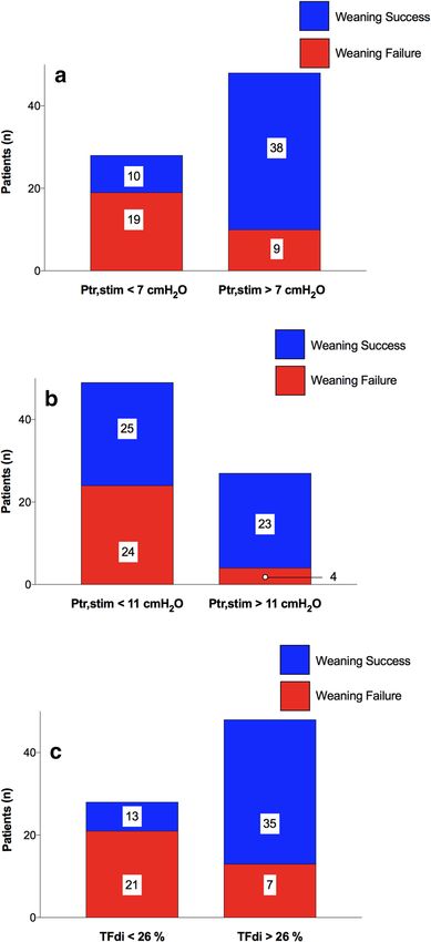
Fig. 2 Receiver operating characteristics curves of endotracheal
pressure induced by a bilateral phrenic nerve stimulation (Ptr,stim)
and diaphragm thickening fraction (TFdi) to predict failure of the
spontaneous breathing trial
Discussion
This study reports a dual assessment of diaphragm
function and its relationship with weaning outcome in
mechanically ventilated medical patients undergoing
a first spontaneous breathing trial. Our findings can be
summarized as follows: (1) a lower value of Ptr,stim (i.e.,
7.0 cmH2O) than the value commonly accepted value to
define diaphragm dysfunction (i.e., 11.0 cmH2O) is more
reliable to predict SBT failure, (2) Ptr,stim and TFdi are
equivalent to predict SBT failure.
Diaphragm function and weaning from mechanical
ventilation
The negative impact of diaphragm dysfunction on suc-
cessful weaning from mechanical ventilation has been
established by several investigations in critically ill
Fig. 1 Patients with successful spontaneous breathing trial and failed patients [3, 4, 11, 22]. At the time of weaning, diaphragm
spontaneous breathing trial according to 7 cmH2O (a) and 11 c mH2O dysfunction is highly prevalent [1] with reported rates
(b) thresholds of endotracheal pressure induced by a bilateral phrenic
ranging from 25–30% [11, 22] to 60–80% [3, 4]. To our
nerve stimulation (Ptr,stim) and 26% (c) threshold of diaphragm
thickening fraction (TFdi). Numbers indicate the number of patients knowledge, only three studies have assessed diaphragm
in each category dysfunction at the time of attempted liberation from
mechanical ventilation using the gold standard tech-
nique, namely the phrenic nerves stimulation [3, 4, 23].
SBT failure was 25.8%. Figure 1c shows the number of However, none of them provided any threshold values
patients with SBT success and SBT failure according to for Ptr,stim to predict weaning outcome. Of note, these
25.8%-TFdi threshold. Predictive performances of TFdi studies including ours indicate that a substantial propor-
are shown in Table 2. The comparison of AUC-ROC of tion of patients (up to 44%) can be successfully weaned
Ptr,stim and TFdi is displayed in Fig. 2. Ptr,stim and TFdi from the ventilator despite having diaphragm dysfunc-
had similar AUC-ROC (p = 0.83). tion defined as Ptr,stim < 11 cmH2O [3, 4]. Therefore,Dres et al. Ann. Intensive Care (2018) 8:53 Page 5 of 7
normal diaphragm function according to a definition Strengths and limitations of our study
established in healthy subjects [12] is not a prerequisite This study is the largest to report a dual approach pro-
for a successful SBT. This finding is not altogether sur- viding comparison between the gold standard evaluation
prising as many patients with chronic diaphragm dys- method of diaphragm function and diaphragm ultra-
function do not require mechanical ventilation [24, 25]. sound during the weaning phase. However, this study
While diaphragm dysfunction might limit exercise capac- has limitations. First, the generalizability of our findings
ity, the clinical consequences of diaphragm dysfunction may be limited by the characteristics of the patients of
in successfully liberated patients are uncertain. However, our cohort. Accordingly, our study might be viewed as a
the impact of respiratory muscles dysfunction (not spe- hypothesis generator and further trials are warranted to
cifically the diaphragm) after critical illness may be of confirm the clinical relevance of our findings. Second,
importance since it is associated with worse long-term while we obtained good inter- and intra-reproducibility
outcomes [26, 27]. Overall, our findings are of impor- in the measurements of diaphragm ultrasound, centers
tance since they highlight that presence of diaphragm employing the technique must also demonstrate ade-
dysfunction at the time of weaning should not discourage quate technical skill (based on reproducibility) before
clinicians from attempting liberation from ventilation. By implementing the technique for clinical purposes. Third,
contrast, not all patients (23/27) with a Ptr,stim higher we performed diaphragm ultrasound while patients were
than 11.0 cmH2O had a successfully SBT. As a matter of ventilated with pressure support and not during the SBT.
fact, the 11.0 cmH2O threshold of Ptr,stim was associated While this approach is easier to implement (no change in
with a lower specificity but a higher sensitivity than the ventilator setting) and less stressful for patients, it could
7.0 cmH2O threshold in the prediction of SBT failure. underestimate diaphragm thickening [33]. However, the
The lower 7.0 cmH2O Ptr,stim threshold provides the amount of pressure support was standardized in order
optimal combination of sensitivity and specificity in the to target a tidal volume between 6 and 8 ml/kg predicted
prediction of SBT failure. body weight. Reassuringly, any effect of ventilatory sup-
port on TFdi is likely to introduce ‘noise’ in its correla-
Diaphragm ultrasound in the prediction of SBT failure tion with weaning outcome and this would tend to bias
The use of diaphragm ultrasound is growing in the ICU the observed association toward the null. Fourth, we have
[28, 29]. It has many advantages over phrenic nerve assessed diaphragm function by using the changes in tra-
stimulation, which requires costly equipment and exten- cheal twitch pressure rather than the changes in trans-
sive technical expertise. Ultrasound is a noninvasive and diaphragmatic twitch pressure. This last measurement is
highly feasible bedside imaging modality, and ultrasound more specific to the diaphragm function but requires the
devices are widely available in ICUs. Several studies have placement of two balloons, which make it more invasive.
proposed various ultrasound-derived markers aiming at Although the two twitch pressures are not interchange-
assessing diaphragm function. Importantly, in our study, able, they are well correlated [15].
Ptr,stim and TFdi demonstrated similar performance in
the prediction of weaning. Of note, the optimal TFdi cut- Conclusions
off (26%) identified in our study is very close to the cutoffs Diaphragm ultrasound is a reliable surrogate of the
reported in previous investigations [16, 30, 31]. Consid- phrenic nerve stimulation method in the assessment of
ering ultrasound as a substitute of the phrenic nerves diaphragm function to predict weaning outcome. A mul-
stimulation technique, it will make diaphragm evaluation ticenter investigation is now required to confirm whether
much easier at the bedside. However, the indication of the 26% value of TFdi cutoff could or could not be used
diaphragm ultrasound during the weaning process is not widely to predict SBT outcome. Diaphragm ultrasound
yet clearly defined. In addition, it is important to remind could be combined with cardiac echo or lung ultrasound
that the majority of patients are shortly and safely sepa- to tailor post-extubation management according to the
rated from the ventilation. As it happens, the place of dia- risk of weaning failure. Although diaphragm dysfunction
phragm ultrasound might be viewed as a complementary did not systematically impair weaning outcome, it may
investigation and not as a surrogate of clinical judgment. behave as a marker of severity and poor prognosis. Future
It may be used as a screening tool to identify patients studies should address this hypothesis and investigate
who are at high risk of SBT failure (before conducting the mid- and long-term consequences of diaphragm dysfunc-
SBT) or as a diagnostic method to determine the cause of tion on patient functional status and quality of life.
SBT or extubation failure [32].Dres et al. Ann. Intensive Care (2018) 8:53 Page 6 of 7
Additional file de Paris, Paris, France and the Fondation pour la Recherche Médicale, Paris,
France (FDM 20150734498).
Additional file 1. Full description of the Methods. Figure E1. Flow chart
of the patients. Publisher’s Note
Springer Nature remains neutral with regard to jurisdictional claims in pub‑
lished maps and institutional affiliations.
Abbreviations
Received: 2 February 2018 Accepted: 16 April 2018
SBT: Spontaneous breathing trial; Ptr,stim: Tracheal pressure in response to
bilateral magnetic stimulation of the phrenic nerves; TFdi: Diaphragm thicken‑
ing fraction; ICU: Intensive care unit; AUC-ROC: Area under the curve of receiv‑
ing operating characteristics.
Authors’ contributions References
MD and AD designed the study. MD, AD, BPD and TS coordinated the study. 1. Dres M, Goligher EC, Heunks LMA, Brochard LJ. Critical illness-associated
MD, BPD, DR, EM, LD and JM were responsible for patient screening, enroll‑ diaphragm weakness. Intensive Care Med. 2017;43:1441–52.
ment, and follow-up. MD, AD, EG and TS analyzed the data. MD, AD, EG and TS 2. Demoule A, Jung B, Prodanovic H, Molinari N, Chanques G, Coirault C,
wrote the manuscript. All authors had full access to all study data, contributed et al. Diaphragm dysfunction on admission to the intensive care unit.
to drafting the manuscript or critical revision of it for important intellectual Prevalence, risk factors, and prognostic impact-a prospective study. Am J
content, approved the final version of the manuscript, and took responsibility Respir Crit Care Med. 2013;188:213–9.
for the integrity of the data and the accuracy of the data analysis. All authors 3. Dres M, Dubé B-P, Mayaux J, Delemazure J, Reuter D, Brochard L, et al.
read and approved the final manuscript. Coexistence and impact of limb muscle and diaphragm weakness at
time of liberation from mechanical ventilation in medical intensive care
Author details unit patients. Am J Respir Crit Care Med. 2017;195:57–66.
1
UPMC Univ Paris 06, INSERM, UMRS1158, Neurophysiologie Respiratoire 4. Jung B, Moury PH, Mahul M, de Jong A, Galia F, Prades A, et al. Diaphrag‑
Expérimentale et Clinique, Sorbonne Universités, Paris, France. 2 Service de matic dysfunction in patients with ICU-acquired weakness and its impact
Pneumologie et Réanimation Médicale (Département “R3S”), AP-HP, Groupe on extubation failure. Intensive Care Med. 2016;42:853–61.
Hospitalier Pitié-Salpêtrière Charles Foix, 47‑83 boulevard de l’Hôpital, 5. Jaber S, Petrof BJ, Jung B, Chanques G, Berthet J-P, Rabuel C, et al. Rapidly
75013 Paris, France. 3 Interdepartmental Division of Critical Care Medicine, progressive diaphragmatic weakness and injury during mechanical
University of Toronto, Toronto, Canada. 4 Division of Respirology, Department ventilation in humans. Am J Respir Crit Care Med. 2011;183:364–71.
of Medicine, University Health Network and Mount Sinai Hospital, Toronto, 6. Levine S, Nguyen T, Taylor N, Friscia ME, Budak MT, Rothenberg P, et al.
Canada. 5 Département de Médecine, Service de Pneumologie, Hôpital Rapid disuse atrophy of diaphragm fibers in mechanically ventilated
Hôtel‑Dieu, Centre Hospitalier de l’Université de Montréal (CHUM), Montréal, humans. N Engl J Med. 2008;358:1327–35.
QC, Canada. 7. Hermans G, Agten A, Testelmans D, Decramer M, Gayan-Ramirez
G. Increased duration of mechanical ventilation is associated with
Acknowledgements decreased diaphragmatic force: a prospective observational study. Crit
None. Care Lond Engl. 2010;14:R127.
8. Vassilakopoulos T, Petrof BJ. Ventilator-induced diaphragmatic dysfunc‑
Competing interests tion. Am J Respir Crit Care Med. 2004;169:336–41.
Alexandre Demoule has signed research contracts with Medtronic, Maquet 9. Heunks LMA, Doorduin J, van der Hoeven JG. Monitoring and preventing
and Philips; he has also received personal fees from Medtronic, Maquet, Res‑ diaphragm injury. Curr Opin Crit Care. 2015;21:34–41.
med, Fisher & Paykel and MSD. Martin Dres received personal fees from Pulsion 10. Supinski GS, Callahan LA. Diaphragm weakness in mechanically venti‑
Medical System and Lungpacer Inc. Bruno-Pierre Dubé has received honoraria lated critically ill patients. Crit Care Lond Engl. 2013;17:R120.
from GlaxoSmithKline, Boehringer Ingelheim, Astra Zeneca and Roche. Rel‑ 11. Kim WY, Suh HJ, Hong S-B, Koh Y, Lim C-M. Diaphragm dysfunction
evant to the present study, Thomas Similowski has received personal fees from assessed by ultrasonography: influence on weaning from mechanical
Lungpacer Inc and is a member of the board of a research association that has ventilation. Crit Care Med. 2011;39:2627–30.
received, over the past 10 years, unrestricted research grants from Maquet, 12. American Thoracic Society/European Respiratory Society. ATS/ERS
Hamilton, Covidien, and Philips; he is the head of a research unit (UMRS 1158) statement on respiratory muscle testing. Am J Respir Crit Care Med.
that has signed research contracts with Air Liquide Medical Systems, France; 2002;166:518–624.
he is listed as inventor or co-inventor on several patents, granted or pending, 13. Mills GH, Kyroussis D, Hamnegard CH, Polkey MI, Green M, Moxham J.
describing a brain–ventilator interface. The other authors have no conflict of Bilateral magnetic stimulation of the phrenic nerves from an anterolateral
interest relevant to this study. approach. Am J Respir Crit Care Med. 1996;154:1099–105.
14. Mills GH, Ponte J, Hamnegard CH, Kyroussis D, Polkey MI, Moxham J,
Availability of data and materials et al. Tracheal tube pressure change during magnetic stimulation of the
The datasets used and/or analyzed during the current study are available from phrenic nerves as an indicator of diaphragm strength on the intensive
the corresponding author on reasonable request. care unit. Br J Anaesth. 2001;87:876–84.
15. Watson AC, Hughes PD, Louise Harris M, Hart N, Ware RJ, Wendon J,
Consent for publication et al. Measurement of twitch transdiaphragmatic, esophageal, and
Patients or their next of kin gave informed consent. endotracheal tube pressure with bilateral anterolateral magnetic phrenic
nerve stimulation in patients in the intensive care unit. Crit Care Med.
Ethics approval and consent to participate 2001;29:1325–31.
Human research ethics committee approval for the study was provided by the 16. DiNino E, Gartman EJ, Sethi JM, McCool FD. Diaphragm ultrasound as a
Comité de Protection des Personnes—Ile de France 6. predictor of successful extubation from mechanical ventilation. Thorax.
2014;69:423–7.
Funding 17. Dubé B-P, Dres M, Mayaux J, Demiri S, Similowski T, Demoule A. Ultra‑
M.D. was supported by the French Intensive Care Society, Paris, France (bourse sound evaluation of diaphragm function in mechanically ventilated
de mobilité 2015); The 2015 Short Term Fellowship program of the European patients: comparison to phrenic stimulation and prognostic implications.
Respiratory Society, Lausanne, Switzerland; The 2015 Bernhard Dräger Award Thorax. 2017;72:811–8.
for advanced treatment of acute respiratory failure of the European Society of
Intensive Care Medicine, Brussels, Belgium; the Assistance Publique HôpitauxDres et al. Ann. Intensive Care (2018) 8:53 Page 7 of 7
18. Boles J-M, Bion J, Connors A, Herridge M, Marsh B, Melot C, et al. Weaning 27. Medrinal C, Prieur G, Frenoy É, Robledo Quesada A, Poncet A, Bonnevie
from mechanical ventilation. Eur Respir J Off J Eur Soc Clin Respir Physiol. T, et al. Respiratory weakness after mechanical ventilation is associated
2007;29:1033–56. with one-year mortality—a prospective study. Crit Care Lond Engl.
19. Similowski T, Yan S, Gauthier AP, Macklem PT, Bellemare F. Contractile 2016;20:231.
properties of the human diaphragm during chronic hyperinflation. N 28. Zambon M, Greco M, Bocchino S, Cabrini L, Beccaria PF, Zangrillo A.
Engl J Med. 1991;325:917–23. Assessment of diaphragmatic dysfunction in the critically ill patient with
20. Bossuyt PM, Cohen JF, Gatsonis CA, Korevaar DA, STARD group. STARD. ultrasound: a systematic review. Intensive Care Med. 2017;43:29–38.
updated reporting guidelines for all diagnostic accuracy studies. Ann 29. Haaksma M, Tuinman PR, Heunks L. Ultrasound to assess diaphrag‑
Transl Med. 2015;2016(4):85. matic function in the critically ill-a critical perspective. Ann Transl Med.
21. DeLong ER, DeLong DM, Clarke-Pearson DL. Comparing the areas under 2017;5:114.
two or more correlated receiver operating characteristic curves: a non‑ 30. Ferrari G, De Filippi G, Elia F, Panero F, Volpicelli G, Aprà F. Diaphragm ultra‑
parametric approach. Biometrics. 1988;44:837–45. sound as a new index of discontinuation from mechanical ventilation.
22. Jiang J-R, Tsai T-H, Jerng J-S, Yu C-J, Wu H-D, Yang P-C. Ultrasonographic Crit Ultrasound J. 2014;6:8.
evaluation of liver/spleen movements and extubation outcome. Chest. 31. Dres M, Demoule A. Diaphragm dysfunction during weaning from
2004;126:179–85. mechanical ventilation: an underestimated phenomenon with clinical
23. Laghi F, Cattapan SE, Jubran A, Parthasarathy S, Warshawsky P, Choi implications. Crit Care Lond Engl. 2018;22:73.
Y-SA, et al. Is weaning failure caused by low-frequency fatigue of the 32. Mayo P, Volpicelli G, Lerolle N, Schreiber A, Doelken P, Vieillard-Baron
diaphragm? Am J Respir Crit Care Med. 2003;167:120–7. A. Ultrasonography evaluation during the weaning process: the
24. Manders E, Bonta PI, Kloek JJ, Symersky P, Bogaard H-J, Hooijman PE, heart, the diaphragm, the pleura and the lung. Intensive Care Med.
et al. Reduced force of diaphragm muscle fibers in patients with chronic 2016;42:1107–17.
thromboembolic pulmonary hypertension. Am J Physiol Lung Cell Mol 33. Blumhof S, Wheeler D, Thomas K, McCool FD, Mora J. Change in diaphrag‑
Physiol. 2016;311:L20–8. matic thickness during the respiratory cycle predicts extubation success
25. Kelley RC, Ferreira LF. Diaphragm abnormalities in heart failure and aging: at various levels of pressure support ventilation. Lung. 2016;194:519–25.
mechanisms and integration of cardiovascular and respiratory patho‑
physiology. Heart Fail Rev. 2017;22:191–207.
26. Adler D, Dupuis-Lozeron E, Richard J-C, Janssens J-P, Brochard L. Does
inspiratory muscle dysfunction predict readmission after intensive care
unit discharge? Am J Respir Crit Care Med. 2014;190:347–50.You can also read