Corneal dystrophy in a cocker spaniel dog: a case report - CAB
←
→
Page content transcription
If your browser does not render page correctly, please read the page content below
APMB - Atti della Accademia Peloritana dei Pericolanti                                                                       ISSN 1828-6550
       Classe di Scienze Medico Biologiche
                  Vol. 105(2) 2017
                                                                                            DOI: 10.6092 / 1828-6550 / APMB.105.2.2017.A7

Clinical Case Seminar                                                                                 A7(1-5)

Corneal dystrophy in a cocker spaniel dog: a case report.
Monica Ragusa1a, Michela Pugliese1b, Domenico Britti2, Ernesto Palma2,
Antonio Pugliese1b
1a
 Scuola di Specializzazione in "Fisiopatologia della Riproduzione degli Animali Domestici",
1b
 Dipartimento di Scienze Veterinarie Università degli Studi di Messina, 2Dipartimento di Scienze
della Salute Università di Catanzaro

Abstract
A 1-year-old female Cocker Spaniel dog was examined at the ophthalmology service of the Veterinary
Teaching Hospital - University of Messina (Italy) for evaluation of symmetrical white spots in both
corneas and “red eyes”. Dog was clinically healthy, haematological and biochemical examination were
unremarkable, Leishmania PCR was negative. After a complete ophthalmic examination, the clinical
diagnosis was corneal stromal dystrophy with uveitis-induced. In dogs, corneal stromal dystrophy is a
primary, inherited, bilateral opacity of the corneanot associated with ocular inflammation or systemic
disease. Detailed description of corneal dystrophy are available only for few breeds. This lesion is not
progressive and treatment is not usually recommended unless vision is impaired or the deposits become
irritating.

KEYWORDS: cornea, dystrophy, inherited, eye, dog

IntroducingMember: Antonio Pugliese
Corresponding Author: Monica Ragusa, m.ragusa@hotmail.com

Introduction
In dogs, corneal dystrophy is an inherited, non-inflammatory, bilateral, symmetrical condition not
associated to systemic disease.1-3. Corneal stromal dystrophy is a stromal metabolic defect that
result in accumulation of extracellular and intracellular lipid in dog between 6 months and 5
years of age.4-9In most breeds, corneal dystrophy appears as gray-white, crystalline or metallic
deposits in central or paracentral cornea. The opacities involving anterior stroma are usually oval
or round,sometimes doughnut-shaped, mostly progressive.1,2. Their progression may be very
slow and may or may not lead to blindness (e.g. Cocker Spaniels, Poodles, Samoyeds, and
Bichon Frises) or may be rapid and lead to blindness (e.g. Airedale Terriers, Boston Terriers,
Chihuahuas and Dachshunds). The mode of inheritance varies among breeds and is unknown in
many breeds.

              APMB - Atti della Accademia Peloritana dei Pericolanti - Classe di Scienze Medico-Biologiche (2017), 105(2):A7(1-5)
                                             DOI: 10.6092 / 1828-6550 / APMB.105.2.2017.A7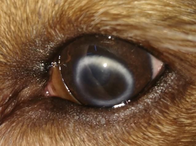
APMB - Atti della Accademia Peloritana dei Pericolanti ISSN 1828-6550
Classe di Scienze Medico Biologiche
Vol. 105(2) 2017
DOI: 10.6092 / 1828-6550 / APMB.105.2.2017.A7
Case Report
A 1.3 year-old female English Cocker Spaniel was examined at the Veterinary Teaching Hospital
of University of Messina for “red eyes” and white spots in both corneas appearing from about a
year.
The dog was previously treated with dexamethasone eyedrops q12h for 7days and cyclosporine
ophthalmic ointment applied twice a day on both eyes(OU). Cyclosporine was interrupted after
few days, for local side effects (hyperemia, corneal irritation).
At the presentation, the dog was in good health conditions clinically healthy. Haematological and
biochemical exams were within normal ranges, Leishmania PCR was negative.Vision
assessment and neuro-ophthalmic examination were normal. Eye exam showed conjunctival
hyperemia, epiphora, and no eyelid anomalies. Ring corneal opacities, composed by a myriad of
fine, white and small particles in axial portion of the cornea in right eye(OD) were located. In left
eye(OS), an arc-shaped opacity of the same density as OD was evident in the subepithelial and
posterior stroma of the axial cornea. The epithelium was intact and no neovascularization was
observed(OU). Schirmer Tear Tests were 22mm/min(OU). Fluorescein dye test was
negative(OU). Light aqueous flare was present(OU). Intra-ocular pressure measured with
Tonovet® was 8mmHg(OD) and 6mmHg(OS). Any anomalies were present in lens, vitreous and
fundus.
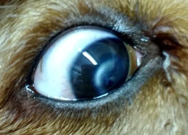
Figure 1. Right eye at the presentation. Arc-shaped Figure 2. Left eye at the presentation. Donught-shaped opacities
opacities in the corneal stroma and conjunctival in the corneal stroma and conjunctival hyperemia.
hyperemia
APMB - Atti della Accademia Peloritana dei Pericolanti - Classe di Scienze Medico-Biologiche (2017), 105(2):A7(1-5)
DOI: 10.6092 / 1828-6550 / APMB.105.2.2017.A7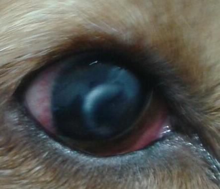
APMB - Atti della Accademia Peloritana dei Pericolanti                                                                       ISSN 1828-6550
       Classe di Scienze Medico Biologiche
                  Vol. 105(2) 2017
                                                                                            DOI: 10.6092 / 1828-6550 / APMB.105.2.2017.A7

Figure 3. Right eye post-treatment                                      Figure 4. Left eye post-treatment.

Final diagnosis was corneal stromal dystrophy with uveitis-induced. A treatment with both oral
Prednisone 1mg/kg/twice a day and topical Dexamethasone 1 drop/four times a day was
prescribed to resolve the uveitis and relieved eye discomfort. Atropine eye drop 1% was topically
applied to decrease ciliary spasm. Furthermore, EDTA in ocular solution was applied in OU q6h.
After a month, eye discomfort and inflammation were resolved and IOP were 13mmHg(OS) and
14mmHg(OD).

Discussion.
The diagnosis of corneal stromal dystrophy was formuled on signaling and clinical
manifestations. Differential diagnoses includednot inherited or familial, non-symmetrical, and
not necessarily bilateral ocular diseases: corneal fibrosis, corneal degeneration, and lipid
keratopathies. No alterations on blood examinations suggested the presence of systemic diseases
that could be related to corneal opacities.
No tear or eyelid abnormalities were present and the corneas were not irritated.
The lesion found in this case report was suggestiveof Schnyder's crystalline stromal dystrophy.In
crystalline stromal dystrophy inflammatory element and no vascularization are present. The
dystrophy is associated with accumulation of lipid within the corneal fibroblasts, but typical foam
cells are absent, the crystalline opacity involves the coolest part of the cornea, correlates with
local fibroblast death, and is always bilateral. This condition is described in both the Cavalier
King Charles Spaniel and Rough Collie8 and not was reported in English Cocker Spaniel In this
breed, two different kind of corneal dystrophy has been decribed only two different type of
corneal dystrophy:
- epithelial/stromal: non-inflammatory corneal opacity (white to gray) present in one or more of
the corneal layers;

              APMB - Atti della Accademia Peloritana dei Pericolanti - Classe di Scienze Medico-Biologiche (2017), 105(2):A7(1-5)
                                             DOI: 10.6092 / 1828-6550 / APMB.105.2.2017.A7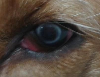
APMB - Atti della Accademia Peloritana dei Pericolanti                                                                       ISSN 1828-6550
       Classe di Scienze Medico Biologiche
                  Vol. 105(2) 2017
                                                                                            DOI: 10.6092 / 1828-6550 / APMB.105.2.2017.A7

- posterior polymorphous: multifocal, non-pigmented, vesicular to linear posterior corneal
opacities at the level of endothelium. The condition affecting dogs from 1 to 7 y.o. is bilateral
and differs from endothelial dystrophy for absence of corneal edema.10,11
Corneal dystrophy is not primarily associated with corneal vascularization; however, with
chronicity, the lipid accumulation may cause cell death and induce inflammation with subsequent
development of corneal vascularization.12
In this case report, the lipid accumulation in subepithelial layer may have caused the painfull and
a subsequent secondary anterior uveitis.
The prognosis for corneal stromal dystrophies is excellent. They do not tend to cause visual
disturbance and they are usually not associated with ocular pain.13
No medication "dissolves" the opacity and surgery is not usually recommended. The lesions can
be removed by keratectomy if the opacity is obstructing vision significantly. Often, the opacities
will reform in the healed cornea.1,14
Reduction of opacities has been reported with topical cyclosporine and tacrolimus.15In this case
report an initial topical treatment with cyclosporine was performed, but discontinued for ocular
discomfort. The arc and donught-shaped opacities did not impair the vision, the rest of the cornea
was transparent in both eye and prognosis for this dog is good for the function.
Topical EDTA, in artificial tears may be effective in removing calcium,3,16,17reducing mineral
accumulation and facilitating re-epithelialization16 in corneal degeneration. In this case, this
therapy was added to the treatment without significant amelioration of opacities.

Conflicts of Interest: There is no potential conflict of interest, and the authors have nothing to disclose.
This work was not supported by any grant.

References

1. Cooley, P.L., Dice, P.F(1990) Corneal dystrophy in the dog and cat. Vet Clin North Am 20:681-692.
2. Crispin, S.M.(1988) Crystalline corneal dystrphy in the dog: histochemical and ultrastructural study.
Cornea,7,149-161.
3. Crispin, S.M., Barnett,C.(1983) Dystrophy, degeneration and infiltration of the canine cornea. J Small
AnimPract; 24:63–83
4. Elkins, M., Warring, G.O., Harris, R.R. (1980) Oval lipid corneal opacities in beagles part II. J Am
AnimHosp Ass16,601-605.
5. MacMillan, A., Warring, G.O., Spangler, W.L., Roth, A.M. (1979) Crystalline corneal opacities in the
Siberian husky. J Am Vet Med Ass. 175,829-832.
6. Morrin, A., Warring, G.O., Spangler, W.L.,(1982) Oval lipid corneal opacities in Beagles:
ultrastructure of the abnormal Beagle cornea. Am J Vet Resear, 43,443-453
7. Rubin, L.F.(1989) Inherited Eye Diseases in Purebred Dogs. Baltimore, MD: Willams and Wilkins.
8. Spangler, W.L., Warring, G.O., Morrin, A.(1982) Ovallipid corneal opacities in Beagles: V.
Ultrastructure. Vet Comp Ophthalmol19,150-159.
9. Warring, G.O., MacMillan, A., Reveles, O.(1986) Inheritance of crystalline cernealdystrophy in
Siberian Huskies. J Am AnimHospit Ass, 22, 655-658.
              APMB - Atti della Accademia Peloritana dei Pericolanti - Classe di Scienze Medico-Biologiche (2017), 105(2):A7(1-5)
                                             DOI: 10.6092 / 1828-6550 / APMB.105.2.2017.A7
APMB - Atti della Accademia Peloritana dei Pericolanti ISSN 1828-6550
Classe di Scienze Medico Biologiche
Vol. 105(2) 2017
DOI: 10.6092 / 1828-6550 / APMB.105.2.2017.A7
10. ACVO Genetics Commitee 2014
11. Gwin, R. M., Cunningham D. E., Shaver R. P. (1983) Posterior polymorphous dystrophy of
the cornea in Cocker Spaniel: preliminary clinical & specular microscopic findings. Trans. Am.
Coll. Vet. Ophthalmol. 14, 154-164.
12. Crispin, S.M. (1987) Lipid keropathy in the dog. In : The Veterinary Annual ( Grunsel, C.S.G., Hill,
F.W.G. &Raw, M.E.), 27th issue, pp. 196-208.Bristol: John Wright and Sons.
13. Summers,J.F, O’Neill, D.G., Church, D.B., Thomson, P.C., McGreevy P.D., Brodbelt D.C. (2015)
Prevalence of disorders recorded in Cavalier King Charles Spaniel attending primary-care veterinary
practices in England. Canine Genetics and Epidemiology;2:4.
14. Whitely, D.(1991) Canine cornea. In. Gelatt KN, editor. Vet Ophthalmol 2nd ed. Pages 307-356
15. Balicki, I., Trbolova, A. (2010),Clinical evaluation of tacrolimus eye drops for chronic superficial
keratitis treatment in dogs.bull vet instpulawy 54, 251-258
16. Whitley R.D., GilgerB.C.(1999) Diseases of the canine cornea and sclera. In:VetOphthalmol, 3rd edn.
(ed. Gelatt KN) Lippincott Williams & Wilkins, Philadelphia; 655–663;
17. Glaze M.B.(1997) Ophthalmic disease and its management. Vet Clin North Am: Small AnimPract;
27:1505–152
©2017 by the Author(s); licensee Accademia Peloritana dei Pericolanti (Messina, Italy). This article is an open
access article distributed under the terms and conditions of the Creative Commons Attribution 4.0
International License (https://creativecommons.org/licenses/by/4.0/).
Communicated and received November 15, 2017, Revised November 22, published on line December 20, 2017
APMB - Atti della Accademia Peloritana dei Pericolanti - Classe di Scienze Medico-Biologiche (2017), 105(2):A7(1-5)
DOI: 10.6092 / 1828-6550 / APMB.105.2.2017.A7You can also read