OCULUS Pentacam Pentacam HR - The Gold Standard in Anterior Segment Tomography
←
→
Page content transcription
If your browser does not render page correctly, please read the page content below

OCULUS Pentacam®
       Pentacam® HR
              The Gold Standard in
              Anterior Segment Tomography

   We focus on progress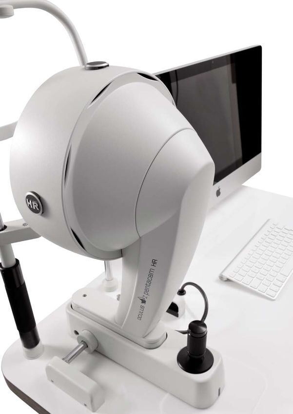
Providing the Best Care                                                                       Establish the basis for long-
to Your Patients                                                                              term relationships
> Sight is the most important sense.                                                          with your patients.
    Loss or impairment of sight is something your patients
    would rather not even imagine.

             > Fast, non-contact measurements will find
               favour with your patients.
                   Establish good doctor-patient relationships by showing your patients the
                                                                                              Glaucoma screening
                   intuitive Pentacam® displays and discussing treatment options with them.
                                                                                              I went to my eye doctor to get contact lenses. A
                                                                                              quick Pentacam® exam revealed very small anterior
                                                                                              chamber angles and showed that I was a glaucoma
> Your staff will appreciate the ease of use and seamless                                     risk patient. According to my doctor this early
  connectivity to the EMR System.                                                             detection was crucial in managing the condition
                                                                                              and preserving my eyesight!

             > The OCULUS Pentacam® wins patient trust.

                                                                                              Cataract surgery                                    Refractive surgery
                                                                                              Following a Pentacam® examination my doctor         I was tired of wearing eye glasses and went to
                                                                                              used the Pentacam® images on his iPad® and          a refractive surgeon to explore the options for
                                                                                              showed me my cloudy crystalline lens. I was         refractive surgery. A Pentacam® exam confirmed
                                                                                              surprised that I could see anything at all! My      that I was a good candidate for LASIK. My doctor
                                                                                              doctor used the Pentacam® information to select     used my Pentacam® images and explained the
                                                                                              and recommend a premium IOL. I am delighted         surgery in detail. I was happy with the competent
                                                                                              with the result of my cataract surgery and          consultation and careful evaluation prior to
                                                                                              recommend my doctor to all my friends.              surgery.
Pentacam® Pentacam® HR
The Affordable Model The Professional Model
The Pentacam® measures the cornea from limbus to limbus. It The Pentacam® HR is our high resolution model, measuring up to
measures up to 25,000 true elevation points. During the rotating 138,000 true elevation points. It is equipped with a high resolution
scan that takes max. 2 seconds, up to 50 Scheimpflug images of CCD chip and optimized optics.
the anterior eye segment are captured with outstanding precision.
Based on these images the Pentacam® provides topographic data While performing a rotating scan the Pentcam® HR captures up
on elevation and curvature of the entire anterior and posterior to 100 high-resolution Scheimpflug images to provide a detailed
corneal surfaces. The corneal thickness (pachymetry) is measured analysis of the cornea. IOLs and pIOLs, corneal rings, corneal
and presented graphically over its entire surface. A topography injuries and corneal opacities such as in Fuchs dystrophy are
based keratoconus detection and quantification are performed. The thus brought into view in brilliant detail. The crystalline lens is
anterior chamber depth, chamber volume (size) and the chamber displayed from anterior to posterior capsule, even with opacities
angles are automatically calculated and presented for glaucoma present.
screening. The illumination of the eye using blue LED light makes
corneal and lens opacities (cataract) visible. The anterior chamber The Pentacam® HR is optionally available with a unique 3D pIOL
can be visualised and displayed with the virtual tomography model. simulation software including aging prediction.
After measurements have been taken and the quality specifications
of the scan have been checked by the user the Pentacam® can
generate an Indices Report. This Indices Report summarises the
abnormalities found during the scan to direct doctor’s attention
to the areas of concern. This report is based on published clinical
studies and articles that define abnormalities.
The standard model is affordable and is suitable for pre-exam
lanes, satellite offices and for quick screenings.
Basic Software: This is What Makes the HR Special:
Indices Report to detect abnormalities in the anterior Now with a new High resolution CCD camera and high end optical design Now with a new
eye segment iris camera lens system for to generate brilliant Scheimpflug images to display iris camera lens system for
for glaucoma screening automatic HWTW measurement. corneal opacities, corneal implants and rings automatic HWTW measurement.
for refractive screening IOLs and pIOLs in terms of position and orientation
General Overview Display PCO (posterior capsular opacification)
4 Maps Refractive Special scan mode to display pIOLs
Qualitative assessment of the cornea Precise measurement of the cornea with up to
Topography and elevation maps of the anterior and 100 Scheimpflug images
posterior corneal surface Movable fixation target
Overall pachymetry, absolute and relative 3D pIOL simulation software including aging prediction
Iris camera and automatic HWTW
Topography based keratoconus detection and classification
3D anterior chamber analysis
Comparison and differential analysis of 2 exams
Comparison and superimposition of Scheimpflug images
Overview of all captured Scheimpflug images
Anterior segment tomography
These functions are unique and are only available
with the OCULUS Pentacam® 
Data Network An efficient practice means
more time for your patients.
The Pentacam® comes with a free patient data management software
The front desk staff checks in the
Patient Check-in
Local Network
that ensures increased efficiency and networking capabilities. patient and enters the patient‘s personal
information into the OCULUS Patient Data Patient
Management (PDM) or the EMR system. data
EMR Compatibility DICOM Compatibility (Optional) PDM: Patient Data Management
The Pentacam® software is compatible with many The Pentacam® software is fully DICOM compatible. The
commercially available EMR systems. This allows a quick Pentacam® software receives information from the DICOM
and paperless transfer of examination results and reports Modality Work List (MWL) from the Hospital Information
Select the patient‘s name in the PDM.
Pre-examination
to all workstations. System (HIS) and transmits the results to the Picture
Archiving and Communication System (PACS) for storage Explain the testing procedure to the
and further evaluation. patient and perform the required Measurement
examinations. The results will be saved data
Centerfield® 2 PARK 1® Pentacam®
and available on every workstation
connected to the server.
Software Licenses on a
Keratograph 5M Corvis® ST
Floating License Key
Server
The Pentacam® results can be displayed as static pages
The physician reviews the pre-examination
Examination
on workstations in a network. To have full functionality
of the Pentacam® software and to be able to interactively data. If an additional slit lamp examination
work with it, additional analysing software options may is performed, it can be recorded and
be required. documented with the ImageCam® 2.
ImageCam® 2
The new OCULUS Floating License Key is designed to work
in a local network and is plugged into the central server. It Pre examination
is equipped with the basic software and provides parallel data and additional
access to the Pentacam® basic software results from data
every workstation. The Floating License Key can support
multiple instruments such as Pentacam®, Keratograph 5M
and other OCULUS devices simultaneously. In addition
to the basic software other optional software modules Patient Consultation Patient consultation sessions can be
can be purchased and added to the floating license key. optimised. The scans can be interactively
This allows viewing the additional software features on assessed and images can be shown to the
parallel workstations. For example, the Holladay Report patient on the monitor or the iPad®.
can be used on 2 and the Belin/Ambrósio Enhanced Ectasia
software on three workstations. “A picture is worth a thousand words.”
Complete patient
PDM: Patient Data Management information and
examination data
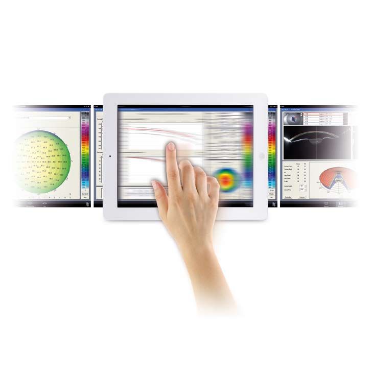
Cutting Edge Technology
The Free Pentacam® iPad® App
Modern, Easy and Intuitive
Optional iPad®
The invisible WIFI access point for the iPad connection
®
is integrated in the OCULUS lifting tables. OCULUS sets new standards in patient consultation and
information. A free Pentacam® Viewer App allows easy
transfer of Pentacam® exams to an iPad®.
The Comprehensive Package
The Pentacam® Viewer App
Show your patients how you utilise cutting-edge
technology in your practice. Download the free
Pentacam® Viewer App from the Apple® store
to your iPad®.
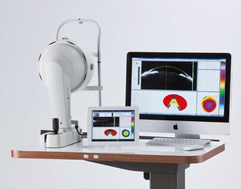
Sleek and Ergonomic Design Illuminated Up/Down Buttons
The iMac stand is attached to the table and the monitor Illuminated “Up” and “Down” keys allow easy control of
can swivel, providing a shapely, functional solution with the lift table even when the Pentacam® is operated in a
no more tangled cables. dark or dimmed room.
Clinical Applications
The OCULUS Pentacam® offers many clinical
applications for anterior segment analysis.

                                                                                                                              > Glaucoma Screening                                          > Cornea
General Screening
                                                                                                                              The Pentacam® provides comprehensive, complete                The Pentacam® stands out by virtue of its high-resolution
The Indices Display combined with the brilliant Scheimpflug images permit intuitive screening for close angle glaucoma in
                                                                                                                              and automatic analysis of the anterior chamber. The           images. Surgical success is documented completely from
the anterior segment. Treatment follow-up, documentation and patient education are supported qualitatively as well as
                                                                                                                              instrument displays whether the patient has an increased      start to finish. Even the smallest irregularities in the
quantitatively by various comparison displays.
                                                                                                                              risk of glaucoma. It shows alterations of the anterior        healing process are detected early.
                                                                                                                              chamber after, for example, an iridectomy or other
                                                                                                                              surgical interventions.
Refractive Screening and Surgery
Refractive screening and early detection of corneal abnormalities can be performed quickly and intuitively with the Belin/
Ambrósio Enhanced Ectasia Display. The OCULUS Pentacam® can help plan for corneal refractive surgery and corneal ring
implantation.

Cataract Screening, IOL Power Calculation, Premium IOL Selection
The opacity of the crystalline lens is measured quantitatively and can be shown to patients. The Cataract Pre-Op Display
helps in selecting the most suitable premium IOL. Various links to modern IOL power calculators such as Phaco Optics,
OKULIX or the BESSt II formula are available to assist in challenging cases of post-refractive IOL power calculation and
improve the outcome of the toric IOL procedures. The keratometry overlay in the iris image helps with the orientation of
toric IOLs.

Cataract Refractive
Planning cataract refractive surgery with phakic IOL implantation becomes a routine process with the OCULUS Pentacam®.        > Cataract                                                    > Cataract Refractive
A special simulation software including aging prediction is available for iris supported phakic IOLs (pIOLs). It visualises
the fit of the pIOL after surgery. The OCULUS Pentacam® provides all necessary information for pre-op planning for ICL
                                                                                                                              The Scheimpflug images produced by the Pentacam® offer        The Pentacam® data helps the doctor decide whether the
implants.
                                                                                                                              a clear representation of lens opacity. The 3D cataract       patient is a suitable candidate for a certain procedure,
                                                                                                                              analysis combined with PNS (Pentacam® Nucleus Staging)        such as premium IOL implantation. The Pentacam® images
                                                                                                                              is a unique feature. The centre of the cornea and its         can be used as a patient education tool to explain the
                                                                                                                              anterior and posterior surfaces are measured precisely        condition and the surgical procedure. The Pentacam®
                                                                                                                              for optimal calculation of corneal refractive power. This     simulates the position of iris fixated phakic IOLs in 3D.
                                                                                                                              translates into better IOL power calculation. The Pentacam®   Moreover, the Pentacam® simulates the future position of
                                                                                                                              supports the selection of the optimal premium IOL for the     the pIOL.
                                                                                                                              respective patient. All measurements are performed with
                                                                                                                              the same high precision device and during the same eye
                                                                                                                              examination.

10                                                                                                                                                                                                                                                  11
Brilliant Images

                                                                                                                > Corneal inlay for presbyopia correction                > Endothelian detachment after Descemet Membrane
                                                                                                                                                                           Endothelium Keratoplasty (DMEK)

                                                                                                                > High keratoconus causing an extreme central thinning   > Cornea with Fuchs’ endothelial dystrophy

                                                                                                                > Pterygium – beginning growth of the conjunctiva        > Descemet stripping and automated endothelium
                                                                                                                                                                           keratoplasty (DSAEK) after Fuchs’ endothelial dystrophy

                                    Enhance your diagnosis
       The real images of the anterior segment obtained with the Pentacam® can enhance your diagnosis.
     These images provide valuable insight into the condition of the cornea, iris, anterior chamber and the
      crystalline lens or intraocular lense. It is easy to detect narrow chamber angles or early cataracts by
      looking at these high resolution images. The best part is that these images are documented and can
            be shown to the patient to describe the medical findings and discuss treatment options.

                                                                                                                > Penetrating keratoplasty – corneal transplant after    > Pellucid marginal degeneration – an inferior band of
                                                                                                                  Fuchs’ endothelial dystrophy                             corneal thinning

12                                                                                                                                                                                                                                13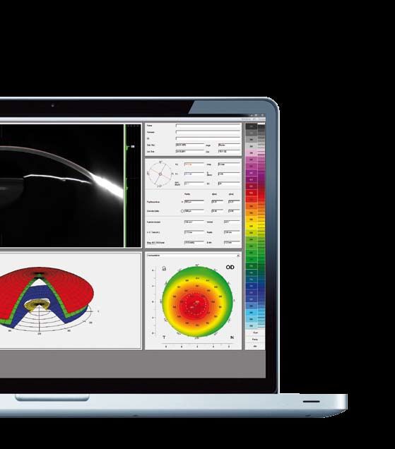
Basic Software
Discover the Possibilities
Indices Display General Overview
Applications: Applications:
Quick screening of new patients Comprehensive clinical overview
Detection of abnormalities Patient information
Intuitive guide through the Pentacam® / Pentacam® HR
software Details:
The Overview Display provides important clinical
Details: information on the anterior eye segment.
Important parameters of the anterior eye segment The Scheimpflug image provides physician and patient
that are displayed with the Pentacam® / Pentacam® HR an intuitive representation of opacities of the cornea and
software are analysed and validated in published papers lens (cataract) or of the position of an IOL. The corneal
and articles. Their normal (green) and pathological (red) keratometry, pachymetry and asphericity are displayed.
distribution is displayed in diagrams. This presentation The anterior chamber is described in terms of anterior
provides an intuitive, quick but comprehensive overview. chamber depth, volume and angle. When combined
The literature source used is also mentioned, permitting with tonometry readings the IOP can be corrected with
detailed reference to corneal thickness to permit an assessment
of possible glaucoma risks. It also allows the display of
Based on individual results the software recommends all colour maps.
further displays to review additional details.
These functions are unique and are only available
14 with the OCULUS Pentacam® 15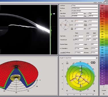
Basic Software
Discover the Possibilities
Anterior Segment Tomography 3D Anterior Chamber Analysis Topography Maps of the Anterior Pachymetry Maps
and Posterior Corneal Surface
Applications: Applications: Applications: Applications:
Fast overview of gathered data Glaucoma screening Keratoconus detection Pre-surgical planning of corneal refractive surgery
Changes which become visible in the Scheimpflug Pre- to post-operative comparison of changes in Pre-surgical planning of corneal refractive surgery Absolute and relative presentation
image are represented amazingly well in a 3D model. anterior chamber, e.g. after iridectomy Follow-up after corneal surgery Glaucoma screening
Tomographic representation, virtual model of anterior Calculation of IOL refractive power Relative pachymetry map for early keratoconus
Details: segment Planning of astigmatism of limbal relaxing incisions (LRI) detection
The rotatable and movable 3D model of the anterior Automatic calculation of Follow-up after refractive surgery IOP correction taking into account measured corneal
eye segment proves an enormous help in patient Anterior chamber angles (ACA) Pre-post LASIK, PRK, PKP, LKP, DSEK thickness based on various correction formulas
education. The patient can see his or her eye from all Anterior chamber volume (ACV) (for e.g. Ehler, Shah, Dresden etc.)
sides. Irregularities and scars can be explained easily. Internal or external anterior chamber depth (ACD) Details:
The rotating measurement principle guarantees high Details:
Details: resolution of the measuring points in the central cornea. An overview representation in colour shows the corneal
The high resolution images and the 3D Anterior Chamber Topographic analysis of the anterior and posterior corneal thickness from limbus to limbus. The measured values
Analysis are used to assess the anterior segment for surfaces is based on measured real height data. These can be displayed in a pre-determined grid or represented
abnormalities. This information can be easily shared provide the basis for: manually at any point via mouse click.
with the patient to explain the condition and discuss Sagittal (axial), tangential (local) curvature maps, Automatic representation of:
treatment options. refractive power maps of the anterior and posterior Corneal thickness in the centre of the pupil
corneal surface Corneal thickness in the apex
Elevation maps of the anterior and posterior The thinnest point of the cornea
corneal surface
Four colour coded maps display for refractive
assessment
Topometric display for detailed corneal shape
assessment including true net power
Topography based keratoconus detection and
classification
Comparison and differential displays
These functions are unique and are only available
16 with the OCULUS Pentacam® 17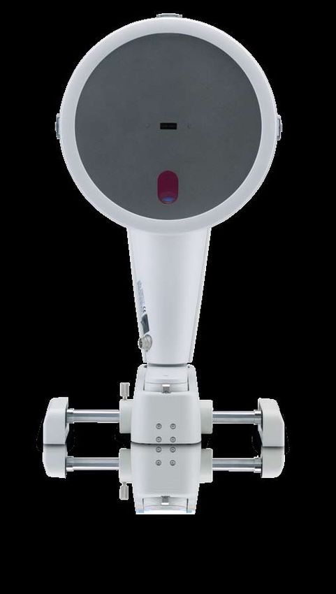
Basic Software
Discover the Possibilities
Topometric Iris Camera Comparison and Differential Comparison and Superimposition
Analysis of 2 Exams of Scheimpflug Images
Applications: Applications: Application: Applications:
Corneal shape assessment Overview image of the iris to recognise landmarks Objective changes and progression analysis Comparison of the Scheimpflug images of two
Detection and classification of corneal abnormalities Automatic determination of the corneal diameter different exams
(HWTW) Details: Visualisation of changes after surgical intervention
Details: Determination of pupil location and shape Two exams can be loaded and analysed. The difference Patient education
The early detection and follow-up of corneal shape between the two loaded exams is shown. The colour maps
abnormalities such as keratoconus and others is a Details: and parameters can be assessed and compared. Details:
vital part of the eye specialist‘s everyday practice. The The Pentacam® and the Pentacam® HR are equipped with The display consists of three parts: The superimposition of the Scheimpflug images includes
software performs an objective classification of anterior an improved iris camera optic. The corneal diameter Numerical, for pure data comparison a blending function to qualitatively visualise and analyse
corneal shape abnormalities based on various indices. (HWTW) is calculated automatically from the iris photo. It Combined, with maps and values changes in the anterior chamber.
The anterior and posterior corneal curvature are displayed can be used for the selection and calculation of IOLs and Colour maps only Examples:
in order to: pIOLs as well as for contact lenses. Cornea, before and after LASIK
Detect differences in terms of the axis of the Anterior chamber condition before and after iridotomy
astigmatism Progression of lens opacifications (cataract)
Early detection of posterior shape abnormalities.
These functions are unique and are only available
18 with the OCULUS Pentacam® 19Additional Software
Refractive Software Package
This Package Contains:
Overview Display for refractive surgeons
Corneal, optical densitometry analysis in different
layers and zones qualitatively and quantitatively
Corneal thickness progression analysis for early
keratoconus detection
Fourier Analysis Display
Freely selectable Four Maps Display
Extended comparative and differential analysis of
up to four examinations
Side-by-side comparison of two examinations
Side-by-side comparison of topometric and
pachymetric data
Freely selectable reference bodies for elevation Overview Display for Refractive Corneal Optical Densitometry Corneal Thickness Progression
maps Surgeons
Applications: Applications: Applications:
Comprehensive corneal shape assessment Objective analysis of corneal optical density. For Early detection of corneal abnormalities like
General printout example before and after CXL, PRK, LASIK, DSEK etc. keratoconus or Fuchs dystrophy
Detection of corneal diseases like Fuchs dystrophy or
Details: corneal scars Details:
The keratometry and corneal pachymetry are displayed Corneal thickness progression is analysed on concentric
as colour maps or presented in terms of important Details: rings around the thinnest point towards the periphery.
parameters like the thinnest location. The corneal In the past, objective evaluation of the corneal optical It is displayed in two diagrams and numeric parameters.
thickness progression combined with the topographic densitometry was not possible. Now the new Pentacam® / Corneal thickness progression is different in normal
evaluation of the anterior corneal shape gives a first Pentacam® HR software allows customised and corneas than it is in a cornea with keratoconus or Fuchs
hint of a possible irregular cornea. The sagittal (axial) standardised evaluation of corneal densitometry. dystrophy. Often corneal thickness progression gives the
curvature map, together with corneal asphericity data, first hint of an abnormality.
provides a detailed analysis of the corneal shape. The The corneal optical densitometry
posterior elevation and pachymetry maps complete the is displayed as a colour coded map over its full depth
evaluation of early signs of keratoconus or post LASIK and area.
ectasia. can be assessed individually in different layers and
zones. The respective layer depth and zone size are
displayed in the two Scheimpflug images as well as
in a coloured or grey scaled map.
is displayed in fixed layers and zones for study
purposes in the table chart.
These functions are unique and are only available
20 with the OCULUS Pentacam® 21Additional Software Additional Software
Refractive Software Package Cataract Software Package
Cataract Software Package
Cataract Pre-Op Display, developed in
collaboration with Prof. Naoyuki Maeda, MD, for
the pre-op assessment of corneal optical quality to
select premium IOLs
PNS and 3D Cataract Analysis
Corneal Wavefront and Zernike Analysis of the
total cornea
True measurements in Scheimpflug images
Power Distribution Display / Total Corneal
Refractive Power for improved IOL power
calculation
Orientation of toric IOls
Corneal Rings Fourier Analysis True Net Corneal Power Evaluation of Corneal Optical
Extended comparative and differential analysis Quality for Premium IOLs
of up to four examinations
Side-by-side comparison of two examinations
Applications: Applications: Side-by-side comparison of topometric and Applications:
Pre-op planning Assessment of differences in the astigmatism axis pachymetric data 4 steps in assessing corneal optical properties:
Minimum corneal thickness is shown according to the Assessment of achievable optical outcome Four maps, anterior chamber Evaluation of corneal irregular astigmatism
operation method Four maps, topometric Detection of abnormal corneal shape
for manual dissection technique Details: Anterior Chamber Depth Map Evaluation of corneal spherical aberration
for Femto laser treatments The shape of the cornea is broken down into its spherical, Anterior chamber angle in 360°, automatic Evaluation of corneal cylinder
Orientation of the corneal rings with regard to astigmatic and its remaining irregular components. The
keratometry or coma axis regular astigmatic component shows the difference in Details:
Qualitative and quantitative assessment ofcorneal axis orientation between the centre and the periphery. Thanks to the availability of new IOL designs such as toric,
properties This information is essential when fitting contact lenses multifocal or aspheric, surgeons are today better able to
for highly irregular corneas. improve the visual outcome of their patients. To support
Details: The remaining irregularities give an indication of the surgeons in the selection of the best premium IOL a new
The Pentacam® can measure very irregular corneas retinal image quality which can be achieved with glasses display called “Cataract Pre-Op” was developed in co-
with advanced keratoconus. This assists the surgeon or contact lenses. operation with Prof. Naoyuki Maeda, University Medical
pre-operative planning for corneal ring implantation. School, Osaka, Japan.
Essential data include the minimum corneal thickness
and its axis, but how these are best presented will depend
on the operation technique. In the Pentacam® they are
shown for manual dissection technique as well as for
Femto laser controlled treatments. The following data are
shown to assist in the orientation of the corneal ring:
Keratometry
3rd and 5th order coma aberration.
These colour maps provide a comprehensive overview of
corneal conditions.
22 23Additional Software
Cataract Software Package
PNS and Corneal Wavefront Analysis Keratometry Overlay on Corneal Power Distribution
3D Cataract Analysis the Iris Image
Applications: Applications: Applications: Applications:
Objective quantification of lens opacities Selection of aspheric IOLs for correction of corneal Assessing pupil size and shape with regard to Comparing anterior sagittal power with total corneal
(densitometry) in 2D and 3D spherical aberrations (Z4.0) corneal power refractive power
Graduation of lens opacities (PNS) Fitting of corneal rings according to the axis of coma Evaluation of differences between anterior corneal Detecting differences in axis orientation of corneal
Visualisation of lens opacities Determination of low and high order aberrations power and total corneal refractive power astigmatism between different zones taking pupil
Visualisation of posterior capsular opacities (PCO) Defining a pointer to an iris mark to compensate for diameter under scotopic and mesopic conditions into
Representation of Bowman’s membrane Details: cyclo-rotation account
Zernike Analysis as performed by the Pentacam® consists Evaluation of virtual pupil size as compared to true Calculation of corneal power in different zones or for
Details: of two parts: pupil size rings of definable diameter around the corneal apex or
Opacities of the natural lens are made visible by Calculation of the corneal wavefront for the entire pupil centre
blue light illumination. The excellent quality of the cornea (anterior and posterior surface) is performed via Details: Assessment of corneal power distribution
Scheimpflug images allows automatic and objective ray tracing and is thus independent of the shape of the The keratometry of the anterior sagittal power as well as
quantification of lens opacities. cornea (e.g. post LASIK, PRK, LKP, PKP etc). the total corneal refractive power are projected onto the Details:
Surface based Zernike Analysis is performed with iris image. This allows, e.g., pre-op evaluation for toric IOL The Corneal Power Distribution Display allows a detailed
respect to, e.g., an ideal corneal ellipse (ecc = 0.751). implantation to see possible differences in the axis of the assessment of corneal power in different zones and
This can be shown separately for the anterior and astigmatism. The keratometry is calculated with reference rings from 1mm up to 8mm in the table chart. Data is
posterior surface of the cornea. to the corneal apex or to the pupil centre. presented in reference to the pupil centre or the corneal
A marker can be defined for prominent landmarks to apex. This display makes it easy to determine the amount
compensate for cyclo-rotation. and axis of posterior corneal astigmatism and compare
Finally the colour maps are projected onto the pupil the result with anterior surface conditions.
area or apex for comprehensive pre-op assessment and Corneal power can be individually calculated in every
planning. zone/ring around the corneal apex.
These functions are unique and are only available
with the OCULUS Pentacam® 25Additional Software Modules
Contact Lens Fitting Holladay Report and Belin/Ambrósio Enhanced 3D pIOL Simulation Software
Holladay EKR Report Ectasia Display and Aging Prediction
Applications: Applications: Applications: Applications:
Automatic display of all measurement data required for Comprehensive clinical comparative representations Minimises “false positives” and “false negatives“ Pre-surgical planning of an iris-fixated pIOL
contact lens fitting EKRs (Equivalent Keratometer Readings) for optimised Detection of keratoconus in very early stages Simulation of the post-operative position of the
Automatic suggestion of contact lenses IOL power calculations for post-refractive patient eyes Includes a database for myopic and hyperopic eyes iris-fixated pIOL
Realistic fluorescein image simulation in cases where pre-op data are not available Simulation of age-related lens growth and the position
Integrated and expandable contact lens database with Details: of the iris-fixated pIOL resulting from this
over 560,000 lens geometries Details: The Belin/Ambrósio Enhanced Ectasia display is the Patient selection
The Holladay Report was developed in collaboration with first screening tool which combines elevation data of
Details: Jack T. Holladay, MD, USA. It supplies data for IOL power the anterior and posterior corneal surface with corneal Details:
Dynamic fluorescein image simulation makes it possible calculation for patients who have undergone refractive thickness progression analysis. It was developed in The examiner enters the subjective refraction result.
to view the fit of contact lenses in advance. The corneal surgery such as LASIK or RK. This is especially collaboration with Prof. Michael W. Belin, MD, USA and The software calculates the refractive power required
integrated expandable contact lens database contains useful in cases where pre-op data are not available. The Renato Ambrósio Jr., MD, Brazil. for the selected pIOL type using the “van der Heyde
over 560,000 lens geometries. The user can draw up his Holladay Report calculates the true relationship between Originally designed for myopic eyes the Belin/Ambrósio is formula”. The examiner accordingly selects a pIOL from
own rating list and add further contact lenses to the the posterior and anterior corneal surfaces. The overall now also available for hyperopic eyes. In addition to its the current database. The position of the pIOL in the
database as required. The inclination and position of the refractive power of the cornea is calculated and described overall more precise keratoconus detection this screening anterior chamber is automatically calculated in 3D and
contact lens can be customised manually. in various zones using EKRs. These can be used in IOL facilitates early detection in particular. Corneal thickness represented in the Scheimpflug images. The minimal
formulas, e.g. the Holladay 2 Formula. progression analyses are calculated using concentric distances between
rings, starting at the thinnest point and extending to the the pIOL and the crystalline lens
periphery. Evaluation of deviations from the standard the pIOL and the endothelium
elevation map and the enhanced elevation map is are calculated automatically in 3D and displayed
facilitated by displaying the results in green, yellow and numerically and with a colour map. The results can be
red. shown to the patient. This software module is available
Several indices are calculated individually and then for the Pentacam® HR only.
combined into one global index and displayed colour
coded.
These functions are unique and are only available
26 with the OCULUS Pentacam® 27Technical Data
All Pentacam® Models
Feature Pentacam® Pentacam® HR
Camera digital CCD camera digital CCD camera
Light source blue LEDs (475 nm UV-free) blue LEDs (475 nm UV-free)
Processor DSP with 400 mil. operations/s DSP with 400 mil. operations/s
Speed 50 images in 2 seconds 1) 100 images in 2 seconds 2)
Dimensions (HxWxD) 535 x 280 x 360 mm 535 x 280 x 360 mm
Weight 9 kg 9 kg
PC minimum requirements Pentium® IV, 1.5 GHz, Windows® XP, 2 GB RAM, Pentium® IV, 1.5 GHz, Windows® XP, 2 GB RAM,
VGA graphic card 1024 x 768 true color, VGA graphic card 1024 x 768 true color,
SB interface SB interface
Measurement Range Pentacam® Pentacam® HR
Precision ± 0.2 D ± 0.1 D
Reproducibility ± 0.2 D ± 0.1 D
Operating distance 80 mm 80 mm
1)
Scheimpflug image of the entire anterior segment
2)
Cornea fine scan
in accordance with Medical Products Directive 93/42/EEC
Specifications and design are subject to change without notice and may vary depending on region.
7.99 inch 10.71 inch
203 mm 272 mm
19.88 – 21.06 inch
505 - 535 mm
14.17 inch 11.02 inch
360 mm 280 mm
WWW.OCULUS.DE OCULUS Optikgeräte GmbH
Postfach • 35549 Wetzlar • GERMANY
Tel. +49-641-2005-0 • Fax +49-641-2005-295
15/0313/EN/Ha
Email: export@oculus.de • www.oculus.de
P/70700/EN
• OCULUS USA, sales@oculususa.com
• OCULUS Asia, info@oculus.hk
• OCULUS Czechia, oculus@oculus.cz
Oculus is certified by TÜV according to • OCULUS Iberia, info@oculus.es
DIN EN ISO 13485/DIN EN ISO 9001 • OCULUS Poland, biuro@oculus.plYou can also read