Periocular CO2 laser resurfacing: severe ocular complications from multiple unintentional laser impacts on the protective metal eye shields ...
←
→
Page content transcription
If your browser does not render page correctly, please read the page content below
Periocular CO2 laser resurfacing: severe ocular complications
from multiple unintentional laser impacts on the protective
metal eye shields
Citation for published version (APA):
van Gemert, M. J. C., Bloemen, P. R., Wang, W-Y., van der Geld, C. W. M., Nuijts, R. M. M. A., Hortoglu, H.,
Wolkerstorfer, A., de Bruin, D. M., van Leeuwen, T. G., Martino Neumann, H. A., & Jager, M. J. (2018).
Periocular CO2 laser resurfacing: severe ocular complications from multiple unintentional laser impacts on the
protective metal eye shields. Lasers in Surgery and Medicine, 50(10), 980-986.
https://doi.org/10.1002/lsm.22951

DOI:
10.1002/lsm.22951

Document status and date:
Published: 01/12/2018

Document Version:
Accepted manuscript including changes made at the peer-review stage

Please check the document version of this publication:
• A submitted manuscript is the version of the article upon submission and before peer-review. There can be
important differences between the submitted version and the official published version of record. People
interested in the research are advised to contact the author for the final version of the publication, or visit the
DOI to the publisher's website.
• The final author version and the galley proof are versions of the publication after peer review.
• The final published version features the final layout of the paper including the volume, issue and page
numbers.
Link to publication

General rights
Copyright and moral rights for the publications made accessible in the public portal are retained by the authors and/or other copyright owners
and it is a condition of accessing publications that users recognise and abide by the legal requirements associated with these rights.

 • Users may download and print one copy of any publication from the public portal for the purpose of private study or research.
 • You may not further distribute the material or use it for any profit-making activity or commercial gain
 • You may freely distribute the URL identifying the publication in the public portal.

If the publication is distributed under the terms of Article 25fa of the Dutch Copyright Act, indicated by the “Taverne” license above, please
follow below link for the End User Agreement:
www.tue.nl/taverne

Take down policy
If you believe that this document breaches copyright please contact us at:
openaccess@tue.nl
providing details and we will investigate your claim.

Download date: 08. Aug. 2021
1

OcularComplicationsAfterPeriocularCO2LaserResurfacingRevisionsIndicatedSubmittedLSM.docx 31-3-2018 10.45 h

Periocular CO2 laser resurfacing: severe ocular
complications from multiple unintentional laser
impacts on the protective metal eye shields
Short title: Ocular complications of CO2 laser resurfacing
Martin J.C. van Gemert, PhD,1* Paul R. Bloemen, BSc,1
Wei-Yong Wang, MD,2,‡ Cees W.M. van der Geld, PhD,3
Rudy M.M.A. Nuijts, MD, PhD,4 Hayri Hortoglu, MD,5
Albert Wolkerstorfer, MD, PhD,6 Daniel M. de Bruin, PhD,1
Ton G. van Leeuwen, PhD,1 H.A. Martino Neumann, MD, PhD,7 and
Martine J. Jager, MD, PhD2

1Department of Biomedical Engineering & Physics, Academic Medical Center,
University of Amsterdam, Amsterdam, The Netherlands
2Department of Ophthalmology, Leiden University Medical Center, Leiden, The

Netherlands
3Department of Chemistry, Eindhoven University of Technology, Eindhoven, The

Netherlands
4Department of Ophthalmology, Maastricht University Medical Center, Maastricht,

The Netherlands
5Amstelzijde Clinic, Amsterdam, The Netherlands
6Department of Dermatology, Academic Medical Center, University of Amsterdam,

Amsterdam, The Netherlands
7Department of Dermatology, Erasmus Medical Center, Rotterdam, The

Netherlands
‡. Wei-Yong Wang is now at the Department of Ophthalmology, Haaglanden
Medical Center, The Hague, The Netherlands
Correspondence:
Martin J.C. van Gemert, Department of Biomedical Engineering & Physics,
Academic Medical Center, University of Amsterdam, Meibergdreef 9, 1105 AZ
Amsterdam, The Netherlands.
Email: m.j.vangemert@amc.uva.nl
The authors have stated explicitly that there are no conflicts of interest in
connection with this article.
Informed consent was obtained from the patient.
Conflict of Interest Disclosures: All authors have completed and submitted the
ICMJE Form for Disclosure of Potential Conflicts of Interest and none were
reported.
2

Background and Objectives: A 36-year old woman underwent CO2 laser
resurfacing for periocular rhytides using protective stainless steel Cox II ocular
shields. Immediately after the treatment, corneal lesions were seen in both eyes.
The left eye subsequent developed corneal ulceration and scarring, a deformed
iris, cataract and lower eye lashes showing signs of acute burns. The right cornea
had a small inferior mid-peripheral superficial lesion and concomitant lower mid-
peripheral burned eye lashes. Our objective was to determine the most likely cause
of these ocular complications.
Study: We estimated temperature-time combinations that could induce corneal
injury and cataract. Heat conduction effects from a heated cornea to the lens and
from a heated ring of periocular skin to the cornea were computed. The
temperature response of a shield following CO2 laser irradiation was determined.
Results: We computed that cataract can develop when the corneal temperature
reaches e.g. 80 °C for 14 sec. A periocular ring of heated skin contributes little to
the corneal temperature. After 7 pulses of consecutive CO2 laser bursts in 7.5 s,
the total shield area already reached a homogeneous temperature of 63 °C.
Conclusion: Despite uncertainties in procedural details and modeling of cataract
temperatures, the eye injuries were caused beyond doubt by heating of tear-
covered metal eye shields by at least 10 consecutive but unintentional laser
impacts.

Key words: CO2 laser; periocular skin resurfacing; ocular complications;
temperature-time predictions of cataract formation; corneal melting; burned eye
lashes; metal eye shields
3

INTRODUCTION
 Fractional carbon dioxide (CO2) laser treatment has become a common
procedure to treat various cutaneous conditions, including periocular rhytides.
Sporadically, dermal side effects and complications occur [1] whereas very few
publications address ocular complications [2-8]. We present a patient who was
treated for periocular rhytides with a CO2 laser. Immediately after the treatment,
her vision was decreased and the eyes were irritated. Although both eyes had
been protected by stainless steel Cox II shields, corneal clouding was seen in
both eyes, with subsequent corneal melting and scarring in the left eye, with a
deformed iris and the development of cataract. The lower eye lashes showed
signs of acute burns. The right eye was less severely damaged.
 Our objective was to determine whether heating of the metal eye shields by
absorption of unintentional CO2 laser impacts and/or heat conduction towards the
cornea and the lens from a heated ring of periorbital skin by intentional CO2 laser
impacts could explain the complications. Our 2nd aim was to review the literature
on this complication.
 To achieve the 1st objective requires calculating: (1) temperature-time
combinations that result in (a) irreversible injury of the cornea, and (b) lens injury
that leads to cataract; (2) temperature effects of heat conduction from (i) a hot
cornea to the lens and (ii) a hot ring of periorbital skin to the cornea; And,
measuring the temperature response of a Cox II eye shield to CO2 laser impacts.
 Challenging was that neither sufficient clinical details were available nor metal
eye shield temperatures when hit by laser impacts, and that temperature histories
of thermal lens cataract were unknown. This complication was handled by
combining measured shield temperatures after laser impacts with analysis of
existing cataract data and considering scenarios that comply with the clinical
procedure.

MATERIALS AND METHOD
Calculating heat diffusion from a hot cornea to the lens and from a hot ring
of periorbital skin to the center of the cornea

 For the thermal representation of an eye we used a sphere of water with a
diameter of 2 cm and a thermal diffusivity of 1.5x10⁻⁷ m2/sec, very close to the
value of water (1.43x10⁻⁷ m2/sec) and the value often used for tissue (1.77x10⁻⁷
m2/sec [9]). We assumed that the anterior surface of the lens is 2 mm away from
the corneal surface. Because heating of the cornea occurred by a heated metal
eye shield and/or by heat conduction from the heated periorbital skin, the
temperature of the cornea is unknown. Therefore, we used the corneal
4

temperature as a parameter that we varied between 60 and 100 °C. The initial lens
temperature was 37 °C (Table 3 of [10]). The temperature rise of the anterior lens
capsule caused by heat conduction from the hot cornea follows by solving the bio-
heat diffusion equation in this cornea-lens geometry using commercial package
Comsol®.
 The effect of heat diffusion from a series of laser spots on the lower orbital
rims was (over)estimated by taking a fully filled circumferential ring of periorbital
skin that is instantaneously put at temperature levels between 60 and 100 °C,
kept constant during 30 s; after that, the eye cooled down (Fig. 1). We used
Comsol® again to calculate the temperature of the corneal center (Fig. 1).
Because actual laser bursts are given consecutively and placed mainly on the
lower rim, this approach obviously leads to overestimated temperatures
compared to the original treatment. This approach was used for convenience to
show (below) that even these exaggerated corneal and thus also on lens
temperatures are small.

Measured temperature response of a Cox II eye shield to CO2 laser impacts
 We measured the temperature response of a Cox II stainless steel eye shield
at the concave side in response to CO2 laser impacts on the convex side. First,
freshly-clotted blood was placed on the convex side of the metal shield to mimic
the absorption of laser impacts by the tear film that exists on the shield during
actual treatment. Tear has a thermal diffusivity close to that of freshly-clotted
blood which is for 84% made up of water [11]. Second, we applied a honeycomb
CO2 laser pattern of 9 mm width and length, 1.3 mm spot diameter, 0.225 Joule
per individual pulse, a pulse frequency during formation of the honeycomb pattern
of 100 Hz, spot density 4 (i.e. 100% surface density of the laser spots), with a
close to 1 Hz repetition frequency for consecutive honeycomb patterns. Third, we
measured the temperature during and after multiple CO2 laser impacts on the
Cox II eye shield using a thermal camera (Seek Thermal, Comact XR). We
included a black matted surface (Thorlabs BFP1) at the concave side of the metal
to increase the emissivity of that surface. Finally, calibration was performed by
testing at temperatures of 0 and 100 °C.

RESULTS
Clinical case
 A 36-year old woman was treated with the UltraPulse CO2 laser (Lumenis,
Dreieich, Dreieichenhain, Germany) for periocular rhytides at a cosmetic clinic
according to a standard protocol. The patient was placed horizontally, and an
5

anesthetic cream (lidocaine 23 mg/ml, tetracaine 3.5 mg/ml) was applied to the
periocular skin for 45 minutes. After removal of the cream, oxybuprocaine 0.4%
eye drops and oculentum simplex ointment were applied to both eyes.
Subsequently, stainless steel ocular shields (Type Cox II, size 25.5 x 22 x 1 mm,
Oculo-Plastik Inc, Montréal, Québec) were placed, covering each eye, which
caused distress to the patient. She was placed in a more supine position. For our
analysis below it is essential that the eye lids under such circumstances never
spontaneously close and hence do not cover the total area of the metal shield.
The periocular skin was then treated with the laser in the Active FX mode using
the CPG-scanning hand piece. The laser beam had a honeycomb pattern with an
individual spot size of 1.3 mm, with energy per pulse of 0.1 J, frequency of 100
Hz and pulse duration of
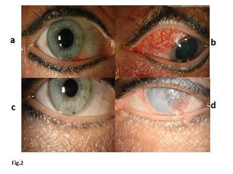
6

distance) in the left eye, with an intense white diffuse stromal scar with corneal
vascularization and central stromal contraction and thinning, corneal flattening, a
permanently curled-up pupillary rim and dilated pupil. For her left eye she
received a cosmetic contact lens with iris coloring. Seven years later (Fig. 2C,D),
the only treatment of her left eye consists of tear substitutes (Fig. 2D).

Temperature-time combinations that irreversibly injure the cornea and that
produce a thermal cataract
 Assuming a normal corneal temperature (35 °C, [12]), Fig. 3 shows the
temperature-time behavior for corneal injury as measured in rabbit eyes [13].
Data from 181 humans [14-16] showed a similar corneal temperature of 34.8 °C
and, although the size of the eyes and the eye lids are quite different, we have
used Fig. 3 as a reasonable approximation for human corneas.
 In the literature, we found only two temperature histories that produced a
cataract in rabbits (Fig. 2 of [17]). Luckily, this allows deriving a generic
temperature history of cataract formation by Arrhenius theory (Appendix A), with
the results also shown in Fig. 3.

Heat diffusion computations from the hot cornea to the lens and from a hot
ring of periorbital skin to the center of the cornea
 A curled-up iris and the typical aspect of the anterior lens implied that a
thermal cataract had developed. From Fig. 3, the duration of the corneal
temperature that predicts a cataract (open circles in Fig. 4) can be estimated. The
computed anterior lens temperatures of cataract development in response to
cornea temperatures of 60 to 100 °C are also shown in Fig. 4. The corneal
temperature-time combinations found are: 60 °C-30 s; 67 °C-23 s; 70 °C-20 s; 80
°C-15 s; 90 °C-11 s and 100 °C-10 s. The combination 67 °C-23 s will be used in
the Discussion (Study limitations) to estimate the effects of using 0.1 J per pulse
instead of 0.225 J.
 The computed temperature increase at the center of the cornea in response
to an instantaneous hot ring of periorbital skin, of 3 mm thickness (Fig. 1), with
exposure to temperatures between 60 and 100 °C during 30 s and cooling
thereafter, varied between 4.4 and 11.5 °C. As stated earlier, these corneal
temperature effects overestimate the actual behaviors since laser bursts are
given consecutively and mainly on the lower rim. Therefore, periorbital laser
impacts along the lower rim cause a few degrees increased corneal temperature
only, hardly affecting the anterior lens temperature.

Measured temperature response of a Cox II eye shield to CO2 laser impacts
 The presence of a tear film on the metal shield is an essential aspect of this
7

laser treatment, because CO2 laser light significantly absorbs in water. Local
irritation and oxybuprocaine eye drops will stimulate tearing and the formation of a
tear film. Therefore, experiments have been conducted with blood clotted on the
metal shield; blood mimics the water film and is obviously easier to handle. Figure
5 shows the measured temperatures of a Cox II eye shield in response to 19 CO2
laser honeycomb impacts (“plus” signs on the horizontal axis). First, we used a
sterile Cox II eye shield. Second, we added blood drops to the convex part of the
shield, waited 1 minute until the drops were clotted, and irradiated the eye shield
area with the clotted blood on its surface. As blood contains about 84% of water
[11], we estimated the temperature response to 19 laser bursts of the eye shield
with water (representing a tear film), by multiplying the data with clotted blood by
1/0.84 = 1.19. After 7 pulses in 7.5 s, the total shield area already reached 63 °C.
Subsequently, the shield area retained a uniform temperature, also during cooling
(137 sec).

DISCUSSION
Most likely scenario that caused the corneal injuries
 We questioned how 10-20 consecutive CO2 laser pulses could have had such
an impact on the left eye. The pulses were part of a normal procedure and we
hypothesized that the 2nd pass on the left lower orbital rim had a more upward
directed CO2 laser hand piece, perhaps because the patient’s head was directed
slightly more upward than normal, and that every CO2 laser burst impacted on the
eye shield. The exceedingly large absorbance of CO2 laser light by tear-covered
stainless steel would have caused a significant temperature rise of the metal
shield. The shield’s excellent thermal properties would lead to spreading of the
heated area over the total metal (and thus corneal) surface within 7.5 s (Fig. 5,
dashed line “% Heated Area”). This hypothesis implies that the lower eye lashes
should be burned. Because examination of the eyelids showed that this was
indeed the case (Figs. 2A,B), we believe that placement of multiple unintentional
CO2 laser impacts on the metal eye shields is the most likely scenario that
caused the ocular complications in this patient. Heat conduction from the hot
cornea also heated up and injured the iris and caused a cataract. We emphasize
that intraocular damage following this CO2 laser procedure has not been
described before.
 This paper must create awareness for all physicians who perform periocular
CO2 laser resurfacing procedures that unintentional CO2 laser irradiation of metal
eye shields, despite protecting the cornea against direct laser impact, has the
capability of significantly heating up these shields (see e.g. [18]). This effect is
8

even intensified by the tear film (Fig. 5), a mechanism that has not been described
before.

Study limitations
 Unfortunately, actual treatment details are unknown, e.g. whether the 2nd pass
around the left lower orbital rim occurred immediately after the 1st pass and
whether the 1st pass of laser bursts also hit the eye shield. If that were the case,
the 2nd pass would have started with an already increased shield temperature. For
simplicity, we assumed that the first pass laser bursts missed the eye shield.
 The way we modeled laser impacts on the periorbital skin led to overestimated
responses. Since the resulting temperature rises were found to be negligible, the
extent of overestimation is irrelevant. Also, the Arrhenius parameters for thermal
cataract formation from only two experimental temperature-time combinations
(Appendix A) have limited accuracy. However, the derived temperature-time curve
(Fig. 3) is the only one available to date that describes cataract formation from an
increased lens temperature. The outcome, that these cataract-causing
temperatures are below those of corneal damage temperatures suggest that the
prediction is meaningful; the opposite would have contradicted the observed
damage events.
 Cooling of the metal eye shield held in air (Fig. 5) is (much) faster than in the
clinical situation, placed on the eye ball and intermittent layer of oculentum
simplex, which has isolating properties (a 2.5 times smaller thermal diffusivity,
Appendix B). Thus, it takes quite a long time before a temperature rise of the
cornea has diffused away, contributing to corneal thermal damage and cataract
formation at sufficiently high temperatures (Figs. 3 and 4).
 The CO2 experimental laser parameters (0.225 J) differed from those used
clinically (0.1 J). However, the estimated temperatures of the Cox II shield with
added water (Fig. 5) in response to 0.225 J laser pulses, can be easily transformed
into temperatures from 0.1 J pulses by dividing temperature increase by 2.25. After
19 pulses, this would give a temperature of about 67 °C instead of 107 °C. Then,
70 °C would have been reached after 24.5 s, thus after 21 pulses. However, a
longer time duration also lowers the critical temperature for corneal injury and
cataract formation (Fig. 3). From Fig. 4, one can see that development of a
cataract occurs when the cornea temperature is about 67 °C during about 23 s.

Incidence of this complication
 This thermal complication either seems to be extremely rare or has been
underreported in the literature as we found only five papers on this subject. In
three, the authors described the thermal cause [2], one written in Korean [6], the
3rd [3] gave a 0.3% incidence of corneal injury after CO2 laser blepharoplasty,
9

however, without details. In the other two [5,8], we believe the cause was thermal
but the authors proposed other mechanisms.

Other possible causes
 The review by Blanco et al. [4] did not refer to Widder et al. [2] and Christian et
al. [5] but included many other mechanisms. Chemical damage can occur when
the cream to anesthetize periorbital skin reaches the cornea, or when corneal eye
shields are cleansed with chlorhexidine prior to insertion [5]. Thermally-burned eye
lashes, eyelid skin and eyebrow hair have been described in a patient who was
wearing mascara while cautery was applied to her eyelid lesion after excision [7].
Although our patient has tattooed eyeliners (Fig. 2), these lines were not damaged.

CONCLUSION
 Despite uncertainties concerning procedural and cataract modeling details,
our experiments on the Cox II shield (Fig. 5), the burned lower eye lashes (Fig.
2A,B), and the supporting heat diffusion computations have shown beyond doubt
that consecutive CO2 laser impacts hit both eye shields caused the left total
shield area to reach a high temperature, probably over 70 °C, followed by a slow
cooling rate. Heat conduction (Fig. 4) caused curling-up of the iris and a thermal
cataract. The right shield also received CO2 laser impacts but less than the left
shield and only around the central part of the eye, shown by mid-peripheral
corneal damage and burned lower middle eye lashes.
 Few treatment options exist for thermal ocular injury. Our case thus
emphasizes the importance of knowing the pitfalls of periocular CO2 laser
resurfacing procedures and, especially now that fractional CO2 laser resurfacing
is gaining in popularity, suggests adapting the safety guidelines with respect to
the use of metal eye shields. Particularly, the eye shields should not only be
positioned properly but the position of the CO2 laser beam, visible by the pilot
beam, should also be cautiously monitored as long as the laser is switched on.

REFERENCES
1. Alster TS, Lupton JR. Prevention and treatment of side effects and complications of cutaneous
 laser resurfacing. Plast Reconstr Surg 2002;109:308-316.
2. Widder RA, Severin M, Kirchhof B, Krieglstein GK. Corneal injury after carbon dioxide laser
 resurfacing. Am J Ophthalmol 1998;125:392-394.
3. Apfelberg DB. Summary of the 1997 ASAPS/ASPRS Laser Task Force Survey on Laser
 Resurfacing and Laser Blepharoplasty. Plast Reconstr Surg 1998;101:511-518.
4. Blanco G, Clavero A, Soparkar CNS, Patrinely JR. Periocular laser complications. Semin Plast
 Surg 2007;21:74-79.
5. Christian MM, Cox DO, Smith CV, Onouye T, Moy RL. Ocular damage due to chlorhexidine
 versus eyeshield thermal injury. Dermatol Surg 2001;27:153-157.
10

6. Kim JH, Ahn MW, Lee JS. Corneal burn caused by Carbon Dioxide Ultrapulsed laser for nevus
 removal J Korean Ophthalmol Soc 2014;55:1376-1379.
7. Al Raqqad N, Liu C. Mascara: a cause of thermal burn after cautery for eye lid lesion excision; a
 case report. J Clinic Experiment Ophthalmol 2010;1:105.
8. Litzinger TC, Vastine D. Bilateral corneal opacities in a LASIK patient after the use of titanium
 eyeshields. J Cataract Refract Surg 2011;37:1160-1164.
9. van Gemert MJC, Welch AJ. Approximate solutions for heat conduction: Time constants. In:
 Welch AJ, van Gemert MJC (Editors). Optical-Thermal response of laser-irradiated tissue,
 Chapter 13, page 425, Plenum Press, New York, 1995.
10. Rahman A, Al-Ghadyan A, Cotlier E. Rise in temperature on exposure to sunlight or high
 ambient temperature. Br J Ophthalmol 1986;70:421-426.
11. Wahl HG. How accurately do we measure blood glucose levels in intensive care units (ICU)
 patients? Best Practice Res Clin Anaesthesiol 2009;23:387-400.
12. Mikesell GW. Corneal temperatures – A study of normal and laser injured corneas in the Dutch
 Belted Rabbit. Am J Ophthalmol 1978;55:108-115.
13. Bargeron CB, Deters OJ, Farrell RA, McCally RL. Epithelial damage in rabbit corneas exposed
 to CO2 laser radiation. Health Phys 1989;56:85-95.
14. Mapstone R. Measurement of corneal temperature (1968) Exp Eye Res 7:237-243.
15. Fabiani C, Li Voti R, Rusciano D, Mutolo MG, Pescosolido N. Relationship between corneal
 temperature and intraocular pressure in healthy individuals: A clinical thermographic analysis. J
 Ophthalmol 2016;3076031. http://dx.dox.org/10.1155/2016/3076031.
16. Giannaccare G, Fresina M, Agnifili L, Versura P Ocular-surface temperature modification by
 cataract surgery. J Cataract Refract Surg 2016;42:983-989.
17. Kramar, Emery AF, Guy AW, Lin JC. The ocular effects of microwaves on hypothermic rabbits: a
 study of microwave cataractogenic mechanisms. Ann NY Acad Sci 1975;247:155-165.
18. Ries WR, Clymer MA, Reinisch L. Laser safety features of eye shields. Lasers Surg Med
 1996;18:309-315.
19. Thomsen S. Pearce JA. Thermal damage and rate processes in biological tissues. In: Welch AJ,
 van Gemert MJC, editors. Optical-thermal response of laser-irradiated tissue. Second Edition.
 Springer, Dordrecht, 2011, Chapter 13, pp 487-549.

APPENDIX A. ARRHENIUS THEORY OF CATARACT
FORMATION
 We combined the two temperature-time ( − ) combinations that produced a
cataract in rabbits (Fig. 2 of [17]): 43.5 °C during 10 min and 41.5 °C during 20 min
with Arrhenius kinetic rate theory ([19], Eq. 13.42), using
 ⁄
 ln( ) =
 − ln( ) (1)
 is activation energy [J/mole], the universal gas constant [8.3143 J/mole/K],
the temperature in Kelvin (0 °C is 273.15 K), and the frequency factor [s-1]. From
the two data points the two unknowns in Eq. (1) follow as ⁄ = 34,530.5 K and
ln( ) = 102.65. The ( − ) curve for a cataract is also given in Fig. 3 (lower
curve).

APPENDIX B. THERMAL DIFFUSIVITY OF OCULENTUM
SIMPLEX
11

 We used the available chemical composition and thermal data of the
components, i.e., 40% paraffin (thermal conductivity = 0.25 W/m/°C, thermal
diffusivity = 0.081x10⁻⁶ m2/s), 51.5% vaseline ( not found, = 0.18 W/m/°C), 6%
lanoline ( = 0.053x10⁻⁶ m2/s), and 2.5% cetostearyl alcohol (here we used
glycerol, = 0.01x10⁻⁶ m2/s). Assuming that is proportional to (implying
identical product of density and heat capacity) gives = 0.064x10⁻⁶ m2/s for
vaseline. Then, based on the composition, an estimated thermal diffusivity of
oculentum simplex is: ≈ (0.4x0.081 + 0.515x0.064 + 0.06x0.053 +
0.025x0.143)x10-6 = 0.07x10⁻⁶ m2/s. Tissue has about 0.18x10⁻⁶ m2/s thus
oculentum simplex has about a 2.5 times lower thermal diffusivity than tissue.

CAPTION TO FIGURES
Fig. 1. Eye (blue sphere) with a ring-shaped skin layer of 3 mm thickness and 9 mm length around
the orbital rim representing the periorbital skin (indicated in green), which was kept during the
simulations at 60 to 100 °C during 30 s (indicated by a reddish line), and the metallic shield of 1 mm
thickness in the middle area (dark blue layer).
Fig. 2. (A) Right eye, directly after treatment. Light burning of the lower middle lashes and
perilimbal redness. (B) Left eye, directly after treatment. Severe burning of the lower eye lashes at
the side of the nose and slight burning of all other lashes. The central cornea shows epithelial and
stromal clouding. (C) Right eye seven years after treatment. (D) Left eye seven years after
treatment. N.B. This patient has tattooed eyelines around both eyes.
Fig. 3. Temperature-time duration curves derived from rabbit experiments that predict (a)
irreversible thermal corneal injury, using 35 °C as normal corneal temperature [10], and (b) a
thermal cataract, Eq. (1).
Fig. 4. Temperature of the anterior lens, at 2 mm inside the eye, assumed at 37 °C, as a function of
the time of heat conduction duration for several corneal temperatures (60 °C to 100 °C). The
temperature-time points that will predict the development of a cataract are indicated as circles.

Fig. 5. Measured temperatures during heating and cooling of the Cox II shield in air in response to
19 CO2 laser bursts (black lozenges) at 0.225 Joule, about 1 Hz per burst, 1.3 mm spot diameter, 9
mm width/length honeycomb pattern, about 100 Hz for individual pulses. Time points of the bursts
are indicated by “plus” signs on the horizontal axis. At the right side (black dashed line, “% Heated
Area (19 pulses with blood”), we plotted the area of the shield (in %) with an increased temperature.
Also shown are measurements without added blood (black circles), and the computed estimated
curve of the eye shield with water added (thick black dashed line), assuming whole blood contains
84% of water [11].
12
13
14

REPLY TO THE COMMENTS OF THE 2 REVIEWERS

Reviewer 1:

I read, with distress, the manuscript, Periocular CO2 laser resurfacing: severe
ocular complications from multiple unintentional laser impacts on the protective
metal eye shields. This manuscript must be published. It is a shame that it has
taken seven years to get this information out.

Thank you. We agree.

One can question the assumptions made in the simulations. However, the clinical
results are real. The authors are careful to state that they are simulating events
long after they happened with a lack of detail. Overall, the work is carefully and
conservatively presented.

Thank you.

There are a couple of questions that I have:

1. In the Materials and Methods, the thermal diffusivity of the eye is given as 1.5 x
10-7 m2/sec. The source for this value should be cited. If this is not a measured
quantity, how the authors estimated this value should be explained.

Thank you. We changed it as: “--- we used --- a thermal diffusivity of 1.5x10⁻⁷
m2/sec, very close to the value of water (1.43x10⁻⁷ m2/sec) and the value often
used for tissue (1.77x10⁻⁷ m2/sec [9]).”

The new Ref 9 is: “van Gemert MJC, Welch AJ. Approximate solutions for heat
conduction: Time constants. In: Welch AJ, van Gemert MJC (Editors). Optical-
Thermal response of laser-irradiated tissue, Chapter 13, page 425, Plenum Press,
New York, 1995.”

2. In the Results and under “Temperature-time combinations that irreversibly injure
the cornea and the produce a thermal cataract” the authors make the conclusion
that the time dependence of a temperature variation measured in a rabbit is the
same as the time dependence of a temperature variation in a human is the same
because the initial temperatures are the same. This conclusion cannot be made.
The geometry (size) of the eyes are quite different, as are the sizes of the eye lids.
I can accept using the rabbit eye parameters as a reasonable approximation…but
it should not be stated that the initial temperatures prove a correlation between the
two different eye systems.

Thank you.

We changed it as: “Assuming a normal corneal temperature (35 °C, [11]), Fig. 3
shows the temperature-time behavior for corneal injury as measured in rabbit eyes
15

[13]. Data from 181 humans [14-16] showed a similar corneal temperature of 34.8
°C and, although the size of the eyes and the eye lids are quite different, we have
used Fig. 3 as a reasonable approximation for human corneas.“

Otherwise, this was a very carefully done evaluation and I complement the authors.

Thank you very much indeed.

Reviewer: 2

I have carefully studied this interesting accident report and the authors' attempts at
an experimental simulation. However, I think the authors have made some
seriously erroneous assumptions that places the validity of their study into serious
doubt. Since lids of the patient should have been closed and 10.6 μm laser
radiation should not penetrate the lids.

No, the lids of the patient were NOT closed.

We added (RESULTS, Clinical case, page 5): “For our analysis below it is essential
that the eye lids under such circumstances never spontaneously close and hence
do not cover the total area of the metal shield.”

One of the dermatologic authors (AW) regularly uses eye shields and he states
that the eye lids tend to open spontaneously except if the patient purposely pushes
them to become closed but even then eye lids often reopen. Even companies that
produce eye shields don’t state the necessity of keeping eye lids closed just
because they assume that eye shields always protect adequately.

The Cox II shields are meant to be under the lids and not directly irradiated.

We agree.

Certainly, multiple (19 bursts) direct aiming of a beam at the shield is simply not
plausible by a trained operator.

The operator was actually very experienced and we obviously don’t know how this
became possible.

As with all too many accident cases, there certainly could be a misrepresentation
of the facts the laser user who does not wish to seem to be incompetent or at
serious fault.

Incompetent: no. At serious fault: yes.

The authors should wonder why routine sauna exposures for 15 minutes at moist
air temperatures of 90 C (good conditions for heat transfer) do not result in
cataracts of millions of Scandinavians.
16

Interestingly, an article published on February 2nd in the NRC (a Dutch major
newspaper) with title: “Why don’t you get burned in a sauna of 90 degrees” referred
to a physician in one of the Dutch burn centers who stated that surviving these air
temperatures is possible because of the huge difference that exist between the
thermal properties of air and water. Our body can handle high air temperatures due
to the very small heat conduction of air combined with cooling of the skin by
vaporization of its sweat. Also, inhaling of hot air is possible because of the cooling
by passing through the nose and upper airways. However, if some water is thrown
on the hot stones you immediately experience this as heating. They ended with the
example of a contender of the World Sauna Championship who died following a
stay of a few minutes in a sauna of 110 degrees where every 30 sec some water
was thrown on the stones.

Because this subject is unrelated to our case we have not tried to include it in the
manuscript.

However, I think the authors have made some seriously erroneous assumptions
that places the validity of their study into serious doubt.

We would have expected that a reviewer who makes this statement (actually
incorrect, see our 1st reply), would have provided an alternative mechanism that
also explains the total thermal destruction of the full area of the cornea, the clinical
reality here.
You can also read