PERSPECTIVES IN EYE CARE - VIRTUAL - Monday May 24th, 2021 - Minnesota Eye Foundation
←
→
Page content transcription
If your browser does not render page correctly, please read the page content below
The Minnesota Eye Foundation
proudly presents

                VIRTUAL
                PERSPECTIVES
                IN EYE CARE

Monday
May 24th, 2021

COPE Activity ID #121548
COPE CREDITS
As a virtual event, we are using the following to verify
attendance for this program.

QR Code
During each presentation, a QR code will be displayed on the
screen. Please use ARBO’s tracker app to scan this QR code
as it appears. If you’re unable to scan for any reason, simply
inform an event coordinator.

ZOOM Link
Each registrant will receive his or her own ZOOM link prior to
the program. This link is unique to each participant, so please
do not share this with anyone else. ZOOM tracking is a cross
reference for us as we award credits.

COPE SURVEY
As in years past, you will receive an email following the event
asking you to complete our online Post-Event Feedback
Survey. Your feedback is incredibly important to us, so please
take a few minutes to complete this.

OE TRACKER ACCOUNT
ARBO will update your OE Tracker account once these credits
have been issued. It may take a few weeks before you notice
these credits in your account.

QUESTIONS?
Contact us at info@mneyefoundation.com.

                                           1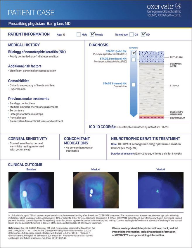
OE TRACKER Mobile App by ARBO
Instructions for Optometrists Attending CE Courses
(for Apple v 1.2 and Android v 1.2)
Description
Optometrists can use the OE TRACKER mobile app to record attendance at continuing education courses
and receive instant course credit. Not only is it easy, but the app is FREE and can be used by anyone with
an OE TRACKER number. The OE TRACKER app is available for iPhones/iPads and Android phones.
How to Get the OE TRACKER App:
iPhone/iPad: Go to the app store on your iPhone or iPad and search for “OE TRACKER.” Find the OE
TRACKER app and touch to download. Alternatively, you can also download the OE TRACKER app from
iTunes.
Android Phone: Download the app from Google Play. Go to Google Play on your Android phone and
search for “OE TRACKER.” Find the OE TRACKER app and touch the install button.
How to Use the OE TRACKER App:
Once you have downloaded the app, open it by simply touching the app icon. You will see the Welcome
Screen, which will ask you to select one of two roles to login as:
Course Attendee
Course Provider
(iPhone) (Android)
2
Logging into the OE TRACKER app as a Course Attendee:
1. Touch “Course Attendee” if you are an optometrist that is attending a course and you want to
   record your attendance using the OE TRACKER app.
       a. You will need your OE TRACKER username and password to log into the OE TRACKER app. If
           you don’t have a username and password, touch “Create User” at the bottom of the Login
           screen to set one up. (See photo below.) Or call ARBO at 704-970-2710 or 866-869-6852
           and we can do it for you. If you have forgotten your username and/or password, touch
           “Forgot Username or Password?” at the bottom of the Login screen (See photo below.) Or
           call ARBO and we’ll tell you what they are.

                                        (iPhone)                                          (Android)

2. Enter your username and password and touch “Log In”. Note: These fields are case sensitive. Many
   phones will capitalize and self-correct what you type, so be sure you entered your username and
   password correctly.

                                                     3
3. When you have logged in, the Main screen
                                                              will open. This screen will display your:
                                                                   a. First and last name at the top of the
                                                                      screen
                                                                   b. OE TRACKER Number
                                                                   c. E-mail address

                                                           IMPORTANT: Before doing anything else, please
                                                           make sure the correct e-mail address is listed in
                                                           your account so we can e-mail you confirmation
                                                           of course attendance. If you need to update your
                                                           e-mail address, touch “Edit Email Address” (See
                                                           photo) and enter your updated e-mail address.
                                                           Then touch “Save” at the top right side of the
(iPhone)                    (Android)
                                                           screen.

Recording Your Attendance at a CE Course:
PLEASE NOTE: In order to record your attendance using the OE TRACKER mobile app, the provider of
the CE course must supply a course-specific QR code created by ARBO. After the course has been
presented, the provider will post the QR code for attendees to scan. Contact the CE provider prior to
attending the course to see if they will be using the OE TRACKER app to record attendance.

1. On the Main screen, after you verify that your personal information is correct, touch “Scan QR
   Code” located below your e-mail address.

                                         (iPhone)                                     (Android)

                                                     4
2. Your phone’s camera will open and you will see “Scan QR Code” at the top of your screen.

3. Center the QR code on your screen and it will automatically scan NOTE: If the code does not
   scan right away, try backing up your phone a little to make sure the entire QR code fits within
   the screen.

4. If you have scanned the QR code correctly, the Confirmation screen will appear telling you that
   your attendance has been recorded in your OE TRACKER account.

                                      (iPhone)                                             (Android)

5. You will also be sent an e-mail from OE TRACKER within the next few minutes advising you that
   your credit for the course has been entered into your account.

6. Touch “Done” at the top right side of the screen to return to the Main screen.

7. To exit, simply close the app. You will stay logged in to the app to scan another QR code. To log
    out of the app touch the “Logout” button.

                                                 5
Viewing Your CE Course History:
If your OE TRACKER subscription fee has been paid, you can view the CE course hours that are
in your account while you are logged into the mobile app.
    1. On the Main screen, touch “View Course History” in the middle of the screen.

                               (iPhone)                                         (Android)

   2. If your OE TRACKER subscription fee has NOT been paid, you will see the “Course History
      Error” screen (See photo below.) If you wish to pay your subscription fee, just touch
      “Pay Fee” and it will direct you to OE TRACKER to complete payment.

                                       (iPhone)                                (Android)

                                                  6
3. If your OE TRACKER subscription fee is paid, you will see a list of CE hours that are
   currently in your OE TRACKER account with the date range listed at the top.

       a. You can change the date range by touching “Filter” on the top right side of the
          screen and selecting your desired start and end date.

                                            7
b. You can see more detailed course information by touching an individual course
              title.

IMPORTANT NOTE: The OE TRACKER mobile app can be used to record attendance of COPE and Non-
COPE courses only at events held by COPE-Approved Administrators/Providers. Credit for other courses
can be submitted to ARBO by CE providers using barcode scanners or submitting attendance on an Excel
spreadsheet. Optometrists who pay their OE TRACKER subscription fee may also submit certificates of
attendance to ARBO to have credits entered into their OE TRACKER account. Simply fax your certificates
to 888-703-4848 or email them to arbo@arbo.org.

                                                   8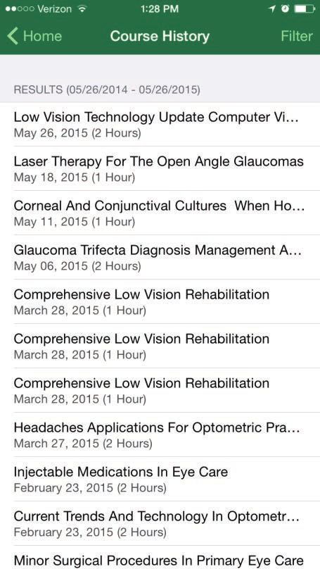
Agenda
Session One   7:55-8:00 – Welcome & Announcements
              8:00-8:50 – New Updates in Oculoplastics
                  William J. Lipham, MD, Jill S. Melicher, MD and Krista J. Stewart, MD
              8:50-9:00 – Break
              9:00-9:50 – Ophthalmic Coding and Compliance Update
                  Leslie Boles, Director of Compliance Audit,
                  Waud Capital Partners Healthcare
              9:50-10:00 – Break
              10:00-12:00 – Glaucoma: What you Need to Know
                  Thomas W. Samuelson, MD, Patrick J. Riedel, MD,
                  Christine L. Larsen, MD, Clara M. Choo, MD and Jefferson Berryman, MD
              12:00-12:30 – Lunch

Session Two   12:30-12:50 – The Vision Project
              12:50-1:40 – Corneal Grand Rounds
                  Sherman W. Reeves, MD
                  Panelists: Elizabeth A. Davis, MD, Omar E. Awad, MD
                  and Mark S. Hansen, MD,
              1:40-1:50 – Break
              1:50-2:40 – Ocular Surface Disease Management
                  Omar E. Awad, MD
                  Panelists: Ahmad M. Fahmy, OD and Noumia Cloutier-Gill, OD
              2:40-2:50 – Break
              2:50-3:40 – Refractive and Keratoconus Surgery Update
                  Richard L. Lindstrom, MD and Mark S. Hansen, MD
              3:40-3:50 – Break
              3:50-4:45 – Hot Topics in Cataract Surgery
                  Elizabeth A. Davis, MD
                  Panelists: Thomas W. Samuelson, MD, David R. Hardten, MD,
                  Patrick J. Riedel, MD and Mark S. Hansen, MD
              4:45 – Adjourn

                                           9
Contents
Presentations
Session One New Updates in Oculoplastics
William J. Lipham, MD, Jill S. Melicher, MD
and Krista J. Stewart, MD .................................................................................... 18
Ophthalmic Coding and Compliance Update
Leslie Boles, Director of Compliance Audit,
Waud Capital Partners Healthcare ..................................................................... 29
Glaucoma: What you Need to Know
Thomas W. Samuelson, MD, Patrick J. Riedel, MD,
Christine L. Larsen, MD, Clara M. Choo, MD
and Jefferson Berryman, MD .............................................................................. 34
Session Two The Vision Project
Corneal Grand Rounds
Sherman W. Reeves, MD
Panelists: Elizabeth A. Davis, MD, Omar E. Awad, MD
and Mark S. Hansen, MD .................................................................................... 48
Ocular Surface Disease Management
Omar E. Awad, MD
Panelists: Ahmad M. Fahmy, OD
and Noumia Cloutier-Gill, OD ............................................................................ 52
Refractive and Keratoconus Surgery Update
Richard L. Lindstrom, MD and Mark S. Hansen, MD ......................................... 56
Hot Topics in Cataract Surgery
Elizabeth A. Davis, MD
Panelists: Thomas W. Samuelson, MD, David R. Hardten, MD,
Patrick J. Riedel, MD and Mark S. Hansen, MD ................................................ 59
10Thank you to our Sponsors
1112
13
14
15
16
17
Session One
                             New Updates in
                             Oculoplastics Part 1:
                             Rapid Fire Oculoplastic
                             Case Series
                             COPE Course ID # 71820-AS
Jill S. Melicher, M.D.
                             Course Description
Minnesota Eye Consultants
Ophthalmic Plastics, Orbit
and Reconstructive Surgery   Oculoplastic Rapid Fire Case Series. This course will
                             provide a series of Oculoplastic cases that assist the learner
                             in identifying common Oculoplastic problems, their most
                             common presentation, differential diagnoses, treatments
                             and outcomes.

                             Course Objective

                             1. Assist the learner in identifying differential diagnoses for
                                the most common Oculoplastic problems.
                             2. Assist the learner in recognizing postoperative problems
                                following the most common Oculoplastic procedures.
                             3. Assist the learner in identifying treatment plans for the
                                most common and some rare Oculoplastic problems.

                                      18
Rapid Fire Oculoplastic Notes
Case Series
Jill S. Melicher, M.D.
1. Case 1
a. 20 something female presents with 1 month
history of right eye redness, blurred vision,
surface irritation and proptosis.
b. Patient Presentation
c. HPI
i. 1 month ago, noted that right eye vision
was blurred and irritated
ii. Over the next 2 weeks, experienced more
irritation, mild crusting in the AM, and
bulging of her right eye
iii. Saw eye care provider at 2-week mark who
prescribed Amoxicillin for 14 days with no
scheduled follow-up
iv. Pt was compliant with treatment for
two weeks, but saw no improvement in
condition
v. Pain slowly progressed – intermittent 5/10
pain over the past few weeks
vi. Called doctor who advised that she be seen
for evaluation
vii. POHx significant for anisometropic
amblyopia of right eye
viii. Denies PMHx
d. Family Hx significant for brother who died
at age 8 from “some kind of leukemia,” and
mother with unknown thyroid disorder.
e. Surgical, Social, Medication histories otherwise
non-contributory
f. Exam
g. Slit Lamp Exam
h. Dilated Fundus Exam
i. Labs
j. CT/MRI Review
k. Differential Diagnoses
l. Infectious
m. Inflammatory/Autoimmune
n. Neoplasia
o. Diagnosis: Rhabdomyosarcoma-
p. Orbital imaging review
q. Treatment options review
192. Case 2
a. 70 something year old male with history of Notes
ptosis
b. HPI
i. 1 year history of progressive onset ptosis
ii. intermittent binocular diplopia while driving
iii. prisms for >20 years due to strabismus
iv. no pain, discomfort
v. no upper or lower extremity weakness
c. Exam:
i. Fatigable ptosis
ii. Cogans lid twitch
d. Review labs
e. CT chest
f. Differential diagnosis
g. Diagnosis: Myasthenia Gravis
h. Work-up: single fiber EMG
i. Treatment: Pyridostigmine, optimal timing
of surgery, IVIg, Immunosuppression,
cardiothoracic surgery for thymectomy
3. Case 3
a. 70 year old female with longstanding >30 year
history of pigmented in lower eyelid lesion
b. Serial photographs obtained
c. Recent growth
d. Exam
e. Differential diagnosis
f. Diagnosis: Malignant Melanoma
g. Review Pathology
h. Treatment: Staging, Sentinel node biopsy,
Breslow’s criteria, Surgical excision and on
going monitoring
4. Case 4
a. 1 week old with acute onset of periorbital
swelling
b. Examination
c. Imaging
d. Relevant clinical anatomy
e. Differential Diagnosis
f. Diagnosis: Dacryocystocele with intranasal cyst
g. Treatment: Hospitalization, Nasolacrimal duct
probe, Removal of intranasal cyst due to
obligate nasal breather
20Session One
New Updates in
Oculoplastics Part 2:
Targeted Monoclonal
Antibody Therapies for
Thyroid Eye Disease
COPE Course ID # 71737-AS
William J. Lipham,
M.D., F.A.C.S.
Minnesota Eye Consultants Course Description
Ophthalmic Plastics, Orbit
and Reconstructive Surgery This lecture will discuss how Monoclonal Antibody IV
infusion therapy with Teprotumumab and Tocilizumab may
be used to treat the inflammatory phase of Thyroid Eye
Disease (TED).
Course Objective
1. Understand the pathophysiology of Thyroid Eye Disease
(TED).
2. Realize that TED can now be treated in the early
inflammatory phase to avoid invasive surgery.
3. Learn the two compounds Teprotumumab and
Tocilizumab that may be used as IV infusions to reduce
the inflammatory phase of TED.
4. Recognize that there are differences in the two
compounds with regard to specificity and cost.
21Targeted Monoclonal Notes
Antibody Therapies for
Thyroid Eye Disease
William J. Lipham, M.D., F.A.C.S.
1. TED Is a Debilitating, Progressive and Vision-
threatening Autoimmune Disease
a. Patients may experience
i. Poor ophthalmic clinical outcomes
ii. Disfigurement
iii. Vision-threatening complications
iv. Psychosocial distress
v. Restrictions in daily activities and ability to work
2. Annual Incidence
a. 16 out of 100,000 Women
b. 3 out of 100,000 Men
3. Leading Risk Factors for TED
a. Smoking increases risk by 8 fold
b. Risk of new onset or worsening of TED is
~20% after RAI treatment
c. Women have higher risk but men have
elevated risk for more severe TED
d. Odds of TED increase by 17% with each
decade of age
4. TED is the Most Common Extrathyroidal
Manifestation of Graves’ Disease
a. TED
i. Immune cells attack orbital tissue
ii. Not directly related to high serum thyroid
hormone concentrations
iii. Treatment of the thyroid gland does not
improve TED
b. Autoimmune disease
i. 90% of patients with TED have concurrent
GD
c. GD
i. Goal of treatment is to inhibit production
of thyroid hormones
ii. Autoantibodies against TSHR trigger
excessive production of thyroid hormones
d. 10% of patients with TED are either
hypothyroid or euthyroid5
e. TED may present before, during, or after the
onset of GD7
5. Inflammation During Progressive TED Advances
to Chronic Fibrosis1
22a. Progressive (active), inflammatory phase of TED
can last up to 3 years1,2 Notes
b. Patients eventually progress to the fibrotic
(inactive) phase of TED, which is characterized
by irreversible fibrosis1
c. Fibrosis begins during the progressive (active)
phase and leads to the lasting sequelae
associated with permanent disfigurement and
functional visual impairment1,3
6. Inflammation, Tissue Expansion, and Eye Muscle
Changes May Lead to the Clinical Manifestations of
TED
a. Healthy Eye and Orbital Tissue
i. Eye is well protected by eyelid
ii. Thin periocular muscles
iii. Orbit contains a small amount of tissue and
fat
b. In the Presence of TED2
i. Eyelid retraction
ii. Eye protrusion
iii. Inflammation of
iv. lacrimal caruncle
v. Eyelid and conjunctival redness
vi. Inflamed and enlarged muscles due to fluid
accumulation
vii. Compression of the optic nerve at orbital
apex
viii. Increase in orbital tissue and fat
7. Invasive Surgery is Currently the Only Option for
Fibrotic TED
a. Orbital Decompression
i. Exposing orbit
ii. Removing bone
iii. Removing adipose tissue
b. Stabismus Surgery
i. Muscle recession
ii. Muscle resection
c. Eyelid Surgery
i. Upper eyelid incision line
ii. Lower eyelid incision line
8. Recognizing the Signs and Symptoms of TED
a. Eyelid
i. Upper eyelid retraction: 91% of patients
affected
ii. Eyelid swelling
iii. Pain
iv. Lagophthalmos (incomplete closure of
eyelid)
23b. Orbital Tissue
i. Exophthalmos (proptosis): Occurs in 62% of Notes
patients
ii. Pain/deep ache
iii. Disfigurement
9. Ongoing Inflammation and Expansion of Orbital
Tissues Leads to Changes in Physical Appearance
a. Conjunctiva and Cornea
i. Chemosis (swelling of the conjunctiva)
ii. Conjunctival hyperemia (redness)
iii. Photophobia (light sensitivity)
iv. Pain
v. Foreign body sensation (grittiness)
vi. Exposure keratopathy
vii. Swollen lacrimal caruncle
viii. Dry eye and tearing
b. Extraocular Muscle
i. Restricted ocular motility: Occurs in ~40%
of patients
ii. Strabismus (misalignment of eye)
iii. Diplopia (double vision)
iv. Pain
v. Retro-orbital ache
vi. Decreased vision and depth perception
10. Clinical Manifestations of TED Are Variable
a. Short Term Inflammation
i. Chemosis
ii. Conjunctival hyperemia
iii. Periorbital and eyelid edema (swelling)
iv. Pain
v. Ocular dryness
vi. Foreign body sensation
vii. Epiphora (watery eyes)
viii. Photophobia
ix. Eyelid retraction
x. Spontaneous orbital pain
b. Long-term Consequences
i. Eyelid retraction
ii. Proptosis
iii. Periorbital ache
iv. Strabismus
v. Diplopia
vi. Optic neuropathy
vii. Visual field defect
viii. Gaze-evoked orbital pain
ix. Ocular dryness
x. Photophobia
xi. Corneal ulceration
2411. Current Management Options for TED
12. TED and Graves Disease Appear to be Driven by Notes
Autoantibody Activation of the IL-6R
a. Both IL-6 and IL-6R are overexpressed in
patients with TED
b. IL-6 Signaling Inhibition Decreases:
i. B-cell activation
ii. Autoantibody production
13. More Specifically, TED is Driven by Autoantibody
Activation of IGF-1R
a. Orbital fibroblasts, which are specialized cells
responsible for tissue repair, are central to the
pathophysiology of TED1-3
b. IGF-1R, a gatekeeper of orbital fibroblast
activation, is overexpressed in TED orbital
fibroblasts4
c. IGF-1R and TSHR form a receptor-signaling
complex and colocalize in orbital fibroblasts4
d. Activation of IGF-1R stimulates release of
inflammatory cytokines and production of
hyaluronan and adipogenesis1,5,6
14. Baseline Assessment and Routine Monitoring Can
Help Identify Active (Progressive) TED
15. Limited Window for Treatment in Progressive
TED1-3
16. Steroids Provide Symptom Relief but are
Associated with a High Adverse Event Profile
17. Teprotumumab is now FDA approved for treating
the Inflammatory phase of TED
18. Tocilizumab is an alternative, off-label, Monoclonal
Antibody that can used to treat the inflammatory
phase of TED
a. Currently used for the Treatment of RA, GCA,
and COVID-19 (cytokine storm).
b. Four IV doses (8 mg/kg) are administered one
month apart for four months.
c. Can be used as a substitute for patients who
initiated a treatment of Teprotumumab which
was halted for COVID-19 vaccine production.
d. Treatment cost for Tocilizumab is less than
$20,000 per course vs $200,000 to $300,000
for Teprotumumab.
e. Must have an internist monitor liver function
studies and white blood cell counts during
infusion course.
2519. Identifying Progressive TED Through Routine
Assessments Notes
a. Initial assessment:
i. Pain assessment
ii. Visual changes
iii. Changes in appearance
1. Patient photos
iv. Impact on QoL
1. Daily activities
2. Psychosocial health
b. Follow-up with a specialist:
i. CAS assessment
ii. Eyelid retraction and proptosis
measurements
iii. Visual function and optic nerve evaluation
iv. Imaging
1. CT scan or MRI
20. A Collaborative Approach is Important for the
Management of TED
a. Early signs and symptoms can be confused with
other conditions, resulting in a misdiagnosis1
i. Delays seen in TED diagnosis and referral
to a specialist suggest that TED may be
underdiagnosed1,2
ii. Co-management by a multidisciplinary
team is important for:
1. Developing a medical management
strategy
2. Frequent monitoring of symptoms
3. Addressing risk factors for TED
progression
4. Managing comorbidities
21. Summary
26Session One
New Updates in
Oculoplastics Part 3:
2021 Advances in
Treatments and Diagnosis
of Ophthalmic Plastic
Krista J. Stewart, M.D. Conditions
Minnesota Eye Consultants
Ophthalmic Plastics, Orbit COPE Course ID # 71900-AS
and Reconstructive Surgery
Course Description
Understand and be aware of new treatment options and
diagnostics of ophthalmic plastic surgery.
Course Objective
1. Discuss new oncologic treatment options for skin and
orbital tumors.
2. Understand options for new cosmetic surgical and
nonsurgical treatments.
3. Updates to insurance coverage for eyelid surgery.
272021 Advances in Notes
Treatments and Diagnosis
of Ophthalmic Plastic
Conditions
Krista Stewart, M.D.
1. Oncologic update—significant advances with
immunotherapy
a. Tissue diagnosis and specific pathologic marker
request
b. Basal cell carcinoma
i. Vismodegib or sonidegib (hedgehog
pathway inhibitors)
ii. Cemiplimab (Libtayo) and pembrolizumab
(Keytruda) PD-1 inhibitors
c. Squamous cell carcinoma
i. Some response to cemiplimab
d. Melanoma
i. PD-1 inhibitor, PD-L1 inhibitor, CTLA-4
inhibitor
e. Radiation therapy for lymphomas/orbital
tumors
i. Gamma knife
ii. Quicker sessions=less ocular damage
2. Cosmetic update
a. Nonsurgical options
i. Pore tightening—microbotox
ii. IPL/laser updates
b. Surgical tweaks
i. Blepharoplasty with brow support or
canthal support
ii. Noninvasive brow lifting
3. Difficult to treat conditions
a. Neurotrophic keratopathy
i. Corneal neurotization surgery
ii. Oxervate drops
b. Synkinesis
i. Botox vs. selective neurolysis
ii. Insurance updates
c. Medicare requirements remain the same, BUT
addition of prior authorization if necessary to
perform in a hospital setting
28Session One
2021 CPT Code Updates
and Coding Compliance
Education - Ophthalmology
and Optometry
COPE Course ID # 71705-PM
Leslie V. Boles, CCS,
Course Description
CPC, CPMA, CHC,
CPC-I, CRC
This coding compliance course will provide an overview
Director of Compliance
of all 2020 CPT code updates in the specialties of
Audit, Waud Capital Partners
Healthcare
ophthalmology and optometry. It also will include a brief
overview of 2021 evaluation & management (E/M) coding
updates.
Course Objective
1. Attendees will be updated on all 2021 CPT, HCPCS and
ICD-10 ophthalmology coding changes.
2. Attendees will learn compliant coding/billing practices
for ophthalmology procedural coding.
3. Attendees will learn the new methodology for evaluation
and management (E/M) coding that will be implemented
in 2021.
292021 CPT Code Updates Notes
and Coding Compliance
Education – Ophthalmology
and Optometry
Leslie V. Boles, CCS, CPC, CPMA, CHC,
CPC-I, CRC
1. False Claims Act
a. Prohibits the submission of false or fraudulent
claims to the Government
2. OPTOMETRY
a. 2020 CPT Code Updates
3. OPTHAMOLOGY
a. 2020 CPT Code Updates
4. Cyclophotocoagulation
a. Revised CPT code(s)
i. 66711 Cyclophotocoagulation, endoscopic,
without concomitant removal of crystalline
lens
ii. (For endoscopic cyclophotocoagulation
performed at same encounter as
extracapsular cataract removal with
intraocular lens insertion, see 66987, 66988)
iii. (Do not report 66711 in conjunction with
66990)
5. Cyclophotocoagulation
a. Complex Cataract with Cyclophotocoagulation
b. Revised CPT code(s)
i. 66982 Extracapsular cataract removal with
insertion of intraocular lens prosthesis
(1-stage procedure), manual or mechanical
technique (e.g., irrigation and aspiration or
phacoemulsification), complex, requiring
devices or techniques not generally used in
routine cataract surgery (e.g., iris expansion
device, suture support for intraocular lens,
or primary posterior capsulorrhexis) or
performed on patients in the amblyogenic
developmental stage; without endoscopic
cyclophotocoagulation
ii. (For complex extracapsular cataract
removal with concomitant endoscopic
cyclophotocoagulation, use 66987)
iii. (For insertion of ocular telescope prosthesis
including removal of crystalline lens, use
0308T) 306. Cyclophotocoagulation
a. Cataract without Cyclophotocoagulation Notes
b. Revised CPT code(s)
i. 66984 Extracapsular cataract removal with
insertion of intraocular lens prosthesis (1
stage procedure), manual or mechanical
technique (e.g., irrigation and aspiration or
phacoemulsification); without endoscopic
cyclophotocoagulation(For complex
extracapsular cataract removal, use 66982)
ii. (For extracapsular cataract removal
with concomitant endoscopic
cyclophotocoagulation, use 66988)
iii. (For insertion of ocular telescope prosthesis
including removal of crystalline lens, use
0308T)
7. New CPT Codes
a. Ophthalmoscopy
b. New CPT code(s)
i. 92201 Ophthalmoscopy, extended; with
retinal drawing and scleral depression of
peripheral retinal disease (e.g., for retinal
tear, retinal detachment, retinal tumor)
with interpretation and report, unilateral or
bilateral
ii. 92202 with drawing of optic nerve or
macula (e.g., for glaucoma, macular
pathology, tumor) with interpretation and
report, unilateral or bilateral
iii. (Do not report 92201, 92202 in conjunction
with 92250)
iv) (92225, 92226 have been deleted. To
report, see 92201,92202)
8. Deleted CPT Codes
a. Ophthalmoscopy
b. Deleted CPT code(s)
i. 92225 Ophthalmoscopy, extended, with
retinal drawing (e.g., for retinal detachment,
melanoma., with interpretation and report;
initial
ii. 92226 subsequent
9. New CPT Codes
a. Complex Cataract with Cyclophotocoagulation
b. New CPT code(s)
i. 66987 with endoscopic
cyclophotocoagulation
ii. (For complex extracapsular cataract removal
without endoscopic cyclophotocoagulation,
31use 66982)
iii. (For insertion of ocular telescope prosthesis Notes
including removal of crystalline lens, use
0308T)
iv. 66988 with endoscopic
cyclophotocoagulation
v. (For extracapsular cataract removal without
endoscopic cyclophotocoagulation, use
66984)
vi. (For complex extracapsular cataract removal
with endoscopic cyclophotocoagulation,
use 66987)
vii. (For insertion of ocular telescope prosthesis,
including removal of crystalline lens, use
0308T)
10. ICD-10 CODING CHANGES
a. Modifiers
i. Modifiers are added to CPT codes to
inform the payer that the procedure
performed has been altered by a distinct
factor or circumstance. Modifiers can
increase or decrease reimbursement.
b. Modifier -25
i. Significant, separately identifiable
evaluation and management service by
the same physician or other qualified
healthcare professional on the same day of
the procedure or other service.
ii. Both the medically necessary E/M service
and the procedure must be appropriately
and sufficiently documented by the
physician or qualified NPP in the patient’s
medical record to support the need for
Modifier -25 on the claim for these services,
even though the documentation is not
required to be submitted with the claim.
c. Modifier - 59
i. Distinct procedural service. Under certain
circumstances, it may be necessary to
indicate that a procedure or service was
distinct or independent from other non-E/M
services performed on the same day.
ii. Modifier 59 is used to identify procedures/
services, other than E/M services, that
are not normally reported together, but
are appropriate under the circumstances.
Documentation must support a different
session, different procedure or surgery,
32different site or organ system, separate
incision/excision, separate lesion, or Notes
separate injury (or area of injury in extensive
injuries) not ordinarily encountered or
performed on the same day by the same
individual. However, when another already
established modifier is appropriate, it
should be used rather than modifier 59.
Only if no more descriptive modifier is
available, and the use of modifier 59 best
explains the circumstances, should modifier
59 be used.
33Session One
Glaucoma: What You Need
to Know Part 1:
Clinical Pearls from Recent
Updates of Glaucoma RCTs
COPE Course ID # 71704-GL
Clara M. Choo, M.D.
Course Description
Minnesota Eye Consultants
Glaucoma & Cataract
Specialist This course will review some highlights from the large,
ongoing randomized clinical trials in glaucoma. Clinical
applications will be highlighted.
Course Objective
1. Review the original design and outcomes of some of the
pivotal glaucoma randomized clinical trials.
2. Highlight updates from those studies in the last three
years (2018-2021).
3. Identify ways to apply this to day-to-day practice.
34Clinical Pearls from Recent Notes
Updates of Glaucoma RCTs
Clara M. Choo, M.D.
1. Pre-test Questions
2. Ocular Hypertension Treatment Study
a. 2020 Retrospective study of disc photographs
obtained during OHTS
b. 161 disc hemorrhages events documented on
disc photographs in 83 subjects
c. Densitometry measurements of disc
hemorrhages compared to adjacent arterioles
and venules
d. Disc hemorrhages more similar to adjacent
arterioles than venules, suggesting arterial
source for disc hemorrhage
3. 2019 retrospective review of inter-raters’
assessment of clinical endpoints in OHTS trial
4. Masked endpoint committee reviewed 267 first
endpoints from 1636 subjects
a. All cause and POAG endpoints incidence in
observation group: 19.5% and 13.2%
b. All cause and POAG endpoints incidence in
medication group: 13.1 and 5.8%
c. Treatment effect: 33% risk reduction of all
cause endpoints, and 56% of POAG endpoints
5. Endpoint committee improved incidence estimates
of POAG and increased statistical power and
treatment effect by 23%
a. Removed confounding effect of other ocular or
systemic conditions
b. May be useful to use in other clinical trials
6. Other OHTS Updates (within 5 years)
a. Budenz DL et al. Thirteen-Year Follow-up
of Optic Disc Hemorrhages in the Ocular
Hypertension Treatment Study. Ocular
Hypertension Treatment Study Group. Am J
Ophthalmol. 2017 Feb;174:126-133.
7. European Glaucoma Prevention Study
a. 2020 post hoc analysis of IOP data from OHTS
and EGPS
b. Long term IOP variability and prediction of
POAG development in 709 ocular hypertension
subjects
i. Mean IOP at follow up visits
ii. Standard deviation of IOP
iii. Maximum IOP
35iv. Range of IOP
c. Long term IOP variability does not contribute Notes
to prediction of POAG development
i. Original prediction factors of age,
baseline IOP, CCT, vertical C:D ratio and
PSD are equivalently accurate of POAG
development
8. Early Manifest Glaucoma Trial
a. 2019 retrospective study analyzing accuracy of
glaucoma diagnosis after 2 visits
b. 117 EGMT subjects (147 eligible eyes) with 15
year follow up data
i. Glaucoma diagnosis made or disqualified
after 2 visits
A. Repeatable VF defects compatible with
glaucoma
B. Glaucoma Hemifield Test would need
to be “outside normal limits” or
“borderline” with corresponding disc
changes
c. 134 out of 147 eyes (91%) showed VF
progression
d. 13 out of 147 eyes without any VF progression
e. 9 out of 13 with a confirmatory event in
subsequent visits:
i. VF progression in one eye by EGMT criteria
ii. Development of glaucoma in fellow eye
iii. Optic disc progression in one eye
iv. Optic disc hemorrhage in one eye
f. Progression detection is not needed in most
cases to make an accurate initial diagnosis
9. Other EMGT Updates (within past 5 years)
a. Detection of glaucoma progression by
perimetry and optic disc photography at
different stages of the disease: results from the
Early Manifest Glaucoma Trial. Öhnell H, Heijl
A, Anderson H, Bengtsson B.Acta Ophthalmol.
2017 May;95(3):281-287.
10. Collaborative Initial Glaucoma Treatment Study
a. Significant association between VF loss and
medication compliance
b. 307 subjects randomized to treatment arm
(topical medications) followed to 7.3 years
c. 142 patients (46%) reported never missed a
dose: MD loss of 0.62 dB (age related loss)
d. 112 patients (37%) reported missing a dose in
up to 1/3 of visits: MD loss of 1.42 dB
e. 31 patients (10%) reported missing a dose at
36f.
1/3 to 2/3 of visits: MD loss of 2.23 dB
21 patients (7%) reported missing a dose at Notes
>2/3 of visits
g. 607 patients with a novel glaucoma diagnosis
assigned to medication or surgery
h. Center for Epidemiologic Studies Depression
Scale
i. 8 item survey, CES-D score > 7 is mild or
worse depression
i. 12.5% reported mild or worse depression at
baseline
i. 6.7% at 1 year
j. 55.3% reported 1 depression symptom at
baseline
i. 38.4% at 1 year
k. Risk factors: Worse vision-related quality of life,
female, lower age, lower level of education
l. Consider screening patients for depression,
provide reassurance and make referrals for
mental health if needed
11. Other CIGTS Updates (within 5 years)
a. Association of Fellow Eye With Study Eye
Disease Trajectories and Need for Fellow Eye
Treatment in Collaborative Initial Glaucoma
Treatment Study (CIGTS) Participants.
Niziol LM, Gillespie BW, Musch DC.JAMA
Ophthalmol. 2018 Oct 1;136(10):1149-1156.
b. Refusal of Trabeculectomy for the Fellow Eye in
Collaborative Initial Glaucoma Treatment Study
(CIGTS) Participants. Gupta D, Musch DC,
Niziol LM, Chen PP.Am J Ophthalmol. 2016
Jun;166:1-7.
c. Development of an 18-Item Measure of
Symptom Burden in Patients With Glaucoma
From the Collaborative Initial Glaucoma
Treatment Study’s Symptom and Health
Problem Checklist. Musch DC, Tarver ME,
Goren MJ, Janz NK.JAMA Ophthalmol. 2017
Dec 1;135(12):1345-1351.
12. Tube versus Trabeculectomy Study
a. 2020 study reviewing VF outcomes
b. 122 eyes of 122 subjects with prior eye surgery
randomized to tube vs. trabeculectomy
i. 436 reliable VFs included, average 3.6 VFs/
eye
ii. Rate of MD change:
A. -0.60 dB/year in tube group
B. -0.38 dB/year in trabeculectomy group
37iii. Not statistically different between surgical
groups Notes
iv. Higher rate of VF loss in diabetics, higher
baseline IOP and more severe baseline VF
loss
13. Other TVT Updates (within past 5 years)
a. Quality of Life in the Tube Versus
Trabeculectomy Study. Kotecha A, Feuer
WJ, Barton K, Gedde SJ; Tube Versus
Trabeculectomy Study Group.Am J
Ophthalmol. 2017 Apr;176:228-235.
14. Primary Tube versus Trabeculectomy Study
a. 2018 randomized clinical trial with two arms
of medically uncontrolled glaucoma patients
without prior ocular surgery:
i. 350 mm2 Baerveldt tube shunt
ii. Trabeculectomy with mitomycin C (0.4 mg/
mL for 2 minutes)
b. 5 year study of the following outcomes:
i. Failure rate (IOP > 21 mm Hg or <
20% reduction from baseline, IOP < 5,
reoperation, or loss of LP vision)
ii. Visual acuity and intraocular pressure
iii. Need for medical therapy
iv. Visual field performance
v. Surgical complications
15. United Kingdom Glaucoma Treatment Study
a. Risk Factors for Visual Field Deterioration in
the United Kingdom Glaucoma Treatment
Study.Founti P, Bunce C, Khawaja AP, Doré
CJ, Mohamed-Noriega J, Garway-Heath DF;
United Kingdom Glaucoma Treatment Study
Group.Ophthalmology. 2020 Dec;127(12):1642-
1651.
b. Treatment of Advanced Glaucoma Study:
a multicentre randomised controlled trial
comparing primary medical treatment with
primary trabeculectomy for people with newly
diagnosed advanced glaucoma-study protocol.
King AJ, Fernie G, Azuara-Blanco A, Burr JM,
Garway-Heath T, Sparrow JM, Vale L, Hudson
J, MacLennan G, McDonald A, Barton K, Norrie
J.Br J Ophthalmol. 2018 Jul;102(7):922-928.
3816. Treatment of Advanced Glaucoma Study
a. Baseline Characteristics of Participants in the Notes
Treatment of Advanced Glaucoma Study: A
Multicenter Randomized Controlled Trial.King
AJ, Hudson J, Fernie G, Burr J, Azuara-Blanco
A, Sparrow JM, Barton K, Garway-Heath DF,
Kernohan A, MacLennan G; TAGS Research
Group.Am J Ophthalmol. 2020 May;213:186-
194.
17. Conclusions
18. Post-test Questions
39Session One
YOUR Glaucoma:
DOCTORWhat Training, YouExpertise,
Need Ex
to Know Part 2:
Christine L. Larsen, M.D., specializes in cataract and glaucoma treatme
Gonioscopy
She completed anddegree
her doctor of medicine Anterior
at the University of Nebraska
DuringSegment
medical school, Dr. Imaging
Larsen received the Bookmeyer Scholarship, the
Schenken, MD Scholarship and the Nebraska Medical Foundation Student R
She obtained
COPEher ophthalmology
Course residency training at the University of Nebr
ID # 71723-GL
Center, where she also served as Chief Resident and was honored with the
Christine L. Larsen, M.D.
Resident Research
Course Award. She completed her fellowship in Glaucoma at th
Description
Minnesota Eye Consultants
Glaucoma and Cataract
Wisconsin in Madison, WI. She has been heavily involved in medical mission
Specialist Thisparticipating
including course will primarily provideOphthalmologist
as an associate a review of gonioscopy
with ORBIS Flying E
basics and findings that may be seen in secondary
glaucomatous disease and angle closure. This will be
Your Minnesota Eye Consultants doctor is highly trained and experienced
followed by a brief overview of anterior segment imaging
technology. We are passionate about patient care and dedicated to impro
and its role in clinical evaluation.
of life through life-changing vision procedures and treatments
Course Objective
1. Review the basics of gonioscopy including angle
landmarks and grading systems.
2. Identify the abnormal angle findings that may aid in
diagnosis of open and closed angle disease.
3. Introduce the basics of anterior segment imaging and
utilization in clinical practice.
40Gonioscopy and Anterior Notes
Segment Imaging
Christine L. Larsen, M.D.
1. Gonioscopy
a. Basics
i. History
ii. The normal angle
iii. Grading systems
A. Scheie
B. Shaffer
C. Spaeth
iv. Technique
A. Lens options
B. Basic exam
C. Difficult angles
2. Angle Pathology
a. Open angles
i. Pigment dispersion
ii. Pseudoexfoliation
iii. Angle recession
iv. Cyclodialysis cleft
v. Retained lens material
vi. High EVP
vii. Hyphema
viii. After glaucoma surgery
b. Closed angles
i. Anatomically narrow angles and angle
closure
ii. Plateau iris configuration and syndrome
iii. Neovascularization of the angle
3. Anterior Segment Imaging
a. Anterior segment OCT
b. Ultrasound biomicroscopy (UBM)
41Session One
Glaucoma: What You Need
to Know Part 3:
Laser Use the Management
of Glaucoma
COPE Course ID # 71706-GL
Patrick J. Riedel, M.D.
Course Description
Minnesota Eye Consultants
Glaucoma, Cataract and
Refractive Specialist This course/presentation will cover the use of laser
technologies in the management of glaucomatous disease.
A review of the clinical and operating room lasers, their
pluses and minuses, and their position on the treatment
algorithm of glaucoma will be discussed. Certain recent
studies involving laser treatments will be presented as well.
Course Objective
1. Glaucoma laser treatments: clinical.
2. Glaucoma laser treatments: surgical.
3. Recent research regarding laser treatments.
4. Glaucoma treatment algorithm: where do lasers fit?.
42Laser Use the Management Notes
of Glaucoma
Patrick J. Riedel, M.D.
1. Glaucoma lasers:
a. Clinical:
i. SLT: selective laser trabeculoplasty
ii. LPI: laser peripheral iridotomy
iii. Argon laser iridoplasty
b. Surgical:
i. Micropulse transscleral cyclophotocoagula-
tion
ii. Diode transscleral cyclophotocoagulation
iii. Endoscopic cyclophotocoagulation
2. SLT
a. How it works
b. Risks and benefits
c. What the patient can expect
d. How to follow the patient
e. Studies:
i. LiGHT
ii. SALT
iii. COAST
3. LPI
a. How it works
b. Risks and benefits
c. What the patient can expect
d. How to follow the patient
4. Iridoplasty
5. Micropulse cyclodestructive laser
a. How it works
b. Risks and benefits
c. What the patient can expect
d. How to follow the patient
e. Studies
6. Diode cyclodestructive laser
a. How it works
b. Risks and benefits
c. What the patient can expect
d. How to follow the patient
7. Endoscopic cyclodestructive laser
8. Lasers in glaucoma treatment algorithms
a. Where do these lasers fit in the algorithm?
b. Miscellaneous
43Session One
Glaucoma: What You Need
to Know Part 4:
The Surgical Management
of Glaucoma
COPE Course ID # 71819-GL
Thomas W. Samuelson,
M.D. Course Description
Minnesota Eye Consultants,
Glaucoma, Cataract and
The glaucoma surgical landscape continues to change
Refractive Specialist rapidly. Newer procedures allow earlier and safer surgical
intervention than before. However, with new options there is
more nuance.
Course Objective
1. Which procedure? Which patient? How much surgical
risk to take?
2. Do we include cataract surgery? What if the patient is
already pseudophakic?
3. Do we combine surgeries? How does surgical selection
change the post-operative care?
4. This talk will address many of these questions based on
the state of glaucoma surgery in May of 2021.
44The Surgical Management Notes
of Glaucoma
Thomas W. Samuelson, M.D.
1. Megatrends in glaucoma management
a. Risk mitigation:
i. We now have a variety of procedures that
vary considerably in terms of efficacy as
well as risk.
ii. The more efficacious procedures are
generally have greater surgical risk, while
the less efficacious procedures are the
safest. Our role is to best match surgical risk
to disease risk.
iii. The most beneficial aspect of the expanded
portfolio of options is risk mitigation.
That is, we work hard to lessen surgical
risk, while still greatly respecting the risk
of vision loss inherent to inadequately
controlled glaucoma.
b. Reducing dependence on compliance
i. depot drug delivery
ii. expanded use of SLT
iii. MIGS and traditional surgeries
c. More surface friendly treatments
i. similar to above
d. Interventional glaucoma is trending
i. there is a trend toward reducing depen-
dency on eyedrop therapy in glaucoma
management as well as in other ophthalmic
surgeries such as cataract surgery
2. Glaucoma surgery in the phakic eye:
a. I believe that the native lens is central to
decision making in surgical glaucoma.
i. Is the patient phakic or pseudophakic?
ii. If phakic, do they have a surgical cataract?
Are they symptomatic?
iii. Can we improve on their refractive error? Is
their angle compromised?
iv. In general, I steer away from transscleral
surgery (tubes and trabs) in phakic eyes if
at all possible. This is primarily because of
the fact that while cataract surgery usually
lowers IOP, one important exception is eyes
with prior trabeculectomy.
v. Such eyes often have higher IOP post
phaco.
45vi. That said, for severe glaucoma and some
forms of inflammatory glaucoma, transceral Notes
surgery is the best option, even for phakic
eyes.
b. Canal based
i. Gonioscopic assisted transscleral
trabeculotomy (GATT) is the most common
canal procedure I perform in phakic eyes.
ii. Labelling for canal devices/stents are only
approved for use when combined with
phacoemulsification, although studies are
underway that may prove them useful in
phakic eyes.
c. Transcleral surgery
i. Gel stent
ii. Traditional trabeculectomy
iii. Aqueous drainage devices
3. Glaucoma surgery coincident with
phacoemulsification
a. Coincident cataract and glaucoma surgery has
become the most common strategy to surgical
intervene in patients with glaucoma.
b. We generally employ the best of drug and
laser therapy to control IOP until patient has a
symptomatic cataract, then surgically intervene
on both problems.
4. Glaucoma surgery in pseudophakic eyes
a. Once the cataract has been removed and
the patient already pseudophakic, I am far
more willing to give up trabecular outflow and
perform transscleral surgery in the form of trab,
tube, or Xen.
b. GATT is also a very good option in some
patients with less advanced disease or in those
at higher risk of hypotony (ex. long axial length,
extreme myopia etc)
5. Preoperative considerations
a. Procedures involving conjunctival manipulation
(for example, trab or Xen) generally require
preoperative steroid to reduce fibrosis
postoperatively.
b. This is generally not needed for canal based
surgery. As well, procedures involving
phacoemulsification require a pristine ocular
surface to ensure favorable biometry and IOL
calculations.
46c. Accordingly, surface toxic medications should
be discontinued if affecting corneal health. In Notes
general, glaucoma medications are continued
until the date of surgery.
6. Postoperative considerations
a. Traditionally, glaucoma surgery has required
intensive postoperative care.
b. For example, it is not uncommon for
trabeculectomy to require 6-8 postoperative
visits during the 3-month global period.
c. Fortunately, the MIGS procedures are far less
labor intensive postoperatively. On the other
hand, unlike trabeculectomy, MIGS procedures
are not titrateable.
d. Another contrast relates to steroid use.
e. Trabeculectomy and Xen generally require
aggressive and prolonged post-operative
steroid use. The concern for steroid response is
far less with these procedures because outflow
is not via the trabecular meshwork.
f. On the other hand, clinicians need to be very
cautious about the duration of post-operative
steroid with canal based procedures as a
steroid response is far more likely, perhaps
even probable.
g. I generally stop steroid after two weeks
following canal surgery. In contrast, I might
continue steroid for 4 months or longer
following trabeculectomy.
47Session Two
Cornea Grand
Rounds
COPE Course ID # 71732-AS
Course Description
This course will present a variety of
Sherman W. Reeves, Elizabeth A. Davis, corneal diagnostic and therapeutic
problems in a case-based, panel
M.D., M.P.H. M.D., F.A.C.S.
discussion format.
Minnesota Eye Consultants Minnesota Eye Consultants
Cornea, Cataract Cornea, Cataract
& Refractive Specialist & Refractive Specialist Course Objective
The diagnosis of corneal dystrophies,
degenerations, infectious keratitis
and anterior segment neoplasms will
be reviewed. The therapeutic and
management options of these conditions
YOUR DOCTOR T
will be discussed.
Mark S. Hansen, M.D., is a
in cornea and external disease,
Mark S. Hansen, M.D. completed his undergraduate c
Omar E. Awad, M.D.,
F.A.C.S. Minnesota Eye Consultants Utah in Salt Lake City. He was
Cornea, Glaucoma, Cataract
Minnesota Eye Consultants
& Refractive Specialist
medical scholarships. After a o
Cornea, Glaucoma,
Cataract & Refractive
Spokane, Washington, he com
Specialist Durham, North Carolina, where
cataract and intraocular lens im
and other surgeries for patients
including laser vision correction
lens implant surgery.
48
Your Minnesota Eye ConsultaCornea Grand Rounds Notes
Sherman Reeves, M.D., M.P.H.
Co-Instructors:
Elizabeth Davis, M.D., F.A.C.S.
Omar Awad, M.D., F.A.C.S.
Mark Hansen, M.D.
1. Cornea Grand Rounds
a. Faculty Disclosures
b. Overview
2. Anterior Segment Neoplasms
a. Case #1: Approach to the Salmon Patch
i. Presentation: 55- year-old patient with pink
subconjunctival mass, 3 months duration
ii. DDX: Conjunctival lymphoma, non-
pigmented melanoma, papilloma, amyloid
deposition, reactive lymphoid hyperplasia
iii. Workup: Biopsy for histopathologic
examination and flow cytometry. Referral
to Oncology for systemic workup if
malignancy confirmed.
iv. Treatment: Depends on pathology.
Radiation therapy curative in most cases of
lymphoma.
v. Follow-up: Regular observation for
recurrences
b. Case #2: Patient with a limbal conjunctival
plaque
i. Presentation: 67 year-old farmer with dry
bump on the eye, uncertain time course.
ii. DDX: pterygium, benign papilloma,
squamous intraepithelial neoplasia (CIN),
squamous carcinoma, benign folliculitis,
pinguecula
iii. Workup: Photos, biopsy – incisional vs
excisional
iv. Treatment: Depending on pathology.
Excision, cryotherapy to margins if
malignancy. Empiric topical interferon may
be offered with close observation.
v. Follow-up. Regular observation for
recurrences.
c. Case # 3: Pigmented iris lesions
i. Presentation: 36 year-old with a spot on her
iris, “been there for years”
ii. DDX: iris freckle, nevus, melanoma, iris
49pigment epithelial cyst, Lisch nodules,
latanoprost therapy, Notes
iii. Workup: Photos with close observation for
change, Shields ABCDEF risk factors for iris
nevi, needle biopsy
iv. Treatment: Depending on pathology.
Radiation for malignancy, excision in some
cases
3. Infectious Keratitis
a. Case #4: Patient with corneal stromal
inflammation
i. Presentation: 66 year-old with hazy vision,
irritation, white spot on the eye over last
weeks, had facial rash last year.
ii. DDX: bacterial keratitis, herpes simplex,
herpes zoster ophthalmicus, non-infectious
inflammatory melt
iii. Workup: History (facial rash?), corneal
sensation, bacterial and viral cultures
iv. Treatment: topical steroids, prevent
bacterial superinfection, oral valacyclovir.
Scarring may require RGP, PK.
v. Follow-up: Recurrences frequent, chronic
low dose steroid
b. Case #5: Acute Corneal Ulceration
i. Presentation: 26 year-old contact lens
wearer, woke up with eye pain and blurry
vision
ii. DDX: contact lens overwear/hypoxia,
bacterial keratitis, fungal keratitis,
acanthamoeba, corneal erosion, viral
keratitis
iii. Workup: corneal cultures
iv. Treatment: Intensive broad spectrum
topical antibiotics until culture results guide
treatment and/ or clinical improvement.
Topical steroid after initial control. RGP for
scarring, PK may be required.
v. Follow-up
4. Corneal Dystrophies & Degenerations
a. Case #6: Patient with corneal stromal deposits
i. Presentation: 46 year-old, white lump
on my eye, “I’ve been told it’s a scar and
nothing I can do about it.”
ii. DDX: anterior basement membrane
dystrophy, Salzmann’s nodules, corneal scar,
fungal keratitis
iii. Workup: Refraction, topography
50iv. Treatment: Superficial keratectomy /
phototherapeutic keratectomy, manage Notes
associated dry eye and ocular surface
disease
v. Follow-up: Salzmann’s nodules may recur
over years
b. Case #7: Patient with corneal edema
i. Presentation: 81 year old, history of
cataract surgery, blurry vision in the
mornings now, takes an hour or two to clear
ii. DDX: iatrogenic endothelial failure, fuchs
dystrophy, Descemet’s detachement
iii. Workup: Pachymetry, specular microscopy
iv. Treatment: DMEK vs DSEK, Descemet’s
stripping only, Rho-Kinase inhibitor trials
v. Follow-up: long term steroid treatment post
endothelial keratoplasty, regular monitoring
5. Questions
51Session Two
Omar E. Awad, M.D., Ahmad M. Fahmy, Noumia Cloutier-Gill,
F.A.C.S. O.D., FAAO, Dipl., O.D., FAAO
Minnesota Eye Consultants ABO Minnesota Eye Consultants,
Cornea, Glaucoma, Minnesota Eye Consultants, Specialty Contact Lenses,
Cataract & Refractive Dry Eye Specialist, Primary Eye Care
Specialist Primary Eye Care
Ocular Surface Disease Management
COPE Course ID # 71736-AS
Course Description
This course will discuss some updates in the diagnosis and treatment of ocular surface disorders,
focusing on the peri-operative refractive and cataract surgery patient, the use of scleral contact
lenses for dry eye, and concomitant glaucoma and dry eye disease.
Course Objective
1. Describe the recent ASCRS Cornea Clinical Committee algorithm for the pre-operative
diagnosis and treatment of OSD for patients undergoing refractive or cataract surgery.
2. Describe various applications for scleral lenses in the setting ocular surface disease, based on
patient characteristics and disease severity.
3. Recognize potential side effects and contraindications related to scleral lens use for ocular
surface disease.
4. Discuss the effects of glaucoma treatments on the ocular surface.
52Ocular Surface Notes
Disease Management
Omar E. Awad, M.D., F.A.C.S.
Noumia Cloutier-Gill, O.D., FAAO
Ahmad M. Fahmy, O.D., FAAO, Dipl. ABO
1. Introduction to dry eye disease, prevalence
2. New terminology
a. Non visually significant ocular surface disease
(NVS-OSD)
b. Visually significant ocular surface disease (VS-
OSD)
3. ASCRS-modified Pre-operative OSD SPEED II
questionnaire
4. Non-invasive objective testing
a. For refractive and IOL measurements
b. For objective signs of OSD
i. Tear osmolarity
ii. Matrix metalloprotein-9 (MMP-9)
5. Optional/additional non-invasive Objective OSD
tests
a. Meibomian gland imaging
b. Lipid layer thickness (LLT)
c. Non-invasive tear break-up time (TBUT)
d. Ocular Scatter Index (OSI)
e. Tear Meniscus Height (TMH)
f. Sjogren’s Disease Antibody testing
6. Clinical Examination – look, lift, pull, push
a. LOOK
i. Eyelids
ii. Conjunctiva
iii. Cornea
b. LIFT and PULL
c. PUSH
d. Vital Dye staining
7. Visually significant OSD (VS-OSD) versus Non-visu-
ally significant OSD (NVS-OSD)
8. Treatments
a. Anti-inflammatories
b. Lid Margin Treatments
c. Treatments for Ocular Surface Staining
d. Treatment of Eyelid Abnormalities
e. Review of systemic medications
9. SCLERAL LENSES for OSD
5310. Review of potential applications for scleral contact
lenses via 3 case presentations that exemplify dif- Notes
ferent degrees of disease severity:
a. Case 1: young patient with mild dry eye, intol-
erance to SCLs.
b. Case 2: moderate-severe dry eye, scleral lens
for improved VA and therapeutic effect.
c. Case 3: PK with neurotrophic ulcer, larger scler-
al lens, autologous serum used to fill lens.
11. Examples of relative contraindications:
a. Case 4: postop patient who has spent a lot
of time and money to get rid of glasses and
contact lenses (s/p LASIK) may prefer more ad-
vanced drop options such as autologous serum
tears or Oxervate before attempting scleral lens
fitting.
b. Case 5: significantly elevated pterygium in a
dry eye patient complicates the fitting of a
scleral lens, so pros and cons must be consid-
ered carefully.
12. GLAUCOMA AND OSD Clinical Case Presentation
13. Clinical Case Presentation:
a. Balancing the impact of glaucoma therapy on
the ocular surface with reliable reduction in IOP.
i. Mounting clinical studies and interest in
OSD and glaucoma
ii. Best practices in appropriate management
of both conditions together
1. No widely approved protocol
2. Early glaucoma
a. Consider treatment options that
preserve the ocular surface
i. SLT, MIGs
b. The inherited patient without Visual
Field defect and suspicious optic
nerve
i. Medication vacation / hiatus to
re-test diagnosis
ii. Risk factor assessment
1. Family history
c. Mild glaucoma and developing
cataract
i. Cataract surgery as a glaucoma
surgery
54b. Key discussion points:
i. Impact of OSD severity level on glaucoma Notes
drop compliance
ii. Conjunctival inflammation and possible
impact on glaucoma surgery down the road
1. ProKeraTM Contraindication status post
trabeculectomy
iii. Where do OSD procedures best compli-
ment glaucoma care?
iv. Steroid use and IOP
v. Do scleral contact lenses increase IOP?
vi. Best to avoid punctal plugs with topical
glaucoma therapy?
14. Conclusions:
a. Take advantage of PF glaucoma drops
i. Review mechanisms and understand poten-
tial impact on OSD
b. High surgical volume glaucoma practice
i. Natural synergy with excellent OSD line of
service
1. Better patient outcomes
c. Growing list of glaucoma and OSD treatment
options
i. Can be complex, confusing
ii. Arrive at the best treatment plan with pa-
tient after discussing options
iii. Monitor appropriately and conservatively
when patient has both conditions
1. Glaucoma progression
2. OSD progression
55Session Two
Refractive & Keratoconus
Surgery Updates
COPE Course ID # 71725-RS
Course Description
This course is designed to improve baseline knowledge of
Richard L. Lindstrom, refractive surgery, determine which patients are candidates,
common complications, and post-operative management.
M.D.
There will be a panel discussion about the 6-8 cases regard-
Minnesota Eye Consultants
ing diagnostic skills, discussion of risk factors, and post-op-
Cornea, Cataract
& Refractive Specialist erative management.
Course Objective
1. Be able to identify which patients are candidates for
refractive surgery
YOUR DOCTOR Training, Ex
2. Understand contraindications for refractive surgery
3. Understand ectasia risks
4. Know post-operative management plan
Mark S. Hansen, M.D., is a board certified opht
in cornea and external disease, cataract, glaucoma
Mark S. Hansen, M.D. completed his undergraduate coursework and Doc
Minnesota Eye Consultants Utah in Salt Lake City. He was awarded the Linda a
Cornea, Glaucoma, Cataract
& Refractive Specialist
medical scholarships. After a one-year residency at
Spokane, Washington, he completed his ophthalmo
Durham, North Carolina, where he held the role of C
cataract and intraocular lens implant surgery, glauc
and other surgeries for patients with corneal diseas
including laser vision correction procedures such a
lens implant surgery.
Your56 Minnesota Eye Consultants doctor is highly tra
technology. We are passionate about patient careYou can also read