A narrative review of intraocular lens opacifications: update 2020 - Annals of ...
←
→
Page content transcription
If your browser does not render page correctly, please read the page content below

Review Article
                                                                                                                                     Page 1 of 24

A narrative review of intraocular lens opacifications: update 2020
Andrzej Grzybowski1,2, Agne Markeviciute3, Reda Zemaitiene3
1
    Department of Ophthalmology, University of Warmia and Mazury, Olsztyn, Poland; 2Institute for Research in Ophthalmology, Poznan, Poland;
3
Department of Ophthalmology, Medical Academy, Lithuanian University of Health Sciences, Kaunas, Lithuania
Contributions: (I) Conception and design: A Grzybowski; (II) Administrative support: A Grzybowski, R Zemaitiene; (III) Provision of study materials
or patients: None; (IV) Collection and assembly of data: A Grzybowski, A Markeviciute; (V) Data analysis and interpretation: A Grzybowski, A
Markeviciute; (VI) Manuscript writing: All authors; (VII) Final approval of manuscript: All authors.
Correspondence to: Andrzej Grzybowski, MD, PhD, MBA. Institute for Research in Ophthalmology, Gorczyczewskiego 2/3, 61-553 Poznan, Poland.
Email: ae.grzybowski@gmail.com.

                  Abstract: The opacifications of intraocular lenses (IOLs) can significantly impact patients visual quality.
                  Despite the identification of specific risk factors, manufacturing changes, opacifications are not eliminated.
                  Likewise, more attention in recent studies was paid to possible new risk factors, however one of the most
                  important purposes of the studies remains opacifications effect on visual performance, which could be
                  disturbed in different aspects. The aim of this review is to discuss the main risk factors of IOLs opacification
                  in particular IOL types, and its impact on vision quality. Different risk factors were discussed in the study,
                  including the material of IOLs, the impact of the breakdown of blood-aqueous barrier (BAB), and certain
                  surgeries that can be associated with opacification formation. Glistenings occur more often in a hydrophobic
                  material, however, the changes in water content of the IOLs can significantly reduce the formation of
                  glistenings. The studies showed a significant effect of intraocular injection of exogenous air or gas during
                  Descemet-stripping endothelial keratoplasty, Descemet-stripping automated endothelial keratoplasty,
                  Descemet membrane endothelial keratoplasty, and pars plana vitrectomy on calcification formation. It
                  raises a concern, as the incidence of these surgeries is increasing. Visual acuity decreases significantly after
                  the calcification in IOLs occurs, and it usually causes IOLs exchange. However, disability glare seems to be
                  more affected in patients with IOLs, which were affected by glistenings than visual acuity. Disability glare is
                  associated with increased levels of straylight, which was widely evaluated in recent studies and it was reported
                  to be a susceptible measurement to detect the presence of IOLs pathology. For future researches, it should
                  be noticed that disability glare and straylight are more appropriate in evaluating IOLs opacification effect
                  on visual quality than visual acuity. While reviewing the main risk factors of IOLs opacifications particular
                  attention must be paid on calcification occurrence in hydrophilic acrylic IOLs after surgeries with intraocular
                  injection of exogenous air or gas.

                  Keywords: Intraocular lens; opacification; glistening; subsurface nanoglistenings; calcification

                  Submitted May 23, 2020. Accepted for publication Jul 10, 2020.
                  doi: 10.21037/atm-20-4207
                  View this article at: http://dx.doi.org/10.21037/atm-20-4207

Introduction                                                                   pseudophakia by 2020 (3). The main purpose of intraocular
                                                                               lenses (IOLs) that are implanted during cataract surgery
Twenty million cataract surgeries are being performed                          is to restore vision (1). Their performance depends on
annually worldwide defining it as the most common                              different factors, including surgical technique, possible
surgery (1). Nearly five million cataract surgeries were                       complications, lens biomaterial and design, and host
performed in Europe in 2017 (2). It was estimated that                         reaction (4). Posterior capsular opacification (PCO) occurs
in the Unites States 9.5 million people would have                             in 10% of patients two years after cataract surgery and is

© Annals of Translational Medicine. All rights reserved.                              Ann Transl Med 2020 | http://dx.doi.org/10.21037/atm-20-4207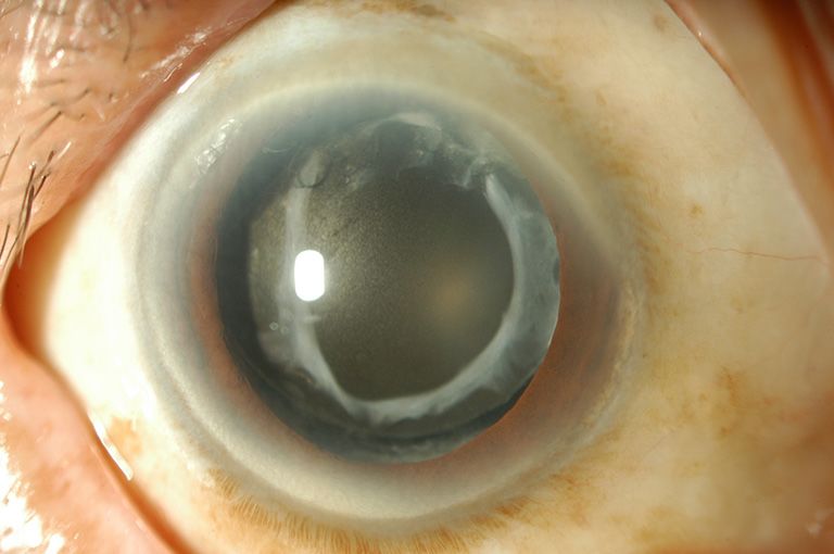
Page 2 of 24                                                    Grzybowski et al. Intraocular lens opacifications: update 2020

the most common of long-term complications (5). After          IOLs with an interrupted sharp optic edge (8,19). However,
5–7 years the incidence may rise to 30–35% (6). Vock et al.    neither of the acrylic lenses is free of material opacification
reported that 42% of the eyes needed Nd:YAG laser              and degradations, which are infrequent but can reduce
capsulotomy due to the PCO 10 years after surgery (7).         visual performance (1).
   It develops from remaining lens epithelial cells, which        Data about IOL opacification impact on visual acuity
proliferate and migrate over the posterior lens capsule (8).   is controversial. While the majority of the peer-reviewed
Other postoperative late complications include cystoid         studies did not show a significant impact of glistenings
macular edema, retinal detachment, endophthalmitis,            and subsurface nanoglistenings on visual acuity, there
lens dislocation, and IOL opacification (5). The IOL’s         are recent data about its significant effect on straylight
biomaterial is one of the most critical factors leading to     (1,12,20). Increased straylight can result in disability glare,
possible post-surgery complications, such as posterior         hazy vision, and loss of contrast (21). The main sources of
and anterior capsule or IOL opacification formation (3).       light scattering in the eye are the crystalline lens, cornea,
The main IOLs materials are hydrophobic, or hydrophilic        fundus reflectance, and light transmittance by sclera and
acrylate, polymethylmethacrylate (PMMA), and silicone (9).     iris (22). Due to the aging processes, including cataract
Different types of material are associated with different      formation, the straylight increases to an average of 1.20
types of IOLs opacifications, which include photochemical      log(s) at 65 years of age, while in youth, the straylight value
material alterations, precipitations and depositions,          is on average 0.90 log(s) (22). Although straylight decreases
glistenings, and discoloration (1). Snowflake degeneration     significantly after cataract surgery, some of the studies
occurred as intraoptic spherical lesions of PMMA material      results show high straylight values in pseudophakic eyes (21).
lenses in the central and midperipheral portion of the         This can be determined by the changes in the IOL material
optic (10). Silicone IOLs were the first foldable lenses       such as calcification, glistenings, subsurface nanoglistenings,
and were known to undergo brownish discoloration and           which can increase the light scattering, therefore having an
central haze within the first 6 weeks postoperatively (11).    impact on visual quality (21,23).
The hydrophobic acrylic lenses have been known to show            The aim of this review is to discuss the recent literature
glistenings, while calcifications develop more often in        on IOL opacifications and their impact on vision quality.
hydrophilic acrylic lenses (11).                                  We present the following article in accordance with the
   Acrylic foldable IOLs have become the most popular          narrative review reporting checklist (available at http://
type of IOLs that are implanted during cataract surgery (9).   dx.doi.org/10.21037/atm-20-4207).
It was reported that Alcon hydrophobic acrylic IOLs are
one of most commonly implanted, and since 1955, over 40
                                                               Methods
million such IOLs have been implanted (12,13). However,
hydrophilic IOLs have different water content, which           PubMed was used for the medical literature search,
makes them more flexible and implantable through smaller       which was conducted up to Apr 7, 2020. The following
incisions than hydrophobic lenses (11,14,15). Hydrophilic      keywords were used in various combinations: cataract
IOLs have better tissue compatibility, though it encourages    surgery, phacoemulsification, intraocular lens, opacification,
lens epithelial cell proliferation and migration, leading      glistening, subsurface nanoglistenings, calcification,
to posterior capsule opacifications (4). The incidence of      snowflake degeneration. Only articles having English
postoperative complications, including posterior capsule       abstracts were reviewed. The reference lists of identified
opacifications, was noticed to be lower with acrylic IOLs      publications were also considered as a potential source
than with other materials, and with the lowest incidence       of relevant articled. Studies were critically reviewed to
in hydrophobic lenses (16,17). Leydolt et al. performed        create an overview and guidance for further search, and no
a randomized controlled study and found that new               attempts to discover unpublished data were made. Emphasis
hydrophobic acrylic Vininex XY1 IOLs had significantly         was placed on articles published since 2010, however, we
lower PCO rates than hydrophobic AcrySof SN60WF                focused mainly on the risk factors for IOL opacification in
IOLs, which were considered to have one of the lowest          particular IOL types. In addition to the literature search,
PCO rates (18). It was reported that anterior capsule          selected chapters from relevant textbooks were included if
opacification and phimosis were significantly less observed    necessary. Due to the large number of studies, in the tables
in the Tecnis IOLs with a continuous edge than in AcrySof      we have presented only original studies and case series, but

© Annals of Translational Medicine. All rights reserved.             Ann Transl Med 2020 | http://dx.doi.org/10.21037/atm-20-4207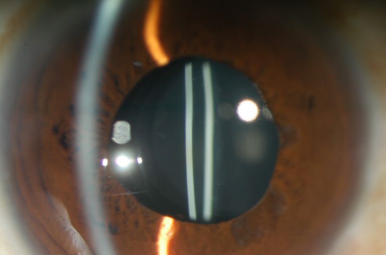
Annals of Translational Medicine, 2020                                                                                     Page 3 of 24

                                                                      of water and loosely packed network chains (25). Saylor
                                                                      and colleagues reported that the water-filled cavities could
                                                                      develop due to osmotic pressure differences between the
                                                                      aqueous solution within the cavity and the external liquid
                                                                      in which the lens is immersed (26). As a result of these
                                                                      mechanisms, water permeates micro-channels within the
                                                                      IOL material and forms small inclusions (1). They can be
                                                                      distributed throughout the entire lens optic, but most often
                                                                      are seen in the anterior and posterior IOL surfaces (27).
                                                                      In addition to these proposed mechanisms , IOLs
                                                                      manufacturing methods and packaging might also have
Figure 1 Light photomicrograph of a hydrophobic acrylic IOL           an impact on glistening formation as well as its material
explanted because of error in power calculation. The presence of      itself (13). It is known that osmotic and temperature changes
microvacuoles (glistenings) can be seen, within the optic substance   are important components in the mechanism of glistening
of the lens (×200). Courtesy: Liliana Werner, MD, PhD, University     formation (1,25,26). However, the breakdown of the blood-
of Utah.                                                              aqueous barrier (BAB) and intraocular inflammatory factors
                                                                      might also have a significant impact on the development
                                                                      of glistening (1). BAB is known to be damaged in diabetes
                                                                      mellitus, uveitis, postoperative inflammation, glaucoma,
                                                                      so that these pathologies may induce the formation of
                                                                      glistening (28,29).
                                                                         Subsurface nanoglistening can be referred to as
                                                                      whitening (Figure 2) of the hydrophobic acrylic IOL
                                                                      affecting the surface or subsurface of the IOL (differently
                                                                      from glistening, which occur within the substance of IOL)
                                                                      (30,31). The formation of these vacuoles is caused by an
                                                                      infiltration of water molecules that can form aggregates
                                                                      within the subsurface of the lens optic (1,12,30,31).
Figure 2 Subsurface nanoglistening (whitening) of the implanted       Nanovacuoles diameter is less than one μm (between 140
IOL. Courtesy: Hiroyuki Matsushima, MD, PhD, Dokkyo Medical           and 185 nm) (1,32). Ong et al. reported that the main source
University.                                                           of hydrophobic acrylic IOL surface light scattering was
                                                                      subsurface nanoglistening (32).
                                                                         Glistening can be found in all IOL materials,
not single case reports.                                              including PMMA, silicone, and hydrophilic acrylic IOLs,
                                                                      but they are observed predominantly in hydrophobic
Glistenings                                                           acrylic lenses, which are one of the most commonly
                                                                      used (24,27,28,33). It was reported that glistening in
Glistenings are small fluid-filled vacuoles in the IOL material       hydrophobic acrylic lenses have higher density (16,33).
(Figure 1), which size usually varies between 1 and 20 μm (24).       However, Łabuz et al. reported that different types of
Microvacuoles refractive indices differ from IOL material, so         hydrophobic acrylic IOLs also differ in their resistance to
when the light redirects and a portion of the light is scattered      glistening formation (20). The majority of studies showed
backward to the observer, it is seen as refractive particles that     a tendency of glistening formation in hydrophobic acrylic
glisten on a slit lamp examination (1,20).                            lenses that were manufactured by Alcon company (1).
   One of the main theories of glistenings formation was              The incidence of glistening in AcrySof IOLs increases
described by Kato et al. in 2001 (25). They concluded                 with time, and it varies from 66% to 100% between
that small changes in temperature could cause the                     published studies results (1). Colin et al. reported the
decompensation of the IOL swollen polymer network,                    formation of glistening in 86.5% of implanted AcrySof
initiating the formation of microvacuoles, which consists             SN60WF IOLs, despite manufacturing changes (34).

© Annals of Translational Medicine. All rights reserved.                    Ann Transl Med 2020 | http://dx.doi.org/10.21037/atm-20-4207
Page 4 of 24 Grzybowski et al. Intraocular lens opacifications: update 2020 However, Miyata et al. reported that the improved (1,16,34,35,41-43). Xi with colleagues, reported that more manufacturing process of AcrySof IOLs suppressed the severe glistening caused the reduction of contrast sensitivity development of surface light scattering up to 3 years at a high spatial frequency and visual field. They suggested postoperatively (35). It was proposed that other types of that the mean deviation (MD) can be considered to be used hydrophobic acrylic IOLs like enVista (manufactured as an indicator for the visual performance of glistening in by Bausch and Lomb) are glistening free clinically (36). IOLs (44). The significant decrease of contrast sensitivity Glistening were not noticed to occur in iMics1 NY- at high spatial frequencies with a higher grade of glistening 60 when compared with the AcrySof SN60WF 3 years severity was reported by Schweitzer et al. (41). The study after surgery (37). It was found that recently developed results also showed a significant association between the hydrophobic acrylic materials that have a higher water incidence of glistening (61.2% including grades 1 and 2) content than the standard (less than 0.5%), which include and the number of topical glaucoma drugs. The disruption the enVista MX60, the Eternity W-60, PodEye IOLs of BAB can be caused by inflammation, chronic use of are glistenings-free in vitro and in vivo (23). Werner et al. glaucoma eye drops (41). These were suggested as possible compared new Clareon CNA0T0 IOLs, which have a water factors that may impact the development of a higher number content of 1.5% with 5 other hydrophobic IOLs (23). The of glistening associated with the daily use of glaucoma authors found that the Clareon showed the lowest levels of drops (41). Godlewska et al. performed a study with 252 surface haze, surface roughness, subsurface nanoglistening, patients undergoing phacoemulsification with AcrySof and glistening (23). IQ IOLs implantation, to evaluate the effect of selected Although hydrophobicity is an important factor in perioperative factors and concomitant diseases to glistening glistening formation, however visual quality may be formation. They reported a significantly higher severity of influenced by glistening properties and their impact on glistening in patients with diabetes, which may influence different optical parameters. the breakdown of physiological intraocular barriers (45). A Philippaki et al. compared glistening formation, their higher refractive power of the intraocular lens and the use of size, and its effect on straylight between the Alcon AcrySof bigger diameter cartridge during phacoemulsification were SN60WF and Santen Eternity Natural Uni NW-60 significantly related to the higher severity of glistening (45). IOLs (13). However, the authors suggested to evaluate The higher refractive power of the IOL can be associated their results carefully, as they had not had the information with the increase in glistening severity due to the if the used IOLs were manufactured before or after the thicker IOL matrix and the higher amount of material changes of manufacturing process that were announced by (24,42). However, other studies did not find a correlation Alcon in 2011 (13). Nevertheless, they found a statistically between the IOLs power and the number of glistening in significantly higher number of glistening produced in Alcon hydrophobic AcrySof IOLs (44,46). AcrySof SN60WF while Eternity Natural Uni NW-60 IOLs Nevertheless, more studies showed that glistening could developed larger glistening (13). Forward light scattering degrade vision by inducing glare symptoms more than for the AcrySof lenses, which produced smaller, but greater lowering visual acuity or contrast sensitivity (20,38). The density glistening, was higher than for the Eternity Natural optical performance of IOL is usually evaluated by using Uni lenses (13). These results were similar to Labuz et al. the modulation transfer function (MTF), which describes study results, which showed that there is a proportional the ability of an IOL to project light from an object onto relationship between the number and the surface portion of the retina for different spatial frequencies (47,48). Light glistening and their effect on straylight (38). More studies scattering from an IOL is quantified as straylight and is found similar results, concluding that glistening of the used to measure a patient’s glare symptoms (46,47). This smaller size and higher density increased the light scattering parameter is becoming an essential aspect of vision quality more (39,40). evaluation (47-49). In laboratory studies, straylight can be Matsushima et al. reported that glistening and quantified using a clinical device (50). It was reported that subsurface nanoglistenings caused decreased vision in 5 straylight is a susceptible measurement to detect the presence patients, leading to IOL explantation, which followed the of IOL pathology, including opacifications. However, MTF improvement of visual acuity (12). However, the majority as the optical quality of IOL deteriorates if the opacification of the studies did not find a significant reduction of visual is severe (51). The correlation between IOL structural acuity caused by glistening or subsurface nanoglistening changes, including opacification, and straylight was found in © Annals of Translational Medicine. All rights reserved. Ann Transl Med 2020 | http://dx.doi.org/10.21037/atm-20-4207
Annals of Translational Medicine, 2020                                                                                 Page 5 of 24

Łabuz et al. study, where IOLs were randomly extracted from       200), grade 3 (201 to 500), and grade 4 (more than 500) (52).
donor’s eyes (21). The results were compared with another         The authors reported that a low number of glistening (
Page 6 of 24                                                        Grzybowski et al. Intraocular lens opacifications: update 2020

                                                                   pars plana vitrectomy (PPV) with silicone oil tamponade,
                                                                   the patient had diabetes mellitus, that could cause the
                                                                   breakdown of BAB as mentioned above, and it was a single
                                                                   case (68). Few studies showed that the hydrophobic surface
                                                                   of hydrophilic acrylic IOLs does not protect them from the
                                                                   development of calcification because it initiates from the
                                                                   hydrophilic subsurface (14,15).
                                                                      Studies performed during the last decade indicated that
                                                                   there is a tendency of centrally localized IOL calcification,
                                                                   that is restricted to the pupillary or capsulorhexis area.
                                                                   This type of calcification occurred after posterior lamellar
Figure 3 Light photomicrograph of a MemoryLens IOL                 keratoplasty: Descemet-stripping automated endothelial
(CibaVision) explanted because of calcification (×40). Courtesy:   keratoplasty (DSAEK), Descemet membrane endothelial
Liliana Werner, MD, PhD, University of Utah.                       keratoplasty (DMEK), and pars plana vitrectomy (PPV),
                                                                   with the intraocular injection of gas or air (51,62). It was
                                                                   reported that direct air or gas contact with the IOL’s surface
                                                                   increases the risk of hydrophilic IOLs opacification (51).
                                                                   However, the incidence of calcification is very low, after
                                                                   DMEK and DSAEK vary between 2.5–5% (51,69,70).
                                                                      Werner et al. proposed possible causes of calcification
                                                                   after surgeries that require exogenous gas or other
                                                                   substances injection into the eye (60). Injected gas, air, tissue
                                                                   plasminogen activator, silicone oil can have direct contact
                                                                   to IOL surface. Secondly, it can be related to a metabolic
                                                                   change in the anterior chamber due to the presence of the
                                                                   exogenous substance and lastly—exacerbated inflammatory
Figure 4 Calcification of the implanted IOL. Courtesy: Hiroyuki    reaction with the breakdown of BAB caused by the surgical
Matsushima, MD, PhD, Dokkyo Medical University.                    procedure itself (1,60).
                                                                      The authors also reported that there is no association
                                                                   between this distinctive calcification localization and IOL
   The anterior segment optical coherence tomography               design or manufacturer (60). A similar conclusion was
(AS-OCT) was reported to be a reliable method to assess            made by Giers et al., who conducted a study of 11 cases
the presence, location and density of IOL’s changes,               of explanted hydrophilic IOLs with calcification from 4
including calcification, however, very superficial changes         different manufacturers after DMEK or DSAEK. The
could not be detected (10,66).                                     authors suggested that calcification occurs irrespective
   The majority of the studies reported IOL calcification          of the manufacturer or the exact composition of the
more commonly in hydrophilic acrylic IOLs (1,62,67).               hydrophilic lens material (71).
Choudhry et al. described two types of hydrophilic acrylic            Most of the single case reports presented calcification
IOLs calcification: first consists of calcium precipitates on      of hydrophilic IOLs after DMEK, DSAEK (72-75). It is
the IOL surfaces. In contrast, the second type includes            important to note that all these cases, except the report by
granular calcium deposits within the substance of the IOL          Lee, had a somehow complicated postoperative course that
optic, beneath the anterior surface and in front of the            led to repeated or different types of surgeries, suggesting
posterior surface of the lens optic, the haptics and the edge      that breakdown of BAB during surgeries might also
of the optic (66).                                                 influence the calcification process (68). Lee reported a case
   Several studies evaluated secondary calcifications, i.e.        when the patient needed the rebulbing after DSAEK (74).
calcifications related with other diseases, pathologies or         It was reported in two studies that rebubbling after
specific intraoperative procedures. Although Ma et al.             DSAEK and DMEK significantly elevates the risk of the
reported calcification of hydrophobic acrylic IOL after            development of IOL’s calcification (69,76). It was proposed

© Annals of Translational Medicine. All rights reserved.                 Ann Transl Med 2020 | http://dx.doi.org/10.21037/atm-20-4207
Annals of Translational Medicine, 2020 Page 7 of 24 that elevated IOP after the injection of intracameral air and hypothesized that rtPA possibly disrupts the BAB (80). might also be a risk factor of the calcification process It is known that calcification in silicone IOLs is (76,77). Although Ahad et al. observed the reduction of the associated with the coexistence of asteroid hyalosis, as opacification rate after reducing the time of high-pressure more than 85% of patients with calcification had clinically (IOP higher than 40 mmHg) air tamponade from 1 h to 10 detectable ipsilateral asteroid hyalosis (10,65,81,82). min, they did not get any statistically significant data (76). Calcification is thought to be caused by the same process Similar cases of centrally localized IOLs calcification as asteroid hyalosis because asteroid bodies are rich of after PPV with intraocular gas/air injection were more calcium and phosphate, and it can occur despite the intact commonly observed in recent years. It was reported by posterior capsule (10,81). Espandar et al. reported three Werner et al. that the possible cause of centrally localized cases of silicone IOLs opacification in patients with asteroid calcification of anterior IOLs surface can be the migration of hyalosis, who underwent Nd:YAG capsulotomy and that gas or silicone oil into the anterior chamber via zonular fiber led to more chalengin IOLs exchange (81). Platt et al. defects (60). Khurana et al. reported a single case of anterior proposed a possible method to remove calcified deposits surface calcification of a hydrophilic acrylic IOL when the from the posterior surface of IOL as the exchange of exposure of air in the anterior chamber was observed the IOLs is associated with intraoperative and postoperative next day after PPV (78). The authors proposed a possible complications (65). The authors presented a surgical mechanism (78). The exposure of gas to the IOL surface technique that included PPV, a lighted pick, and a modified results in an increased hydrolyzation of the polyacrylate, silicone-tipped cannula with successful removal of late forming free carboxylic acid groups that accumulate at the calcium deposition. However, it was a single case, and the IOL surface triggering biomineralization, thereby covering follow-up was limited to 6 months (65). the IOL optic with calcium phosphate deposits (78). These Primary calcification is associated with the problems of mechanisms could explain the fact that calcification was not IOL itself (59). It was reported that some of the different seen in the IOL parts that were covered with the capsule models of Oculentis IOLs, implanted between 2009 and (62,78). However, some of the studies reported cases with 2012, were affected by primary calcification because no no noticeable gas migration to the anterior chamber (61,79). other significant causes were found (11,47). After the efforts Likewise, the opacification of the posterior capsule was to discover possible causes, the company concluded that reported by Marcovich et al. (61). The authors performed a its origin might be multifactorial and published few safety study with detected opacification in 11 hydrophilic acrylic notifications (11,47). Barra et al. performed a study with the IOLs produced by six different manufacturers, 1 month to same design hydrophilic Ioflex IOLs with calcification (67). 6 years after PPV involving the intravitreal gas injection (61). They found the difference of light transmittance at a certain The authors hypothesized that the calcification might be region in explanted and control IOLs, which had different caused by dehydration of the IOL due to slowly dissolving expiration dates (67). The authors noted the importance of gas (61). The dehydration may induce chemical alterations the manufacturing process evaluation because they found on the IOL surface, causing deposition of calcium and that different materials can be used or manufacturing phosphate from the aqueous humor in the exposed areas (61). processes can vary at a different time while manufacturing Yildirim et al. performed a study with one of the most the same type of IOLs (67). extensive series of confirmed calcification in 10 explanted Yildirim et al. performed a study with nine segmented hydrophilic acrylic IOLs after PPV with intraocular gas refractive bifocal Lentis MF IOLs, which were affected by injection (62). They found that calcification was not present primary calcification, as the granular deposits were found only on the surface of the IOL but up to 100 μm within the underneath the anterior and posterior surfaces distributed material (62). In most of the cases, calcification was found in throughout the whole IOL, including the haptics (47). The the anterior central pupillary area (62). The authors reported authors reported that the density of calcium phosphate that there is a strong correlation between the density and size granules affected straylight significantly because the highest of the calcium deposits and the decrease in the IOL's optical values were found in most severe cases (47). They did not quality (47,62). find the correlation between light scattering and the MTF We found one study by Fung et al., who reported 7 cases or visual acuity, suggesting that these are independent of hydrophilic IOLs opacification after treatment with factors, and visual acuity may not be sufficient parameter to intracameral recombinant tissue plasminogen activator (rtPA) quantify the effect of IOLs’ calcification (47). The similar © Annals of Translational Medicine. All rights reserved. Ann Transl Med 2020 | http://dx.doi.org/10.21037/atm-20-4207
Page 8 of 24                                                      Grzybowski et al. Intraocular lens opacifications: update 2020

results were reported by Łabuz et al. (51). A severe increase    glistenings tend to occur more often in AcrySof IOLs than
of straylight was caused by IOLs’ calcification, which           in other hydrophobic IOLs.
occurrence was associated with intraocular gas injection (51).       One of the most important factors in the development
However, the MTF values decreased just in two IOLs.              of opacifications: glistenings and calcification, despite the
These results were similar to other studies results, where       properties of IOL, is the breakdown of BAB, which modifies
MTF values did not show a significant effect too (51). The       the aqueous humor composition (3). It is known to be
authors found a proportional relationship between the            disrupted in diabetes mellitus, and it was the main systemic
straylight parameter and the size and number of calcium          disease associated with opacification formation (12,45).
deposits, suggesting that there is variability in the optical        The significant association between glistenings formation
quality of affected IOLs (51). Tandogan et al. reported          and glaucoma was reported, as the daily use of topical
that MTF values deteriorated significantly in explanted          glaucoma medications may lead to the rupture of BAB (41).
Euromaxx ALI313Y and ALI313 IOLs with the calcification          Although Godlewska et al. did not find a statistically
of the entire optic (64).                                        significant difference, they observed that glistenings
                                                                 formation was more frequent in glaucoma patients
                                                                 when compared to subjects without glaucoma (45). The
Discussion
                                                                 breakdown of BAB can result in uveitis, and it was linked
We presented above main studies on IOL opacifications            to glistenings formation. We found only one study which
based on the review of recent literature. The summary of         assessed patients with uveitis, however, the authors reported
major recent original papers on glistenings is presented in      that they received high intensity steroid therapy, and the
Table 1 and on calcifications in Table 2. In this section we     results showed statistically lower severity of glistenings in
discuss two important and controversial issues related to        these patients (45).
IOL opacifications, which are risk factors and impact on             The majority of the IOLs with confirmed calcification
visual quality.                                                  were hydrophilic in the reviewed studies. However, few
   It is known that the material of IOL plays an important       cases of calcification in hydrophobic acrylic IOLs were
role in opacification formation. Glistenings occurred more       reported, likewise, calcification was observed in silicone
in hydrophobic IOLs and some of the studies observed it          IOLs in association with the asteroid hyalosis, suggesting
despite manufacturing changes (34,82). However, Łabuz            that calcification is not the problem only of hydrophilic
et al. showed that different hydrophobic materials differ in     IOLs (10,65,68,69,81).
their resistance to the glistening formation (20). One of the        Complex or prolonged surgery and postoperative
possible causes is the difference of water content, as it was    inflammation can lead to the breakdown of BAB, and
reported that certain hydrophobic IOLs with higher than          that may induce glistening and calcification formation
ordinary water content was glistening free or its amount was     in the IOLs (1,68,73,75). Although Gurabardhi et al.
showed to be very low (23). It was proposed that the higher      did not find a statistical correlation between ocular or
refractive power of IOL can be associated with higher            systemic comorbidities and primary calcification, they
severity of glistenings. However, we found three studies         found that diabetes, uveitis, and glaucoma were the most
(44,46,83) that did not find the significant association         frequent pathologies (11). Although the incidence of IOLs
between the IOLs power and the severity of glistenings and       calcification is low and studies evaluated not more than
previously reported findings could be caused by the higher       fifteen IOLs explanted due to the secondary calcification,
thickness of the material (24,42,45).                            it showed a significant effect of intraocular injection of
   Two long follow up studies showed that a higher               exogenous air or gas on calcification formation as well as on
amount of glistenings formed in AcrySof IOLs than in             the tendency of central localization. The main surgeries that
ZCBOO IOLs, likewise the anterior capsule opacification          required intraocular gas, air or silicone (86) injection were
and fibrosis was more seen in AcrySof IOLs (8,19). It was        DSEK, DSAEK, DMEK, and PPV. The rebubbling after
suggested that glistenings formation might be associated         DSAEK and DMEK was reported as a significant risk factor
with the occurrence of anterior capsule opacification and        for the development of IOL’s calcification. The summary
tightness of the capsular bag (1). However, the authors did      of risk factors for IOL glistenings and calcifications is
not perform analysis to evaluate this possible association,      presented in Table 3.
and they reported it as an observation. It is also known that        The early controversy related with the influence of IOL

© Annals of Translational Medicine. All rights reserved.               Ann Transl Med 2020 | http://dx.doi.org/10.21037/atm-20-4207
Annals of Translational Medicine, 2020 Page 9 of 24
Table 1 The summary of major original studies on glistenings
Authors IOLs/patients Evaluated parameters/methodology Results
Matsushima 5 explanted IOLs were MA60BM (Alcon) VA was assessed Light transmittance of the IOLs explanted from cases 1, 2, 3, 4 and 5 was 85.0%, 78.2%, 79.1% 80.1% and 76.7% (respectively)
et al. (12)
Unimplanted MA60BM (Alcon) was used as control Light transmission was measured by a double beam spectrophotometer Compared with the light transmittance of a control unused IOL of 88.9%; these values represent a decrease of from 4.4% to 13.7%
Original implantation had occurred over a range of 6–15 All patients VA improved when IOLs were exchanged
years prior to the IOL exchange
Philippaki 5 Alcon AcrySof SN60WF IOLs Glistenings were induced in vitro The median increase in the number of glistenings was 15 for the Eternity and 525 AcrySof IOLs (P=0.012)
et al. (13)
5 Santen Eternity Natural Uni NW-60 IOLs Straylight was assessed Median glistenings diameter was 23.8 μm (AcrySof) and 32.8 μm (Eternity)
All IOLs had same dioptric power (+20.0 D) Four (80%) of the AcrySof lenses had straylight values higher than a 20-year-old CIE standard glare observer and in two cases the
straylight exceeded that of the 70-year-old CIE standard glare observer
None of the Eternity lenses had straylight values that exceeded the value for the 20-year-old CIE standard glare observer
Weindler 38 monofocal hydrophobic acrylic + 21.0 D AcrySof SA60AT Glistenings were induced in vitro The mean glistening diameter in grades 1 through 4 IOLs was 15.31±3.13 mm (range, 7.33 to 24.74 mm).
et al. (52) IOLs
Control group of 20 IOLs A classification was applied based on the glistening number per mm2 The mean glistening numbers ± SD (MV/mm2) in grades 1 through 4 were 74±12.7, 142±22.2, 297±76.2, and 1,509±311.9, respectively.
MTF and Strehl ratio was assessed The mean glistening sizes in grades 1 through 4 were 13.28±3.85, 15.88±2.08, 16.85±3.23, and 15.27±2.25 mm, respectively.
Glistening grades 1 through 3 did not change the optical quality
In grade 4, the MTF and the Strehl ratio were significantly affected, however the effects found were small and are unlikely to affect the VA
Son et al. (58) 4 IOLs: a monofocal AcrySof IQ SN60WF (Alcon); diffractive- An experimental set-up with a water bath containing 0.01% fluorescein solution and Both the diffractive-refractive bifocal IOL and the EDOF IOL showed two defined foci for distance and near vision
refractive bifocal AcrySof IQ Restor SN6AD1 (Alcon); monochromatic green laser light (532 nm) was used
diffractive trifocal AcrySof IQ PanOptix TFNT00 (Alcon);
diffractive extended-depth-of-focus (EDOF) Symfony ZXR00
(Johnson & Johnson)
All studied lenses power was +21.0 D MTF and Through-Focus Response was assessed In the diffractive trifocal IOL, three distinct foci for distance, intermediate, and near vision could be visualized
Łabuz 30 IOLs Glistenings were induced in vitro Glistenings were found in all but one (Avensee) of the studied IOL models
et al. (20)
6 IOL models: CT Lucia 601P (Zeiss); PY60AD (Hoya); Glistening statistics (MV number and size) were derived from image analysis The number of glistenings ranged from 0 to 3,532 MV/mm2
SN60WF(Alcon), MA60AC (Alcon); Aktis SP NS; YG (Nidek);
Straylight was assessed The mean size of glistenings ranged from 5.2 to 10.2 μm
Avansee (Kowa)
The highest density of glistenings was found in the PY-60D IOLs ranging from 3,058 to 4,061 MV/mm2
MA60AC samples demonstrated the largest size of glistenings
The mean straylight parameter (± SD) of the IOLs prior incubation ranged from 0.1 to 1.3 deg2/sr
Straylight of the CT Lucia was 1.09±0.99 deg2/sr, PY-60AD it was 19.30±2.07 deg2/sr, for the SN60WF and MA60AC IOLs it was
1.15±0.15 deg2/sr and 5.95±3.67 deg2/sr respectively, for the Aktis it was 1.71±0.84 deg2/sr, for the Avansee it was 0.95±0.24 deg2/sr
Łabuz 47 monofocal IOLs IOLs extracted from donor pseudophakic eyes The mean straylight at 2.5 degrees and 7.0 degrees was 5.78 deg2/sr±4.70 and 5.06±4.01 deg2/sr, respectively
et al. (21)
Straylight was assessed at a 2.5-degree and 7.0-degree scatter angle, results were 30 of the 74 IOLs (41%) straylight was below the level of that of the 20-year-old crystalline lens
compared with the straylight of a 20-year-old crystalline lens, a 70-year-old crystalline
Straylight was above the level of the 70-year-old crystalline lens in 10 IOLs (14%)
lens, and a lens with cataract
None showed a straylight level close to that of the cataractous lens
Increased straylight was associated with surface deposits, snowflake-like degeneration, and glistenings
34 IOLs (43%) were free of IOL pathology; 40 IOLs showed different levels of opacification
Łabuz et al. 7 AcrySof IOLs: 5 SN60WF; 2 SN60AT Glistenings were induced in vitro The median size of glistenings in was 5.4±2.7 mm (range, 4.6 to 12.5 mm)
(38)
Straylight was assessed The number of induced glistenings ranged from 114 to 12 386 per mm2, the surface portion ranged from 1.4% to 26.9%
At 2.5 degrees, the range in the straylight parameter was 1.49 to 72.49 deg2/sr; at 7.0 degrees, it was 1.72 to 62.87 deg2/sr
Straylight was proportionally related to the total number of glistenings and the surface portion
Table 1 (Continued)
© Annals of Translational Medicine. All rights reserved. Ann Transl Med 2020 | http://dx.doi.org/10.21037/atm-20-4207Page 10 of 24 Grzybowski et al. Intraocular lens opacifications: update 2020
Table 1 (Continued)
Authors IOLs/patients Evaluated parameters/methodology Results
Henriksen 79 pseudophakic patients with visual acuity no worse than All IOLs were photographed, and glistenings were analyzed for size and density All 79 patients had glistenings within 2 diameter groups: 6 to 25 μm and over 25 μm
et al. (39) 0.02 logMAR and no ocular pathology were enrolled
The SN60WF IOL was implanted in 36 eyes (45.5%), the Outcome measures included logMAR CDVA, mesopic 10% contrast logMAR CDVA Linear regression for the non-stratified group was significant for IOL glistening size vs. contrast VA with glare
SN60AT in 36 eyes (45.5%), the SN60T5 in 4 eyes (5.2%), with and without glare, and straylight determination with a straylight meter (C Quant
Linear regression for the 6 to 25 μm group was significant for a measure of severity index (%area) vs. the straylight meter measurements,
the SN60T4 in 1 eye (1.3%), the SN60T3 in 1 eye (1.3%), log)
%area/size vs. straylight meter measurements, IOL age vs. CDVA, IOL age vs. contrast VA, and IOL age vs. contrast VA with glare
and the SN6AD3 in 1 eye (1.3%)
Linear regression for the over 25 μm group was significant for IOL age vs. glistening size and %area/size vs. contrast VA, and density vs.
CDVA and contrast VA with glare
Werner et al. IOLs types: Clareon CNA0T0, Tecnis ZCB00 and Tecnis Glistenings were induced in vitro The surface haze (n=10, PIU) was 4.25±0.87 (CNA0T0), 9.50±1.66 (ZCB00), 39.48±1.97 (ZCB00V), 46.68±3.16 (W-60), 44.70±4.00 (MX60),
(23) OptiBlue ZCB00V, Eternity W-60, enVista MX60, and Vivinex and 4.42±0.71 (XY1) (PAnnals of Translational Medicine, 2020 Page 11 of 24
Table 1 (Continued)
Authors IOLs/patients Evaluated parameters/methodology Results
Chang et al. 80 patients: 40 AcrySof SA60AT (1-piece IOL group); 40 a 5 to 7 years postoperatively, retroillumination images were obtained and the PCO There were no significant differences in PCO between the 2 groups
(83) Sensar AR40e (3-piece IOL group) area and severity were evaluated using computer software
High-contrast (100%) and low-contrast (2.5%) CDVA were measured The 3-piece IOL group had significantly fewer glistenings (PPage 12 of 24 Grzybowski et al. Intraocular lens opacifications: update 2020
Table 1 (Continued)
Authors IOLs/patients Evaluated parameters/methodology Results
Schweitzer 67 glaucomatous eyes (47 patients), who previously had a All eyes underwent a BCVA evaluation, a complete clinical examination, a visual 26 eyes (38.8%) had a grade 0, 12 eyes (17.9%) a grade 1 and 29 eyes (43.3%) a grade 2 of glistening severity grade
et al. (41) phacoemulsification with a hydrophobic acrylic IOL field test, CS evaluation and a wavefront analysis of HOAs using a Shack–Hartmann
aberrometer
Glistening was classified in three groups of severity grade: G0 (Annals of Translational Medicine, 2020 Page 13 of 24
Table 2 The summary of major original studies on calcification
Authors IOLs/Patients Evaluated parameters/methodology Results
Tandogan et al. (64) 6 explanted Euromaxx IOLs: 5 Euromaxx ALI313Y; 1 Euromaxx X-ray spectroscopy Macroscopically, the entire optic was opacified in all IOLs. Numerous fine, granular, crystalline-like deposits, which were always distributed in a line
ALI313 parallel to the anterior and posterior surfaces of the IOLs
Light and scanning electron microscopy X-ray spectroscopy could prove the deposits consisted of Calcium and Phosphate
MTF was assessed The MTF measurements of the IOLs with an intact optic (1 was not able to evaluate) showed a significant decrease in optical quality
Measurements in the optical bench showed significant reduction of MTF values at all spatial frequencies and United States Air Force target pictures
demonstrated a significant reduction of brightness as well as resolution with the opacified IOLs
Barra et al. (67) 7 explanted Ioflex IOLs AS-OCT was used The explanted IOLs demonstrated the presence of granular deposits, which were predominantly located on the surface/subsurface of the IOLs,
particularly the anterior surface
8 control Ioflex IOLs Light scattering was measured with a Scheimpflug Light scattering was 219.71 CCT±2.62 for explanted IOLs and 4.75±2.50 CCT for controls
camera
Light transmittance was assessed with a The mean light transmittance in the visible light spectrum was 75.94% to 87.25% for explanted IOLs and 97.54% to 98.97% for controls
spectrophotometer
A variable degree of light transmittance between 290 nm and 350 nm (ultraviolet-A and B radiation) in the explanted and control IOLs with expiration
dates in 2009/2010 but 0% transmittance in this region in all controls with expiration dates in 2011/2012
Bompastor-Ramos 20 explanted opacified Lentis LS-502-1 (Oculentis GmbH) Slitlamp examination The mean interval between the initial cataract surgery and the diagnosis of opacification of the IOLs was 29.15±9.57 months (range, 6 to 45 months)
et al. (15)
IOLs CDVA Opacification led to a statistically significant reduction in corrected distance visual acuity (mean 0.86±0.76 logMAR; PPage 14 of 24 Grzybowski et al. Intraocular lens opacifications: update 2020
Table 2 (Continued)
Authors IOLs/Patients Evaluated parameters/methodology Results
Stringham et al. (85) 16 lenses were of 8 designs manufactured from different silicone Gross examination The presence of asteroid hyalosis was confirmed in 13 cases (out of 16)
materials
111 hydrophilic acrylic lenses explanted because of calcification Light microscopy The deposits were only on the posterior optic surface of the silicone lenses and were composed of calcium and phosphate
were assessed for comparison
Scanning electron microscopy An Nd:YAG laser posterior capsulotomy was performed in 12 cases a mean standard deviation of 7.57±4.21 years after IOL implantation
EDS A history of asteroid hyalosis was not found in relation to any of the 111 cases of postoperative calcification of hydrophilic acrylic lenses
Giers et al. (71) 13 opacified hydrophilicIOLs from 4 different manufacturers after Optical bench assessment for optical quality Macroscopically, all IOLs showed a more or less circular opacification of the central anterior optical surface, sparing the peripheral optical zone and
posterior lamellar keratoplasty: 8 after DSAEK; 3 after DMEK; 2 after the haptics
both DSAEK and DMEK
Light microscopy Scanning electron microscopy MTF measurements of 10 IOLs that were received with an intact optic showed a significant decrease in optical quality with MTF values deteriorated
at all spatial frequencies
EDS
Schrittenlocher Retrospective review of charts and slit-lamp images of 564 Patients with sufficient documentation during routine IOL calcifications after DMEK occurred in 14 patients (2.5%)
et al. (69) consecutive patients from the prospective Cologne DMEK follow-up examinations with regard to calcifications
database who underwent DMEK in pseudophacic eyes or DMEK in of the anterior surface of the IOL were included in this
combination with cataract surgery(triple-DMEK) analysis
Patients were grouped into affected group and Morphologically, calcifications showed either diffuse clusters of small granular deposits or a denser configuration with sharp edges positioned in the
unaffected group without calcifications of the IOL pupillarycenter of the IOL
VA in affected and unaffected eyes were 0.33±0.24 logMAR and 0.16±0.01 logMAR after 3 months (PAnnals of Translational Medicine, 2020 Page 15 of 24
Table 2 (Continued)
Authors IOLs/Patients Evaluated parameters/methodology Results
Fung et al. (80) 7 hydrophilic acrylic one-piece IOL (Rayner C-flex 570C and Light microscopy Scanning electron microscopy 3 patients had proliferative diabetic retinopathy, 1 had glaucoma
Superflex 620H).
EDS Anterior chamber inflammatory membranes developed between 1 and 4 weeks of surgery and were treated with intracameral rtPA
IOL opacification was noted between 4 weeks and 6 years after rtPA treatment with reduced visual acuity, and IOL exchange was carried out in 3
patients
Diffuse fine granular deposits (confirmed as calcium and phosphate) on the anterior surface/subsurface of IOL optic
Gurabardhi et al. (11) 71 opacified 1-piece or 3-piece hydrophilic acrylic with a Light microscopy Morphological findings were surface, subsurface, or deep calcifications of the IOL material
hydrophobic surface coating IOLs (Lentis) of different designs from
CDVA was assessed The explanted IOLs exhibited a whitish discoloration within the opacified areas
2009 to 2012 (LS-502-1, LS-402-1Y, LS 312-1Y, LS-313-1Y, L-402,
L-312) Opacification was observed in the entire IOLs in some cases (optic and haptics), sometimes with clear localized areas, which were more pronounced
in the optic-haptic junction areas in cases with 3-piece IOLs
Explantation was performed 4 years ±1.2 after initial phacoemulsification
Ocular and systemic comorbidities were found without statistical correlation: the most frequent were diabetes, uveitis, and glaucoma
The preoperative mean corrected distance visual acuity changed from 0.63±0.47 logarithm of the minimum angle of resolution (logMAR) to 0.20±0.28
logMAR postoperatively (PPage 16 of 24 Grzybowski et al. Intraocular lens opacifications: update 2020
Table 2 (Continued)
Authors IOLs/Patients Evaluated parameters/methodology Results
Patel et al. (86) 12 eyes that underwent retinal detachment repair after secondary Post-operative data included characteristics of IOL Major predisposing risk factors for retinal detachment included trauma (42%), prior vitrectomy (33%), and ectopia lentils (17%)
implantation of Akreos AO60 IOL with a subset experiencing the opacification, length of follow-up and complications
The procedure for surgical repair was vitrectomy without scleral buckle in 10 eyes (83%) and combination vitrectomy and scleral buckle in two eyes
complication of IOL opacification
(17%)
Intraocular tamponade used was 5000 centistoke silicone oil (42%), 1000 centistoke silicone oil (25%), C3F8 (25%) and SF6 (8%)
There were 5 cases (42%) of permanent late IOL opacification
Opacification occurred in 2 of 4 eyes (50%) with gas tamponade and 3 of 8 eyes (38%) with oil tamponade
The average time to opacification was 46.2 days (range, 10–104 days)
Two eyes required explantation of the IOL
Photograph of an explanted IOL demonstrated diffuse tan, white calcium deposits on the lens optic and haptics
IOLs, intraocular lenses; MTF, modulation transfer function; AS-OCT, anterior segment optical coherence tomography; CCT, computer compatible tape; CDVA, corrected distance visual acuity; VA, visual acuity; SEM, scanning electron microscopy; EDX/EDS, energy-dispersive x-ray spectroscopy; PMMA,
polymethyl methacrylate, UGH, uveitis-glaucoma-hyphemia; DSEK, Descemet-stripping endothelial keratoplasty; Descemet-stripping automated endothelial keratoplasty, DSAEK; Nd:YAG, neodymium-doped yttrium aluminum garnet; DMEK, Descemet membrane endothelial keratoplasty; PPV, pars plana
vitrectomy; rtPA, recombinant tissue plasminogen activator; UDVA, uncorrected distance visual acuity; UNVA, uncorrected near visual acuity.
Table 3 The summary of risk factors for IOL glistenings and calcifications
Type of IOL opacificiation The material of IOLs The breakdown of BAB Ocular pathologies/surgeries
Glistenings Most often in hydrophobic IOLs Diabetes; preservatives in glaucoma topical medications; postoperative Glaucoma
inflammation; complex or prolonged surgery
Calcifications Most often in hydrophilic IOLs Intraocular injection of gas or air during DSEK, DSAEK, DMEK, PPV; Asteroid hyalosis (in silicone IOLs)
BAB, blood-aqueous barrier; DSEK, Descemet-stripping endothelial keratoplasty; DSAEK, Descemet-stripping automated endothelial keratoplasty; DMEK, Descemet membrane endothelial keratoplasty; PPV, pars plana vitrectomy.
© Annals of Translational Medicine. All rights reserved. Ann Transl Med 2020 | http://dx.doi.org/10.21037/atm-20-4207Annals of Translational Medicine, 2020 Page 17 of 24 glistenings on visual function was focused on visual acuity. subsurface nanoglistenings which were not evaluated by The majority of the reviewed studies that evaluated visual the glistenings detection program (13). Although this study acuity did not find a significant glistening effect on visual presented an important finding of glistenings size effect, the acuity. However, one study, conducted by Matsushima et al. sample of the study is too small, also they did not report the found that glistenings caused significantly decreased visual threshold of glistening size and density which could have a acuity in five patients, leading to IOL explantation (12). significant impact on visual performance. However, they did not perform statistical analysis to The majority of the studies did not present the exact evaluate the changes in visual acuity (12). As well as all levels of glistenings paramaters or straylight values patients had ocular comorbidities: one had a history of that would have a significant impact on certain visual uveitis, one had glaucoma, two had diabetic retinopathy, performances. However, straylight values could be one macular hole, these last three cases had a history of compared to the data found in the literature. The straylight PPV (12). It was reported that a 5-fold increase in light level of a 20-year-old was reported to be s = 2.5 deg 2/ scattering resulted only in a small decrease of 0.1 log units steradian (sr), 70-year-old s = 11.2 deg2/sr, and cataractous s in contrast sensitivity and had no effect on visual acuity = 33.1 deg2/sr crystalline lens (51). It was reported that the (38,56). This finding is similar to ours, as most of the hindrance of straylight could be 1.47 log(s) (54). Van der studies did not find effect on visual acuity, some of the Mooren et al. defined that a lens with significant glistenings studies showed decreased contrast sensitivity (16,39,44). would be the lens that caused straylight levels above those The reduction of contrast sensitivity can be associated of a healthy 20-year-old crystalline lens (85). Increased with the increase of straylight and disability glare. As more straylight by 19.0 deg2/sr was reported to be associated with studies showed decreased contrast sensitivity than visual a 76% increase in halo size and a severe loss in luminance acuity, it could confirm that disability glare and straylight detection threshold (79). are more appropriate in evaluating IOLs opacification Łabuz et al. found a proportional relationship between effect on visual quality. straylight, amount of glistenings, and the surface That was seen in recent years studies that used the portion (38). They concluded that a large number of straylight measurement more commonly to evaluate the glistenings were needed to cause significant straylight visual quality. All of the reviewed studies that measured increase (38). The same group of the authors conducted the straylight values showed a significant increase of straylight other study and reported that 60% of thirty hydrophobic when opacification, including both glistenings and IOLs with in vitro induced glistenings had straylight values calcifications, were detected. The majority of the studies below that for the young lens (20). Only 20% reached reported increased straylight values with a higher amount light scattering levels with an average of 18.1 deg2/sr. that of glistenings (20,38). However, few studies highlighted the could have the potential to hinder visual performance (20). importance of its size. Henriksen et al. performed an in vivo Łabuz et al. evaluated the light scattering in IOLs extracted study with 79 patients and found the correlation between from donors’ eyes (21). They reported that the scattering smaller size glistenings and increased light scattering (39). intensity was higher than in the 70-year-old lens in 14% They also found that the age of the IOL had a negative of the IOLs, and none showed straylight values that could correlation with the contrast visual acuity with glare and correspond to the cataractous lens (21). The median corrected distance visual acuity in patients group with straylight values in donor IOLs were approximately 0.3 to smaller glistening size, suggesting that smaller glistenings 1.0 log(s) (s = 2 to 10 deg2/sr) (21). had a more significant impact on visual function (39). The majority of the studies were performed in vitro. However, they did not include and evaluate IOL Although studies used protocols for glistening induction opacification resulting in surface light scattering in their by the thermal accelerated aging process with varying study, which could also have an impact on the reported temperature and time setting, it was speculated that there is results (39). It was reported by Philippaki and colleagues lack of evidence, if this method produces clinically relevant that the higher amount and smaller glistenings produced results. However, Łabuz et al. compared the highest higher levels of straylight (13). The authors reported that straylight values of 3-piece IOLs from two studies: a study the straylight of two out of five AcrySof IOLs exceeded that with glistenings induced in vitro and another study with of the 70-year-old CIE standard glare observer, however, IOLs extracted from donors’ eyes. They also compared it was proposed that these high values could be caused by the glistening size, and the results were similar. Thus, © Annals of Translational Medicine. All rights reserved. Ann Transl Med 2020 | http://dx.doi.org/10.21037/atm-20-4207
You can also read