Clinical outcomes with a new microincisional diffractive multifocal IOL
←
→
Page content transcription
If your browser does not render page correctly, please read the page content below
Alió et al. Eye and Vision (2015) 2:2
DOI 10.1186/s40662-015-0012-8

 RESEARCH                                                                                                                                      Open Access

Clinical outcomes with a new microincisional
diffractive multifocal IOL
Jorge L Alió1,2,3*, Alfredo Vega-Estrada1,2 and Ana B Plaza-Puche1

  Abstract
  Background: To evaluate the refractive outcomes and the optical performance as well as the quality of life in
  patients implanted with a new diffractive multifocal intraocular lens (IOL).
  Methods: Prospective, clinical study including 41 cases of patients who underwent cataract surgery and were
  divided in two groups: group 1, including 20 eyes implanted with the multifocal IOL SeeLens MF (Hanita Lenses,
  Israel); group 2, 21 eyes implanted with the Acrysof SA60AT IOL. Visual acuity, defocus curve, intraocular aberrations,
  contrast sensitivity function and quality of life were assessed during a follow up period of 6 months.
  Results: Significant improvement was observed in the uncorrected distance visual acuity (UDVA) and corrected
  distance visual acuity (CDVA) in both groups (p < 0.02). The multifocal group showed better results in terms of
  uncorrected near visual acuity (UNVA) and distance-corrected near visual acuity (DCNVA) (p < 0.01). Comparison
  of both groups showed better visual acuities for the multifocal IOL group in defocus levels from -3.0 D to -1.50 D
  (p ≤ 0.01). At 6 months, there was a significant reduction of the internal higher order aberrations (p ≤ 0.04). A
  significant increase in scotopic contrast sensitivity was detected for 6 cycles/° spatial frequency during follow up
  (p = 0.04), but no significant changes were observed for the rest of spatial frequencies (p ≥ 0.06). Visual Functioning
  Index (VF-14) questionnaire showed that patients reported high levels of satisfaction when performing daily tasks.
  Conclusions: The SeeLens MF IOL is able to successfully restore distance, near and intermediate visions after
  cataract surgery. It also provides functional intermediate vision with optimal intraocular optical quality.

Background                                                                           (MICS) together with IOLs that can be implanted through
The outcomes in terms of quality of life in modern cata-                             corneal incisions smaller than 2 mm will show the best
ract surgery depend in part on the type intraocular lens                             outcome in terms of optical performance [10]. There are
(IOL) implanted [1-6]. Recent multifocal IOL technology                              some studies published in the scientific literature that have
emphasizes refractive and optical quality outcomes aim-                              reported good outcomes with diffractive IOLs implanted
ing to provide distance, intermediate and near-spectacle                             through a sub 2 mm incision [10,13,14]. Recently, a new
independence [7-9].                                                                  model of apodized diffractive IOL has been introduced,
  Differences in visual performance achieved with multi-                             with an asymmetrical light distribution and the potential
focal IOLs depend on the optical principles and IOL de-                              to be implanted through a corneal incision smaller than
signs. Diffractive IOLs are one specific type of multifocal                          2.0 mm: the SeeLens MF (Kibbutz Hanita, Israel).
lenses. Many studies [10-17] have reported visual out-                                 The aim of the current investigation is to evaluate the
comes, optical quality, and quality of life with several                             visual and optical quality outcomes, as well as the post-
models of diffractive IOLs. Refractive results are crucial                           operative quality of life and clinical outcomes in patients
for the optimal optical performance of this type of IOLs,                            implanted with the new SeeLens MF apodized diffractive
and control of the refractive astigmatism has a direct im-                           multifocal IOL after cataract surgery .
pact on this factor. Thus, microincision cataract surgery
                                                                                     Methods
* Correspondence: jlalio@vissum.com                                                  Patients
1
 Vissum Corporation, Alicante, Spain
2
 Division of Ophthalmology, Universidad Miguel Hernández, Alicante, Spain            In this prospective pilot consecutive study, we included
Full list of author information is available at the end of the article               20 eyes of 10 bilateral cataract surgery patients aged 58
                                        © 2015 Alió et al.; licensee BioMed Central. This is an Open Access article distributed under the terms of the Creative
                                        Commons Attribution License (http://creativecommons.org/licenses/by/4.0), which permits unrestricted use, distribution, and
                                        reproduction in any medium, provided the original work is properly credited. The Creative Commons Public Domain
                                        Dedication waiver (http://creativecommons.org/publicdomain/zero/1.0/) applies to the data made available in this article,
                                        unless otherwise stated.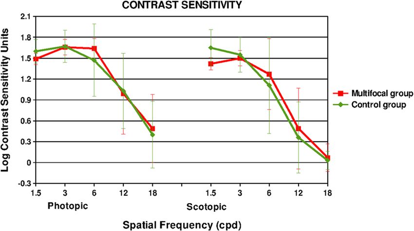
Alió et al. Eye and Vision (2015) 2:2                                                                               Page 2 of 9

to 71 years (mean 67.3 ± 3.8 years) who were implanted           intracameral mydriasis. The main incision was placed on
with the multifocal IOL SeeLens MF. As a control                 the axis of the positive corneal meridian. In the group of
group, we included 21 eyes of 16 cataract patients aged          patients that underwent the procedure with the SeeLens
46 to 80 years (mean 68.4 ± 9.1 years) with a monofocal          MF, the IOL was implanted in the capsular bag through a
IOL implantation. The inclusion criteria of this study           corneal incision of 1.8 mm. In the control group, the
were patients with bilateral visually significant cataract,      monofocal IOL were implanted through an incision of
older than 45 years, with corneal astigmatism less than          2.0 mm. Postoperative topical therapy included a combin-
1.0 D. The exclusion criteria were comorbidities such as,        ation of topical antibiotic and steroid agents (Tobradex®
visually significant corneal scars, and known retinal dis-       Alcon Cusí Inc, Barcelona).
orders. All patients were adequately informed and signed
a consent form. The study adhered to the tenets of the           The intraocular lenses
Declaration of Helsinki and was approved by the Vissum           The SeeLens MF (Kibbutz Hanita, Israel) is a new C-
Alicante Ethical Board Committee.                                loop MICS IOL that consist of an aspheric apodized dif-
                                                                 fractive multifocal IOL. This lens is a single piece IOL
Preoperative examination                                         with an optic diameter of 6.0 mm, and an overall length
Preoperatively, all patients had a full ophthalmologic exam-     of 13.0 mm with a 360° continuous square edge optic.
ination including the evaluation of the refractive status, the   The diffractive steps are located in the 4 mm central
distance and near visual acuities, slit lamp examination, to-    zone, suiting pupil sizes in various lighting conditions.
nometry, and funduscopy. Distance and near visual acuity         The near vision add of this lens is +3.00 D greater than
were measured with the ETDRS charts. Other examina-              the distance power equivalent to +2.4 D at the spectacle
tions included, corneal topography (CSO, Costruzione             plane. This IOL is made from hydrophilic Acrylic
Strumenti Oftalmici), and biometry (IOL Master, Zeiss).          HEMA/EOEMA copolymer and presents as an UV blocker
Finally, in the multifocal IOL group, ocular aberrometry         and violet light filter. It has an open C-loop haptic design
was assessed by means of the KR-1W (Topcon Corp,                 with a 5° haptic angulation. The overall design of this multi-
Tokyo, Japan).                                                   focal IOL allows its implantation through an incision
   The KR-1W [18] system incorporates three different            smaller than 2.0 mm, targeting always an incision size of
technologies for the analysis of optical performance             1.8 mm by using specifically calibrated corneal knives.
of the human eye: wavefront aberrometry using the                  The control IOL used in this investigation was the
Hartmann-Shack principle, Placido-disk corneal topog-            Acrysof SA60AT, which is a 1-piece hydrophobic acryl-
raphy, and standard automatic autorefraction. This sys-          ate–methacrylate copolymer IOL with an anterior asym-
tem has the advantage of performing the measurement              metric biconvex optic, and a posterior sharp-edged optic
of corneal and global wavefront aberrations on the same          interrupted at the optic–haptic junction and neutral
axis, therefore, using the same reference for centration         asphericity. It has an optic diameter of 6.0 mm, an over-
in a relatively short time that avoids misalignments from        all length of 13.0 mm, and supporting haptics of the
the use of more than one device. Standardized Zernike            same acrylic material as the optic, with 0-degree haptic
polynomials were used to reconstruct the wavefront               angulation.
based on the corneal, internal, and whole eye optics. Fi-
nally, we also analyzed the impact of the treatment on           Postoperative examination
the quality of the retinal image as analyzed by the Strehl       Patients were evaluated during the follow up at 1 day,
ratio, which provides objective information on optical           1 week, 1 month, 3 months and 6 months after surgery
quality performance at the retinal plane. The KR-1W              by an experienced optometrist certified in Good Clinical
calculates the Strehl ratio as the relation that exists be-      Practice. Distance-corrected near visual acuity (DCNVA)
tween the maximum point spread function (PSF) of the             and the intermediate visual acuities were only measured
real system and the maximum PSF of the perfect system.           during the postoperative period using the ETDRS test.
   IOL power calculation was performed with optical co-          The postoperative examinations at 1, 3 and 6 months
herence interferometry using the Germany IOL Master              were identical to the preoperative protocol, with add-
(Carl Zeiss Meditec). Target refraction was plano in all         itional measurements at 3 and 6 months of the contrast
cases that were included.                                        sensitivity function in photopic (85 cd/m2) and scotopic
                                                                 (3 cd/m2) conditions (CST 1800; Vision Sciences Re-
Surgery                                                          search Corp, San Ramon, California), and the defocus
The same surgeon (JLA) performed all surgeries using a           curve. To generate defocus curves, the visual acuity was
standard technique of sutureless microincision (MICS) pha-       measured with the ETDRS charts at 4 m. The defocus
coemulsification. All patients received topical anesthesia       curve was obtained in binocular conditions and with
before surgery. Adequate dilation was obtained with              best distance correction by adding plus lenses in 0.50 D
Alió et al. Eye and Vision (2015) 2:2                                                                            Page 3 of 9

steps and recording the visual acuity achieved by the pa-     between 3 and 6 months after surgery (Wilcoxon test,
tient with each type of blur. Next, the procedure was re-     p < 0.01). The uncorrected intermediate visual acuity
peated, but with negative lenses.                             (UIVA) at 63 cm was 0.20 ± 0.13 (range -0.10 to 0.40),
   At 6 months follow up, an additional examination was       0.24 ± 0.14 (range 0.10 to 0.70), and 0.27 ± 0.15 (range
performed in the group of patients implanted with the         0.10 to 0.60) LogMAR at 1, 3 and 6 months after sur-
multifocal IOL: the visual quality of life with the Visual    gery, respectively. Distance corrected intermediate visual
Functioning Index (VF-14) questionnaire. The VF-14 asks       acuity (DCIVA) at 63 cm was 0.23 ± 0.10 (range 0.10 to
patients to rate their subjective functional limitations in   0.40), 0.25 ± 0.14 (range 0.10 to 0.70), and 0.24 ± 0.10
performing 14 vision-dependent activities of daily living     (range 0.10 to 0.40) LogMAR at 1, 3 and 6 months post-
with or without best spectacle corrected vision. Each ques-   operatively, respectively. In addition, the intermediate
tion had five possible responses graded (0–4).                visual acuity was measured at 100 cm and the outcomes
                                                              obtained for this distance were: UIVA 0.22 ± 0.12 (range
Statistical analysis                                          0.00 to 0.40), 0.25 ± 0.17 (range 0.00 to 0.70), and 0.30 ±
The statistical analysis was performed with the SPSS          0.15 (range 0.00 to 0.60) LogMAR; DCIVA 0.22 ± 0.10
software for Windows (version 15.0.1). The average            (range 0.00 to 0.40), 0.23 ± 0.18 (range 0.00 to 0.70),
values and standard deviations were calculated for every      and 0.26 ± 0.12 (range 0.00 to 0.40) LogMAR at 1, 3
parameter during the follow up. A non-parametric stat-        and 6 months after surgery, respectively. No significant
istical test, the Wilcoxon Rank Sum test, was applied to      changes in visual intermediate parameters were ob-
assess the significance of differences between preopera-      served during the postoperative follow-up (Wilcoxon
tive and postoperative data, using in all cases the same      test, p ≥ 0.09).
level of significance (p < 0.05). The Mann–Whitney test         Regarding manifest refraction, no significant changes
was applied to assess the comparative analysis between        were found in the sphere and cylinder 1 month after sur-
groups. For patients who had bilateral surgery, both eyes     gery (Wilcoxon test, p ≥ 0.23). A slight trend towards a
were considered for the statistical analysis following the    positive sphere was observed between 1 and 3 months
recommendations of Karakosta et al. and Armstrong RA          postoperative (from a mean value of -0.04 ± 0.59 D to
[19,20] to elucidate whether data from both eyes could        0.16 ± 0.65 D (Wilcoxon test, p = 0.03), with no signifi-
be used as within-subjects factor. An Intraclass Correl-      cant modifications afterwards (Wilcoxon test, p = 0.89)
ation test was carried out to compare data from pre-          (Table 1).
operative biometric parameters. All parameters studied          Table 2 summarizes the comparative analysis of visual
showed a weak correlation (all ICC r0 ≤ 0.34).                and refractive outcomes between the multifocal IOL,
                                                              and the control group preoperatively and 3 months post-
Main outcome measures                                         operatively. Preoperatively, no statistically significant dif-
Visual, refractive, and defocus curve analyses. The con-      ferences between groups were found in age and IOL
trast sensitivity function, optical quality assessment, and   power, which indicates that both groups are similar. In
the quality of life of the patients were also evaluated at    contrast, statistical significant differences were detected
the end of the follow up period.                              in postoperative UDVA, CDVA, UNVA, and CDNVA
                                                              (Mann Whitney tests, p ≤ 0.02) with better UDVA and
Results                                                       CDVA for the control group, and with better UNVA and
Visual and refractive outcomes                                CDNVA for the multifocal IOL group.
Table 1 summarizes the pre- and post-operative visual
conditions of the eyes. At 1 month after surgery, a statis-   Defocus curve
tically significant improvement was observed in the un-       Figure 1 shows the mean defocus curve of the patients
corrected distance visual acuity (UDVA), corrected            analyzed in the current study. It was found that this
distance visual acuity (CDVA), uncorrected near visual        multifocal IOL provided a bimodal profile showing two
acuity (UNVA) and corrected near visual acuity (CNVA)         peaks of maximum vision, one at distance (around 0 de-
(Wilcoxon test, all p < 0.01). No significant changes in      focus level), and one at near (around -2.5 D defocus
these visual parameters were observed in the remaining        level). Between these two peaks, defocus of approxi-
follow up periods (Wilcoxon test, p ≥ 0.16). DCNVA            mately -1.5 D was deemed to provide acceptable inter-
was 0.22 ± 0.12 (range 0.10 to 0.50), 0.26 ± 0.18 (range      mediate vision (better than 0.3 LogMAR). When the
0.0 to 0.80), and 0.15 ± 0.09 (range 0.0 to 0.30) LogMAR      multifocal IOL group was compared with the monofocal
at 1, 3 and 6 months postoperatively, respectively.           IOL control group, statistically significant differences
No significant change in this parameter was detected          were observed in defocus levels from -3.0 D to -1.50 D
between 1 and 3 months after surgery (Wilcoxon test,          with better visual acuities for the multifocal IOL group
p = 0.35), but a significant improvement was found            (Mann Whitney tests, p ≤ 0.01).
Alió et al. Eye and Vision (2015) 2:2 Page 4 of 9 Table 1 Visual and refractive outcomes comparison between groups Mean (SD) Preoperative 1 Month 3 Months 6 Months P Value Range Pre-1 Month LogMAR UDVA 0.73 (0.38) 0.21 (0.15) 0.22 (0.17) 0.22 (0.20)
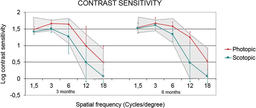
Alió et al. Eye and Vision (2015) 2:2                                                                                           Page 5 of 9

 Figure 1 Defocus curve comparison between groups.                   Figure 3 Contrast sensitivity function comparison between
 Comparison of mean defocus curve between the patients implanted     groups. It shows comparison of the postoperative contrast
 with the SeeLens MF IOL and the monofocal IOL.                      sensitivity function in both groups of patients under photopic and
                                                                     scotopic conditions.

Contrast sensitivity function
Figure 2 shows the mean postoperative contrast sensitivity          for the root mean square (RMS) of the internal high
function (CSF) in logarithmic scale under photopic and sco-         order aberrations and in the coma aberration (Wilcoxon
topic conditions at 3 and 6 months after surgery for the            test, p ≤ 0.04). Also, a significant reduction for the RMS
group of patients implanted with the multifocal IOL. A sig-         for the third and forth order aberrations was detected
nificant increase in scotopic contrast sensitivity was detected     (Wilcoxon test, both p = 0.03). However, no significant
for 6 cycles/° spatial frequency during follow-up (Wilcoxon         changes were observed in the internal trefoil, tetrafoil
test, p = 0.04), but no significant changes were observed           and spherical aberrations (Wilcoxon test, both p ≥ 0.41).
for the rest of spatial frequencies (Wilcoxon test, p ≥ 0.06).        Regarding the optical quality analysis, a significant in-
Figure 2 also shows, in light gray, the normal levels of CSF        crease of the ocular Strehl ratio was observed from 0.11 ±
for patients of the same age. It can be observed that the re-       0.06 preoperative to 0.19 ± 0.11 at 6 months after surgery
sults of the CSF after implantation of the SeeLens MF are           (Wilcoxon test, p = 0.02).
within physiological level for patients of the same age group.
  Figure 3 shows the comparison of the CSF between
both groups of patients in photopic and scotopic condi-             Quality of life outcomes
tions 3 months after the surgery. It was observed that              Table 3 summarizes the achieved mean quality of life
patients implanted with the monofocal IOL for the                   outcomes in patients implanted with the multifocal IOL.
spatial frequencies corresponding to 1.5 cycles/° in sco-           These data were obtained with the VF-14 questionnaire
topic conditions had slightly better behaviors, but these           at 6 months after surgery. Patients had more difficulty
differences were not statistically significant (p > 0.05).          driving at night, and reading small print, such as medi-
                                                                    cine bottle labels, a telephone book, or food labels.
Optical quality assessment
Figure 4 shows the internal aberrometric outcomes. At 6
months after surgery, there was a significant reduction

 Figure 2 Contrast sensitivity function in the multifocal IOL
 group. Mean postoperative contrast sensitivity function (CSF) in    Figure 4 Internal aberrations in the multifocal IOL group. It
 patients implanted with the multifocal IOL in logarithmic scale     shows evolution of the internal aberrations throughout the follow
 under photopic and scotopic conditions at 3 and 6 months after      up period. There is a significant reduction for the root mean square
 surgery. Normal values for the same age group are shown in          (RMS) of the internal high order aberrations six months after
 light gray.                                                         implantation of the SeeLens MF IOL.
Alió et al. Eye and Vision (2015) 2:2 Page 6 of 9
Table 3 Results of the VF-14 quality of life questionnaire in the multifocal IOL group
Items Punctuation
1. Reading small print, such as medicine bottle labels, a telephone book, or food labels 1.00 ± 0.93
2. Reading a newspaper or a book 0.50 ± 0.53
3. Reading a large-print book or large-print newspaper or numbers on a telephone 0.13 ± 0.35
4. Recognizing people when they are close to you 0.33 ± 0.71
5. Seeing steps, stairs or curbs 0.11 ± 0.33
6. Reading traffic signs, street signs or store signs 0.11 ± 0.33
7. Doing fine handwork like sewing, knitting, crocheting, carpentry 0.75 ± 0.89
8. Writing checks or filling out forms 0.63 ± 0.74
9. Playing games such as bingo, dominos, card games, or mahjong 0.00 ± 0.00
10. Taking part in sports like bowling, handball, tennis, golf 0.00 ± 0.00
11. Cooking 0.00 ± 0.00
12. Watching television 0.22 ± 0.44
13. Driving during the day 0.20 ± 0.45
14. Driving at night 1.20 ± 0.45
Mean values of the VF-14 QOL questionnaire items at 6 months postoperatively. Grading scale: 0, no difficulty; 1, a little difficulty; 2, moderate difficulty; 3, quite
difficult; 4, impossible to perform.
Surgical and postoperative complications surgery and refractive lens exchange [10,12,14,15,24]. In
No postoperative complications were observed, specific- the present investigation, we found similar results to those
ally, no significant posterior capsule opacification caus- previously reported by other authors. We observed a sig-
ing visual decrease of 1 or more lines associated with nificant improvement in the different ranges of vision that
visual symptoms and leading to neodymium-doped yt- were evaluated using the SeeLens MF IOL [6,10,12,13].
trium aluminum garnet (Nd:YAG) laser capsulotomy. Although most patients achieved near and distance func-
No significant IOL decentration was detected at the slit tional visual acuity with different models of diffractive
lamp examination either. IOLs, a main limitation is the poor intermediate vision
that this technology provides [22]. Therefore, several
Discussion multifocal IOL manufacturers have developed optics that
Restoring the functional near vision in presbyopic pa- includes aspheric profiles with low addition in order to
tients who undergo cataract surgery has become an im- improve functional intermediate vision [12,15,23]. The
portant outcome goal of refractive surgeons. The loss of SeeLens MF IOL incorporates both an aspheric profile,
near functional vision negatively impacts the quality of and an addition of 3 diopters that allows the patients a
life of presbyopic patients [21]. During the last two de- wide range of reading performance at intermediate dis-
cades, several designs of multifocal IOL technologies tances as the current investigation shows. In addition, the
have been developed in order to provide adequate spec- results derived from the defocus curve show that the de-
tacle independence for patients after cataract surgery. sign of this IOL provides two excellent peaks of maximum
However, some limitations related to the design of the vision for the focus corresponding to the distance and the
optic and the distribution of the light within the differ- near visions with a slight slope between these two peaks,
ent foci have led to the presence of undesirable symp- suggesting that the patients can achieve an adequate and
toms in some cases such as, the presence of photic functional intermediate visual acuity. One of the reasons
phenomena, and the reduction of the contrast sensitivity that could explain this behavior may be related to the fact
function [11,22,23]. that the new design of this IOL is based on an aspheric
The present investigation evaluated the clinical out- refractive-diffractive apodized profile.
comes, as well as the optical performance, and the qual- Measurement of the intraocular aberrations demon-
ity of life in patients that underwent implantation of a strated a significant reduction in the intraocular higher
new type of diffractive IOL for the correction of presby- order aberrations, and in the asymmetric aberrations
opia, the SeeLens MF (Kibbutz Hanita Lenses, Kibbutz, (coma and coma-like aberrations). These results are simi-
Israel). lar to those previously reported by our research group
Previously published studies that have evaluated the re- when analyzing the intraocular optical quality with differ-
sults of multifocal IOLs have reported an improvement ent diffractive surfaces [12,16], and by other studies that
in the visual acuity at different distances after cataract have evaluated the intraocular optical performance ofAlió et al. Eye and Vision (2015) 2:2 Page 7 of 9 multifocal IOLs [24,25]. Although it was not statistically This improvement relates to the quality of the image significant, we found a change towards a more positive perceived, and should be attributed as a possible effect spherical aberration. This might be related to the aspheric of the neuroadaptation process that was observed in profile on the SeeLens MF, which introduces a negative those patients implanted with multifocal IOLs [31]. aspheric factor within the optic of its design. Finally, the present investigation also assesses the qual- This study also evaluated the quality outcomes of the ity of life by analyzing the answer of the patients to the patients by measuring the quality of the image in the ret- VF-14 questionnaire [32]. Even though patients found inal plane. We found that after the implantation of the more difficulty in driving at night, and reading small SeeLens MF IOL, the patients achieved better levels of print, such as medicine bottles or food labels, most of Strehl ratio to those shown in the preoperative period. the answers to the questionnaire showed the high levels In addition, the mean postoperative Strehl ratio was bet- of satisfaction that the patients had when asked about ter than those observed in a normal population of the the tasks that they often perform in their daily lives. same age and was comparable to values obtained in These findings are consistent with those found with the young, healthy patients [26]. Moreover, Strehl ratios in other variables analyzed in the present investigation, patients that underwent implantation of the SeeLens MF confirming the ability of this IOL in improving the qual- were better than those previously reported by our re- ity of life of the patients. Additionally, van der Linden search group with other types of diffractive IOLs [16]. In et al. also showed in their investigation a high rate of addition, a recent study in which also analyzed the out- satisfaction in patients implanted with the SeeLens MF comes of patients implanted with the SeeLens MF, the [27]. In that study, as many as 96% of the subjects that authors reported statistically better results in terms of were evaluated reported to be satisfied with the result of visual quality when comparing with another diffractive the procedure. multifocal IOL [27]. We have to take into account the In the current study, a control group, which under- fact that the intraocular optical quality in the present in- went a monofocal IOL implantation, was also included vestigation was evaluated with a Hartmann-Shack aber- with the aim of comparing the outcomes with those ob- rometer, thus, the outcomes presented in the current served in the SeeLens MF IOL group. Both groups pre- investigation should be taken with caution as this kind sented similar preoperative characteristics and therefore of wavefront technology has shown to be limited when could be compared without significant statistical bias. As evaluating the diffractive surface [28]. expected, an improvement in both, corrected and uncor- Regarding the contrast sensitivity function, it was ob- rected vision were observed after the surgery in the two served that six months after the surgery, the SeeLens groups of patients. In terms of near vision, the SeeLens MF provides results in photopic condition and on the MF provided a better near visual outcome than the higher spatial frequencies that are within the physio- monofocal IOL, confirming the efficacy of this IOL in logical levels for the normal population of the same age restoring near visual function. Regarding the defocus group [29]. Nevertheless, analysis of the results in sco- curve, it was demonstrated that the patients implanted topic condition and in the remaining spatial frequencies with the multifocal IOL were able to achieve two peaks showed that there was a reduction of the CSF after sur- of maximum vision, one at distance and one at near with gery. Similar findings were reported in other studies that a good intermediate vision. On the other hand, those pa- have evaluated the outcomes related to the CSF after im- tients implanted with the monofocal IOL only showed plantation of diffractive IOL models, which have also one peak of maximum vision. These findings demon- found a reduction of the CSF in the spatial frequencies strate that the SeeLens MF IOL provides a range of that were analyzed [15]. The decrease in contrast sensi- functional vision for different distances in comparison tivity in patients implanted with multifocal IOL is due to with those that can be achieved with a monofocal IOL. the dispersed distribution of light energy within the sur- Another aspect that is worth discussing is the one re- face of the optic [30], which is usually more pronounced lated to MICS that is defined by those phacoemulsifica- in low light conditions as our results showed. Another tion procedures that can be performed through an reason that explains the reduction of CSF with this type incision smaller than 1.5 mm [33]. This type of surgery of multifocal IOL is the relationship that exists between offers several advantages such as a stable anterior cham- the optical quality and the near visual performance of ber during surgery, a reduced amount of ultrasound the IOL. Thus, the better the near vision provided by power, less surgical induced astigmatism, among others. the IOL, the greater the limitation that will exist in Nevertheless and despite the aforementioned advan- terms of visual quality [16]. Even though there is a re- tages, one of the limitations of this procedure is that duction of the CSF after the surgical procedure, we there are few multifocal IOLs available in the market found that there is a trend to obtain better contrast per- that can be implanted through an incision of less than ception of the image between 3 and 6 months follow up. 2 mm. On the other hand, stability and centration of
Alió et al. Eye and Vision (2015) 2:2                                                                                                                      Page 8 of 9

multifocal diffractive IOL optics is critical for the ad-                           Received: 10 September 2014 Accepted: 13 January 2015
equate optical performance of this technology. Thus, a
tilt or decentration can decrease the visual quality and
produce optical side effects, causing subjective symp-                              References
                                                                                    1. Espindle D, Crawford B, Maxwell A, Rajagopalan K, Barnes R, Harris B, et al.
toms and patient dissatisfaction [34]. Hence, an IOL de-                                Quality-of-life improvements in cataract patients with bilateral blue
sign is crucial to the optimum optical function, and to                                 light-filtering intraocular lenses: clinical trial. J Cataract Refract Surg.
prevent problems that can lead to vision complaints.                                    2005;31:1952–9.
                                                                                    2. Walkow L, Klemen UM. Patient satisfaction after implantation of diffractive
The SeeLens MF incorporates in its design, an open C-                                   designed multifocal intraocular lenses in dependence on objective
loop haptic that provides stability of the IOL inside the                               parameters. Graefes Arch Clin Exp Ophthalmol. 2001;239:683–7.
capsular bag. Previous reports in the scientific literature                         3. Javitt JC, Steinert RF. Cataract extraction with multifocal intraocular lens
                                                                                        implantation: a multinational clinical trial evaluating clinical, functional, and
have demonstrated that IOLs that showed the best per-                                   quality-of-life outcomes. Ophthalmology. 2000;107:2040–8.
formances in terms of stability inside the capsular bag                             4. Javitt J, Brauweiler HP, Jacobi KW, Klemen U, Kohnen S, Quentin CD, et al.
are those with C-loop haptic designs [35]. To the best of                               Cataract extraction with multifocal intraocular lens implantation: clinical,
                                                                                        functional, and quality-of-life outcomes. Multicenter clinical trial in Germany
our knowledge, the SeeLens MF is currently the only                                     and Austria. J Cataract Refract Surg. 2000;26:1356–66.
multifocal diffractive IOL with available scientific data                           5. Javitt JC, Wang F, Trentacost DJ, Rowe M, Tarantino N. Outcomes of cataract
published in the literature, which can provide both the                                 extraction with multifocal intraocular lens implantation: functional status
                                                                                        and quality of life. Ophthalmology. 1997;104:589–99.
capability to be implanted through an incision smaller                              6. Alió JL, Plaza-Puche AB, Piñero DP, Amparo F, Rodríguez-Prats JL, Ayala MJ.
than 2 mm and a C-loop design of the haptics.                                           Quality of life evaluation after implantation of 2 multifocal intraocular lens
                                                                                        models and a monofocal model. J Cataract Refract Surg. 2011;37:638–48.
                                                                                    7. Bellucci R. Multifocal intraocular lenses. Curr Opin Ophthalmol. 2005;16:33–7.
Conclusions                                                                         8. Keates RH, Pearce JL, Schneider RT. Clinical results of the multifocal lens.
In conclusion, the MICS SeeLens MF IOL can restore                                      J Cataract Refract Surg. 1987;13:557–60.
distance and near visions in presbyopic patients under-                             9. Duffey RJ, Zabel RW, Lindstrom RL. Multifocal intraocular lenses. J Cataract
                                                                                        Refract Surg. 1990;16:423–9.
going cataract surgery. This new IOL also provides func-                            10. Alió JL, Agdeppa MC, Pongo VC, El Kady B. Microincision cataract surgery
tional intermediate vision with an adequate intraocular                                 with toric intraocular lens implantation for correcting moderate and high
optical quality performance. In addition, the results ob-                               astigmatism: pilot study. J Cataract Refract Surg. 2010;36(1):44–52.
                                                                                    11. Montés-Micó R, Alió JL. Distance and near contrast sensitivity function
tained from the quality of life questionnaire confirm the                               after multifocal intraocular lens implantation. J Cataract Refrac Surg.
high levels of satisfaction in the patients implanted with                              2003;29:703–11.
the SeeLens MF IOL. Finally, by providing both, the cap-                            12. Alió JL, Elkady B, Ortiz D, Bernabeu G. Clinical outcomes and intraocular
                                                                                        quality of a diffractive multifocal intraocular lens with asymmetrical Light
ability of being implanted through a 1.8 mm incision,                                   distribution. J Cataract Refract Surg. 2008;34:942–8.
and the stability due to the open C-loop haptic design,                             13. Alió JL, Montalbán R, Peña-García P, Soria FA, Vega-Estrada A. Visual outcomes
the SeeLens MF IOL offers an excellent alternative for                                  of a trifocal aspheric diffractive intraocular lens with microincision cataract
                                                                                        surgery. J Refract Surg. 2013;29(11):756–61.
modern cataract surgical techniques. Further long-term                              14. Alió JL, Elkady B, Ortiz D, Bernabeu G. Microincision multifocal intraocular
studies with a larger sample of patients should be per-                                 lens with and without a capsular tension ring: optical quality and clinical
formed in order to confirm the outcomes observed in                                     outcomes. J Cataract Refract Surg. 2008;34:1468–75.
                                                                                    15. Alfonso JF, Fernández-Vega L, Señaris A, Montés-Micó R. Prospective study
this investigation with the new SeeLens MF IOL.                                         of the Acri.LISA bifocal intraocular lens. J Cataract Refract Surg.
                                                                                        2007;33:1930–5.
Competing interests                                                                 16. Alió JL, Piñero DP, Plaza-Puche AB, Amparo F, Jiménez R, Rodríguez-Prats JL,
The authors have no proprietary or commercial interests in the medical                  et al. Visual and optical performance with two different diffractive multifocal
devices that are involved in this manuscript.                                           intraocular lenses compared to a monofocal lens. J Refract Surg.
                                                                                        2011;27:570–81.
Authors’ contributions                                                              17. Alió JL, Plaza-Puche AB, Piñero DP, Amparo F, Jiménez R, Rodríguez-Prats JL,
JLA, participated in the design of the study, providing the subjects of                 et al. Optical analysis, reading performance, and quality-of-life evaluation
investigation, critical analysis of the manuscript and helped in the redaction          after implantation of a diffractive multifocal intraocular lens. J Cataract
of the manuscript; AVE, helped to collect the data, performed the analysis              Refract Surg. 2011;37:27–37.
and interpretation of the results, redaction of the article, critical analysis of   18. Piñero DP, Juan JT, Alió JL. Intrasubject repeatability of internal aberrometry
the draft; and ABP, helped to collect the data, performed the statistical               obtained with a new integrated aberrometer. J Refract Surg. 2011;27:509–17.
analysis, helped redacting the manuscript and critical analysis of the              19. Karakosta A, Vassilaki M, Plainis S, Elfaal NH, Tsilimbaris M, Moschandreas
manuscript. All the authors read an approved the final version of the                   J. Choice of analytic approaches for eyespecific outcomes: one eye or two.
manuscript before publishing.                                                           Am J Opthalmol. 2012;153:571–9.
                                                                                    20. Armstrong RA. Statistical guidelines for the analysis of data obtained from
Acknowledgements                                                                        one or both eyes. Ophthalmic Physiol Opt. 2013;33:7–14.
This study is supported in part by a grant from the Spanish Ministry of             21. Uusitalo RJ, Brans T, Pessi T, Tarkkanen A. Evaluating cataract surgery gains
Health, Instituto Carlos III, Red Temática de Investigación Cooperativa en              by assessing patients’ quality of life using the VF-7. J Cataract Refract Surg.
Salud “Patología ocular del envejecimiento, calidad visual y calidad de vida”,          1999;25(7):989–94.
Subproyecto de Calidad Visual (RD07/0062).                                          22. Blaylock JF, Si Z, Vickers C. Visual and refractive status at different focal
                                                                                        distances after implantation of the ReSTOR multifocal intraocular lens.
Author details                                                                          J Cataract Refract Surg. 2006;32:1464–73.
1
 Vissum Corporation, Alicante, Spain. 2Division of Ophthalmology,                   23. Alfonso JF, Fernández-Vega L, Puchades C, Montés-Micó R. Intermediate
Universidad Miguel Hernández, Alicante, Spain. 3Avda de Denia s/n, Edificio             visual function with different multifocal intraocular lens models. J Cataract
Vissum, 03016 Alicante, Spain.                                                          Refract Surg. 2010;36(5):733–9.
Alió et al. Eye and Vision (2015) 2:2 Page 9 of 9
24. Marcos S, Barbero S, Jimenez-Alfaro I. Optical quality and depth-of-field of
eyes implanted with spherical and aspheric intraocular lenses. J Refract Surg.
2005;21:223–35.
25. Barbero S, Marcos S, Jimenez-Alfaro I. Optical aberrations of intraocular
lenses measured in vivo and in vitro. J Opt Soc Am A Opt Image Sci Vis.
2003;20:1841–51.
26. Alió JL, Schimchak P, Negri HP, Montés-Micó R. Crystalline lens optical
dysfunction through aging. Ophthalmology. 2005;112(11):2022–9.
27. van der Linden JW, van der Meulen IJ, Mourits MP, Lapid-Gortzak R.
Comparison of a hydrophilic and a hydrophobic apodized diffractive
multifocal intraocular lens. Int Ophthalmol. 2013;33(5):493–500.
28. Charman WN, Montés-Micó R, Radhakrishnan H. Problems in the
measurement of wavefront aberration for eyes implanted with diffractive
bifocal and multifocal intraocular lenses. J Refract Surg. 2008;24(3):280–6.
29. Hohberger B, Laemmer R, Adler W, Juenemann AG, Horn FK. Measuring
contrast sensitivity in normal subjects with OPTEC 6500: influence of age
and glare. Graefes Arch Clin Exp Ophthalmol. 2007;245(12):1805–14.
30. Montés-Micó R, España E, Bueno I, Charman WN, Menezo JL. Visual
performance with multifocal intraocular lenses; mesopic contrast sensitivity
under distance and near conditions. Ophthalmology. 2004;111:85–96.
31. Knorz MC. Multifocal intraocular lenses: overview of their capabilities,
limitations, and clinical benefits. J Refract Surg. 2008;24(3):215–7.
32. Gothwal VK, Wright TA, Lamoureux EL, Pesudovs K. Measuring outcomes of
cataract surgery using the Visual Function Index-14. J Cataract Refract Surg.
2010;36(7):1181–8.
33. Alió JL, Rodriguez-Prats JL, Vianello A, Galal A. Visual outcome of microincision
cataract surgery with implantation of an Acri. Smart lens. J Cataract Refract
Surg. 2005;31(8):1549–56.
34. Alió JL, Piñero DP, Plaza-Puche AB, Rodriguez Chan MJ. Visual outcomes and
optical performance of a monofocal intraocular lens and a new-generation
multifocal intraocular lens. J Cataract Refract Surg. 2011;37:241–50.
35. Chang DF. Comparative rotational stability of single-piece open-loop acrylic
and plate-haptic silicone toric intraocular lenses. J Cataract Refract Surg.
2008;34(11):1842–7.
Submit your next manuscript to BioMed Central
and take full advantage of:
• Convenient online submission
• Thorough peer review
• No space constraints or color figure charges
• Immediate publication on acceptance
• Inclusion in PubMed, CAS, Scopus and Google Scholar
• Research which is freely available for redistribution
Submit your manuscript at
www.biomedcentral.com/submitYou can also read