Expression of microRNA 155 5p in patients with refractory diabetic macular edema and its regulatory mechanism
←
→
Page content transcription
If your browser does not render page correctly, please read the page content below
EXPERIMENTAL AND THERAPEUTIC MEDICINE 22: 975, 2021

       Expression of microRNA‑155‑5p in patients with refractory
         diabetic macular edema and its regulatory mechanism
                        JUNWEN HE1, RUI ZHANG1, SHAN WANG2, LU XIE1, CHENGFENG YU1,
                                       TAO XU1, YANZI LI1 and TAO YAN1

                            Departments of 1Retinal and Vitreous Diseases and 2Ophthalmic Imaging,
                            Aier Eye Hospital of Wuhan University, Wuhan, Hubei 430000, P.R. China

                                         Received March 18, 2021; Accepted June 18, 2021

                                                   DOI: 10.3892/etm.2021.10407

Abstract. Diabetic macular edema (DME) is the main cause            of DME. To further study the molecular mechanism, human
of visual impairment in diabetic patients, but its pathogenesis     retinal microvascular endothelial cells (HRMECs) were
remains unclear. The purpose of the present study was to            cultured and treated with high glucose in vitro. The results
analyze the expression of microRNA (miR)‑155‑5p in patients         showed that miR‑155‑5p expression was significantly upregu‑
with DME and its regulatory mechanism. A total of 72 patients       lated in HRMECs induced by high glucose. After inhibiting
diagnosed with DME and 17 with idiopathic macular hole (MH)         the expression of miR‑155‑5p, cell proliferation, angiogenesis
were recruited. Among samples from patients with DME, 45            and VEGF protein levels were significantly downregulated,
were DME and 27 were refractory DME, whereas patients with          whereas miR‑155‑5p mimics had the opposite effect. In
idiopathic MH served as the control group. Optical coherence        summary, miR‑155‑5p is closely associated with DME and is a
tomography and fundus photograph analysis revealed that part        potential target for refractory DME treatment.
of the retina in the fundus of patients with DME was thickened,
with macular edema occurring simultaneously. In refractory          Introduction
patients with DME, macular edema was associated with
bleeding and a dark cavity between retinal layers. Through          DME is one of the most common causes of vision loss in diabetic
reverse transcription‑quantitative PCR analysis, miR‑155‑5p         patients (1,2). The pathogenesis of DME is multi‑factorial
was highly expressed in the aqueous humor (AH) and plasma           and involves multiple pathways (3), such as poor blood sugar
of patients with DME compared with that in patients with MH,        control (4), high blood pressure (5), hyperlipidemia (6) and
and this was even higher in the refractory DME group. Upon          proteinuria (7), which eventually lead to the thickening of the
analyzing patient clinical data, the difference in miR‑155‑5p       central retina; if untreated in time, it will cause loss of vision.
expression in the AH and plasma was positively associated           Currently, the treatment of refractory DME is still progressing
with disease course, body mass index, fasting blood‑glucose,        slowly (8,9).
glycated hemoglobin, proteinuria and glycosuria. The expres‑            MicroRNAs (miRNAs) are small non‑coding RNAs that
sion of miR‑155‑5p was not significantly different based on         regulate biological networks by regulating gene expression.
hemoglobin, intraocular pressure and sex. The aforementioned        In several studies on DME, miRNAs have been found to play
results indicate that miR‑155‑5p might promote the development      an important role in the pathogenesis of DME (10,11). Studies
                                                                    have shown that miR‑155‑5p is overexpressed in diabetic
                                                                    patients (12), and can be used as a new biological indicator for
                                                                    the diagnosis and evaluation of diabetes (13).
Correspondence to: Mr. Tao Yan, Department of Retinal and               Vascular endothelial growth factor A (VEGF‑A) plays
Vitreous Diseases, Aier Eye Hospital of Wuhan University,           an important role in the occurrence of DME (2,14,15), and is
481 Zhongshan Road, Wuchang, Wuhan, Hubei 430000, P.R. China        the main angiogenesis promoter of endothelial cells mainly
E‑mail: aieryantao99@163.com                                        through its homologous receptor VEGF‑R1. The upregulation
                                                                    of VEGF‑A, inflammatory cytokines and chemokines induces
Abbreviations: DME, diabetic macular edema; OCT, optical            pathological changes in the vascular endothelium, triggering
coherence tomography; RT‑qPCR, quantitative reverse transcription   the destruction of the blood‑retinal barrier, causing fluid to
polymerase chain reaction; NC, negative control; AH, aqueous
                                                                    penetrate into the extracellular space, which is clinically
humor; BMI, body mass index; FBG, fasting blood‑glucose; HbA1c,
                                                                    manifested as macular edema and leads to vision loss (6,16).
glycated hemoglobin; HB, hemoglobin; IOP, intraocular pressure;
CCK‑8, Cell Counting Kit‑8; HRMECs, human retinal microvascular     In vitro molecular studies demonstrated that miRNAs modu‑
endothelial cells; VEGF, vascular endothelial growth factor         late VEGF to promote high‑glucose‑induced apoptosis (17)
                                                                    and angiogenesis (18) of HRMECs. Qiu et al (19) confirmed
Key words: DME, microRNA‑155‑5p, OCT, human retinal                 that miR‑21‑5p inhibitor inhibited the proliferation and angio‑
epithelial cells, regulatory mechanism                              genesis of HRMECs induced by high glucose. miR‑199a‑3p
                                                                    inhibits the angiogenesis of HRMECs induced by high glucose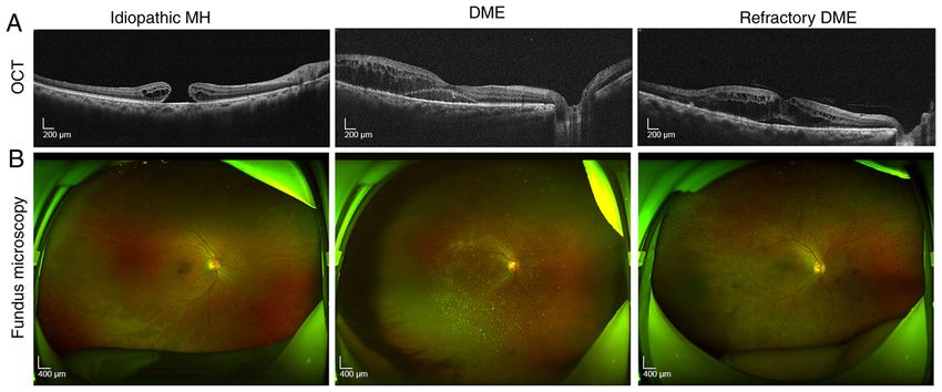
2                                  HE et al: ROLE OF miR-155-5p IN DIABETIC MACULAR EDEMA

by inhibiting VEGF (20). However, in DME, there has been no       recorded. Fasting blood‑glucose (FBG), glycated hemoglobin
report on the effect of miR‑155‑5p targeting cytokines on the     (HbA1c), hemoglobin (HB), proteinuria and glycosuria were
angiogenesis of HRMECs.                                           measured with the Beckman automatic biochemical analyzer
    The present study aimed to evaluate the association           (cobas c311; Roche Diagnostics GmbH) according to the kit's
between miR‑155‑5p and the clinical features of DME and           instructions.
refractory DME, as well as to evaluate the potential mechanism
of miR‑155‑5p on HRMECs induced by high glucose in vitro.         Ophthalmoscopic examination. Retinal thickness was
                                                                  analyzed using optical coherence tomography (OCT;
Materials and methods                                             RTVue XR; Optovue, Inc.) using the software RTVue XR
                                                                  (version: 2018.1.0.43). Images of the fundus were captured
Materials. Antibodies against VEGF‑A (cat. no. ab214424) and      using Optos PLC's Panoramic Ophthalmoscope (200TX;
glyceraldehyde‑3‑phosphate dehydrogenase (GAPDH; cat.             Nikon Corporation) with the software Optos V2®Vantage Pro
no. ab181602) were purchased from Abcam. The miR‑155‑5p           Review (version: 2.11.0.3; Nikon Corporation).
mimics or inhibitors, and their negative controls, as well as
primers for reverse transcription‑quantitative (RT‑q)PCR were     Aqueous humor (AH) sample collection and processing. AH
synthesized by Guangzhou RiboBio Co., Ltd., and the miRNA         was collected from each patient (~50 to 100 µl) through para‑
sequences were as follows: miR‑155‑5p mimics, 5'‑UUAAUG           centesis. The AH samples were collected in 0.5‑ml RNA‑free
CUAAUCGUGAUAGGGGUU‑3'; miR‑155‑5p negative control,               Eppendorf (EP) tubes, stored on ice, and further processed
5'‑GGGUUAGGUAAUGUAAUCCGUUUA‑3'; miR‑155‑5p                        within 1 h after collection. In all cases, sample collection
inhibitor, 5'‑AACCCCTATCACGATTAGCATTAA‑3';                        was non‑invasive, eliminating the risk of blood or cell debris
miR‑155‑5p inhibitor negative control, 5'‑CAGUACUUUUGU            contamination. By centrifuging the AH sample at 1,200 x g for
GUAGUACAA‑3'. The primer sequences for miRNAs for the             30 min at 4˚C, the final cell/cell debris contamination could
RT‑qPCR assay were as follows: miR‑155‑5p forward, 5'‑ACA         be prevented. Then, the supernatant was transferred to a new
CTCCAGCTGGGTTAATGCTAATCGTGATA‑3'; miR‑155‑5p                      sterile EP tube for RNA extraction.
reverse, 5'‑CTCAACTGGTGTCGTGGA‑3'; U6 forward, 5'‑CTC
GCTTCGGCAGCACA‑3'; and reverse; 5'‑AACGCTTCACGA                   Collection and processing of whole blood samples. Firstly,
ATTTGCGT‑3'.                                                      5 ml whole blood was collected from the patients in a
                                                                  BD Vacutainer‑EDTA tube (BD Biosciences) and immediately
Study subjects. Between April 2020 and August 2020,               mixed 5‑8 times. Within 1 h after blood collection, the blood
72 patients diagnosed with DME were selected from the             samples were processed by centrifugation at 1,200 x g for
outpatient inpatients in Wuhan Aier Eye hospital, including       10 min at 4˚C; the collected plasma was centrifuged at 1,200 x g
49 males and 23 females, aged 31‑71 (mean 51.17±9.98) years,      for 20 min at 4˚C to remove contaminant cells and cell debris,
with a disease course of 0.5‑20 years. All patients underwent     and then RNA extraction was performed. Hemolyzed plasma
detailed medical examinations and fundoscopy. According           samples were excluded from the study.
to the staging standard specified by the 2002 Ocular Fundus
Disorders Academic Conference (21), the patients were             Cell culture, transfection and treatments. The HRMECs (cat.
divided into three groups as follows: 45 cases in the DME         no. CP‑H130; Procell Life Science & Technology Co., Ltd.) were
group, including 31 males and 14 females, with an average age     cultured at 37˚C with 5% CO2 in complete culture medium (cat.
of 50.72±11.48 years and an average course of 6.45±1.73 years;    no. CM‑H130; Procell Life Science & Technology Co., Ltd.).
27 cases in the refractory DME group, with 18 males and           Once cells achieved 60‑80% confluence, they were transfected
9 females, average age of 53.55±9.63 years and an average         with RNA using Lipofectamine® 2000 (Invitrogen; Thermo
course of 9.25±3.36 years. The exclusion criteria were as         Fisher Scientific, Inc.) at 37˚C for 4 h, according to the manu‑
follows: Patients with diabetic ketoacidosis, diabetic hyperos‑   facturer's instructions. Then, medium was replaced with fresh
molar coma, and other acute complications of diabetes; patients   culture medium, and cells were incubated for a further 48 h prior
with severe stress; patients with acute or chronic infections;    to RT‑qPCR or western blot analysis. The high‑glucose group was
and patients with liver disease. The patients with idiopathic     pre‑cultured with 30 mM glucose at 37˚C for 48 h, and the normal
macular hole (MH) (17 males and 5 females) served as the          glucose group (control) was pre‑cultured with 5 mM glucose at
control group; their age, sex ratio and body mass index (BMI)     37˚C for 48 h before experiment. The glucose concentration used
were matched, and average age was 50.57±4.28 years. The           in the high glucose group was based on previous studies (23,24).
examination of patients with idiopathic MH excluded hyper‑
tension, heart, liver, kidney disease and other endocrine and     RNA extraction. Using the miRNeasy Mini kit (217004;
metabolic diseases. All patients or their family members          Qiagen GmbH), total RNA was extracted from 50 µl AH or
signed an informed consent form, and the experiment was           200 µl plasma samples. The AH and plasma samples were
approved by the Aier Eye Hospital and conformed with the          centrifuged at 3000 x g for 5 min at 4˚C to completely remove
guiding principles of the Declaration of Helsinki (22).           contaminant cell debris. A total of 50 µl AH and 200 µl plasma
                                                                  per patient was diluted to 400 µl with RNA‑free water (Gibco;
Data measurement. On admission, the patient's height              Thermo Fisher Scientific, Inc.) to avoid protein aggregation.
and weight were measured, and BMI was calculated as               Then, the sample was lysed by adding three times the volume
weight/height2 (kg/m 2). Medical history, age, sex, course of     of TRIzol® (Invitrogen; Thermo Fisher Scientific, Inc.) and
diabetes and blood sugar control, among other parameters were     was finally dissolved in 10 µl RNA‑free water.
EXPERIMENTAL AND THERAPEUTIC MEDICINE 22: 975, 2021 3
Figure 1. Refractory DME exacerbates the symptoms of macular edema. (A) OCT analyses of the retinal symptoms among patients in different groups.
(B) Fundus microscopy to analyze the differences in macular edema area and eyeball characteristics among patients in different groups. OCT, optical coher‑
ence tomography; DME, diabetic macular edema; MH, macular hole.
RT‑qPCR assay. Extracted RNA was reverse transcribed to 10 µl CCK‑8 reagent (Elabscience, Wuhan, China) was added
cDNA using a PrimeScript RT reagent kit (Thermo Fisher at 0, 24, 48 and 72 h and incubated for 60 min. The optical
Scientific, Inc.), according to the manufacturer's instructions. density was measured at 490 nm using a microplate reader
The RT‑qPCR procedure was performed using a SYBR (multiscan MK3; Thermo Fisher Scientific, Inc.). The CCK‑8
Premix ExTaq II kit (Thermo Fisher Scientific, Inc.) according experiment was repeated three times.
to the manufacturer's instructions in conjunction with a
7500 Real‑Time PCR System (Applied Biosystems; Thermo Angiogenesis assay. Transfected HRMECs were pretreated
Fisher Scientific, Inc.). The thermocycling conditions were with different concentrations of glucose (5 or 30 mM) at 37˚C for
as follows: 95˚C for 10 min; 55˚C for 2 min;72˚C for 2 min; 48 h. Next, serum‑starved HRMECs (2x10⁴ cells) were seeded
followed by 40 cycles of 95˚C for 15 sec and 60˚C for 1 min. onto 24‑well plates coated with Matrigel (BD Biosciences) in
Target miR‑155‑5p levels were normalized to those of the endothelial basal medium and incubated at 37˚C with 5% CO2
housekeeping gene U6. Relative miR‑155‑5p expression levels for 24 h. Tubular structures of HRMECs in the Matrigel were
were calculated using the 2‑ΔΔCq method (25), and the RT‑qPCR examined with a light microscope (Olympus Corporation).
experiment was repeated three times. The angiogenesis experiment was repeated three times and
five fields were randomly selected for the quantitative analysis
Western blotting. Transfected HRMECs were harvested of each result.
by trypsin digestion and lysed using ice‑cold RIPA lysis
buffer (Beyotime Institute of Biotechnology). Lysate protein Statistical analysis. All data are expressed as means ± stan‑
concentrations were estimated using a BCA protein assay dard deviations. All statistical analyses were performed using
kit (Beijing Solarbio Science & Technology Co., Ltd.). Equal SPSS version 21.0 statistical analysis package (SPSS Inc.).
amounts of denatured proteins (20 µg) were resolved using The male/female data were analyzed using a χ2 test. The AH,
10% SDS‑polyacrylamide gel electrophoresis (Beijing Solarbio serum and clinical characteristics of patients, the expression of
Science & Technology Co., Ltd.) and protein bands were trans‑ miR‑155‑5p under miR‑155‑5p mimic/inhibitor transfection, cell
ferred to a polyvinylidene fluoride membrane (Beijing Solarbio proliferation, angiogenesis, and western blot data proceeded via
Science & Technology Co., Ltd.). The membrane was blocked a one‑way analysis of variance and Bonferroni post hoc test. The
with 5% BSA (Beijing Solarbio Science & Technology Co., difference of miR‑155‑5p expression before and after HG induc‑
Ltd.) for 1 h at 22±3˚C. Subsequently, the membranes were tion was analyzed using the unpaired Student's t‑test. P4 HE et al: ROLE OF miR-155-5p IN DIABETIC MACULAR EDEMA Figure 2. Differences in the expression levels of miRNA between DME and refractory DME. Reverse transcription‑quantitative polymerase chain reaction analysis of the differences in the expression of miR‑155‑5p in the aqueous humor (A) and serum (B) of patients with DME. *P
EXPERIMENTAL AND THERAPEUTIC MEDICINE 22: 975, 2021                                                          5

Figure 3. Association between miR‑155‑5p and clinical symptoms of patients. The differences in the age (A), course of disease (B), BMI (C), FBG (D),
HbA1C (E), proteinuria (F), glycosuria (G), HB (H) and IOP (I) in each group of patients are shown. *P
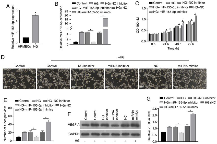
6                                        HE et al: ROLE OF miR-155-5p IN DIABETIC MACULAR EDEMA

Figure 4. miR‑155‑5p reverses the effect of high glucose induction. RT‑qPCR (A) analysis showed that the expression of miR‑155‑5p was different in HRMECs
induced by high glucose. After transfection with miRNAs, RT‑qPCR (B) and Cell Counting Kit‑8 assays (C) were performed to analyze the changes in expres‑
sion of miR‑155‑5p and cell proliferation in each group. Angiogenesis (D and E) was analyzed in each group, and western blotting (F and G) was performed
to analyze the changes in VEGF‑A protein levels. Samples were divided into the control group, HG group, HG+NC inhibitor group, HG+inhibitor group,
HG+NC group and HG+miR‑155‑5p mimic group. *P
EXPERIMENTAL AND THERAPEUTIC MEDICINE 22: 975, 2021                                                   7

Patient consent for publication                                           19. Qiu F, Tong H, Wang Y, Tao J, Wang H and Chen L: Inhibition
                                                                              of miR‑21‑5p suppresses high glucose‑induced proliferation and
                                                                              angiogenesis of human retinal microvascular endothelial cells by
Not applicable.                                                               the regulation of AKT and ERK pathways via maspin. Biosci
                                                                              Biotechnol Biochem 82: 1366‑1376, 2018.
                                                                          20. Wang L, Liu WX and Huang XG: MicroRNA‑199a‑3p inhibits
Competing interests                                                           angiogenesis by targeting the VEGF/PI3K/AKT signalling
                                                                              pathway in an in vitro model of diabetic retinopathy. Exp Mol
The authors declare that they have no competing interests.                    Pathol 116: 104488, 2020.
                                                                          21. Wilkinson CP, Ferris FL III, Klein RE, Lee PP, Agardh CD,
                                                                              Davis M, Dills D, Kampik A, Pararajasegaram R and Verdaguer JT;
References                                                                    Global Diabetic Retinopathy Project Group: Proposed interna‑
                                                                              tional clinical diabetic retinopathy and diabetic macular edema
 1. Tan GS, Cheung N, Simó R, Cheung GC and Wong TY: Diabetic                 disease severity scales. Ophthalmology 110: 1677‑1682, 2003.
    macular oedema. Lancet Diabetes Endocrinol 5: 143‑155, 2017.          22. Mastroleo I: Post‑trial obligations in the Declaration of
 2. Patelli F, Radice P and Giacomotti E: Diabetic macular edema.             Helsinki 2013: Classification, reconstruction and interpretation.
    Dev Ophthalmol 54: 164‑173, 2014.                                         Developing World Bioeth 16: 80‑90, 2016.
 3. Yau JW, Rogers SL, Kawasaki R, Lamoureux EL, Kowalski JW,             23. Lu L, Lu Q, Chen W, Li J, Li C and Zheng Z: Vitamin D3 Protects
    Bek T, Chen SJ, Dekker JM, Fletcher A, Grauslund J, et al;                against diabetic retinopathy by inhibiting high‑glucose‑induced
    Meta‑Analysis for Eye Disease (META‑EYE) Study Group:                     activation of the ROS/TXNIP/NLRP3 inflammasome pathway.
    Global prevalence and major risk factors of diabetic retinopathy.         J Diabetes Res 2018: 8193523, 2018.
    Diabetes Care 35: 556‑564, 2012.                                      24. Gu C, Draga D, Zhou C, Su T, Zou C, Gu Q, Lahm T, Zheng Z and
 4. Fenwick EK, Xie J, Man REK, Sabanayagam C, Lim L, Rees G,                 Qiu Q: miR‑590‑3p inhibits pyroptosis in diabetic retinopathy by
    Wong TY and Lamoureux EL: Combined poor diabetes control                  targeting NLRP1 and inactivating the NOX4 signaling pathway.
    indicators are associated with higher risks of diabetic retinopathy       Invest Ophthalmol Vis Sci 60: 4215‑4223, 2019.
    and macular edema than poor glycemic control alone. PLoS              25. Livak KJ and Schmittgen TD: Analysis of relative gene expression
    One 12: e0180252, 2017.                                                   data using real‑time quantitative PCR and the 2(‑Delta Delta
 5. Stana D, Potop V, Istrate SL, Eniceicu C, Mihalcea AR, Paşca IG,          C(T)) method. Methods 25: 402‑408, 2001.
    Aqel A, Ciuluvică R and Moraru D: Variability of diabetic             26. Bandello F, Battaglia Parodi M, Lanzetta P, Loewenstein A,
    macular edema in correlation with hypertension retinopathy in             Massin P, Menchini F and Veritti D: Diabetic macular edema.
    patients with diabetes mellitus and essential hypertension. Rom J         Dev Ophthalmol 58: 102‑138, 2017.
    Ophthalmol 63: 327‑338, 2019.                                         27. Korobelnik JF, Do DV, Schmidt‑Erfurth U, Boyer DS, Holz FG,
 6. Romero‑Aroca P, Baget‑Ber naldiz M, Pareja‑Rios A,                        Heier JS, Midena E, Kaiser PK, Terasaki H, Marcus DM, et al:
    Lopez‑Galvez M, Navarro‑Gil R and Verges R: Diabetic Macular              Intravitreal af libercept for diabetic macula r edema.
    Edema Pathophysiology: Vasogenic versus Inflammatory.                     Ophthalmology 121: 2247‑2254, 2014.
    J Diabetes Res 2016: 2156273, 2016.                                   28. Michalewska Z, Stewart MW, Landers MB III, Bednarski M,
 7. Shye M, Hanna RM, Patel SS, Tram‑Tran N, Hou J, Mccannel C,               Adelman RA and Nawrocki J: Vitrectomy in the management
    Khalid M, Hanna M, Abdelnour L and Kurtz I: Worsening                     of diabetic macular edema in treatment‑naïve patients. Can J
    proteinuria and renal function after intravitreal vascular                Ophthalmol 53: 402‑407, 2018.
    endothelial growth factor blockade for diabetic proliferative         29. Simunovic MP, Hunyor AP and Ho IV: Vitrectomy for diabetic
    retinopathy. Clin Kidney J 13: 969‑980, 2020.                             macular edema: A systematic review and meta‑analysis. Can J
 8. Kim JE, Pollack JS, Miller DG, Mittra RA and Spaide RF; Isis              Ophthalmol 49: 188‑195, 2014.
    Study Group: ISIS‑DME: A prospective, randomized, dose‑esca‑          30. Jackson TL, Nicod E, Angelis A, Grimaccia F, Pringle E and
    lation intravitreal steroid injection study for refractory diabetic       Kanavos P: Pars Plana Vitrectomy For Diabetic Macular Edema:
    macular edema. Retina 28: 735‑740, 2008.                                  A Systematic Review, Meta‑Analysis, and Synthesis of Safety
 9. Hussain RM and Ciulla TA: Treatment strategies for refractory             Literature. Retina 37: 886‑895, 2017.
    diabetic macular edema: Switching anti‑VEGF treatments,               31. Michalewska Z, Stewart MW, Landers MB III, Bednarski M,
    adopting corticosteroid‑based treatments, and combination                 Adelman RA and Nawrocki J: Response to Vitrectomy in
    therapy. Expert Opin Biol Ther 16: 365‑374, 2016.                         diabetic macular edema. Can J Ophthalmol 54: 403‑404, 2019.
10. Grieco GE, Sebastiani G, Eandi CM, Neri G, Nigi L, Brusco N,          32. Choi MY, Jee D and Kwon JW: Characteristics of diabetic
    D'Aurizio R, Posarelli M, Bacci T, Benedetto E, et al: MicroRNA           macular edema patients refractory to anti‑VEGF treatments and
    Expression in the Aqueous Humor of Patients with Diabetic                 a dexamethasone implant. PLoS One 14: e0222364, 2019.
    Macular Edema. Int J Mol Sci 21: 21, 2020.                            33. Maleki A, Stephenson AP and Hajizadeh F: Topical interferon
11. Chan HW, Yang B, Wong W, Blakeley P, Seah I, Tan QS,                      alpha 2b in the treatment of refractory diabetic macular edema. J
    Wang H, Bhargava M, Lin HA, Chai CH, et al: A pilot study on              Ophthalmic Vis Res 15: 453‑458, 2020.
    MicroRNA profile in tear fluid to predict response to anti‑VEGF       34. Mastropasqua R, Toto L, Cipollone F, Santovito D, Carpineto P
    treatments for diabetic macular edema. J Clin Med 9: 9, 2020.             and Mastropasqua L: Role of microRNAs in the modulation of
12. Wang J, Wang G, Liang Y and Zhou X: Expression profiling                  diabetic retinopathy. Prog Retin Eye Res 43: 92‑107, 2014.
    and clinical significance of plasma MicroRNAs in diabetic             35. Chen CF, Hua K, Woung LC, Lin CH, Chen CT, Hsu CH,
    nephropathy. J Diabetes Res 2019: 5204394, 2019.                          Liou SW and Tsai CY: Expression profiling of exosomal miRNAs
13. Bai X, Luo Q, Tan K and Guo L: Diagnostic value of VDBP                   derived from the aqueous humor of myopia patients. Tohoku J
    and miR‑155‑5p in diabetic nephropathy and the correlation with           Exp Med 249: 213‑221, 2019.
    urinary microalbumin. Exp Ther Med 20: 86, 2020.                      36. Wang G, Wu B, Zhang B, Wang K and Wang H: LncRNA
14. Kim EJ, Lin WV, Rodriguez SM, Chen A, Loya A and Weng CY:                 CTBP1‑AS2 alleviates high glucose‑induced oxidative stress,
    Treatment of diabetic macular edema. Curr Diab Rep 19: 68, 2019.          ECM accumulation, and inflammation in diabetic nephropathy via
15. Lally DR, Shah CP and Heier JS: Vascular endothelial growth               miR‑155‑5p/FOXO1 axis. Biochem Biophys Res Commun 532:
    factor and diabetic macular edema. Surv Ophthalmol 61:                    308‑314, 2020.
    759‑768, 2016.                                                        37. Assmann TS, Recamonde‑Mendoza M, Puñales M, Tschiedel B,
16. Miller K and Fortun JA: Diabetic Macular Edema: Current                   Canani LH and Crispim D: MicroRNA expression profile in
    understanding, pharmacologic treatment options, and developing            plasma from type 1 diabetic patients: Case‑control study and
    therapies. Asia Pac J Ophthalmol (Phila) 7: 28‑35, 2018.                  bioinformatic analysis. Diabetes Res Clin Pract 141: 35‑46, 2018.
17. Zeng Y, Cui Z, Liu J, Chen J and Tang S: MicroRNA‑29b‑3p
    promotes human retinal microvascular endothelial cell Apoptosis
    via blocking SIRT1 in diabetic retinopathy. Front Physiol 10:
    1621, 2020.                                                                            This work is licensed under a Creative Commons
18. Han N, Xu H, Yu N, Wu Y and Yu L: MiR‑203a‑3p inhibits                                 Attribution-NonCommercial-NoDerivatives 4.0
    retinal angiogenesis and alleviates proliferative diabetic reti‑                       International (CC BY-NC-ND 4.0) License.
    nopathy in oxygen‑induced retinopathy (OIR) rat model via
    targeting VEGFA and HIF‑1α. Clin Exp Pharmacol Physiol 47:
    85‑94, 2020.
You can also read