Comparison of a biodegradable ureteral stent versus the traditional double-J stent for the treatment of ureteral injury: an experimental study
←
→
Page content transcription
If your browser does not render page correctly, please read the page content below
IOP PUBLISHING                                                                                                        BIOMEDICAL MATERIALS
Biomed. Mater. 7 (2012) 065002 (10pp)                                                                  doi:10.1088/1748-6041/7/6/065002

Comparison of a biodegradable ureteral
stent versus the traditional double-J stent
for the treatment of ureteral injury:
an experimental study
Wei-Jun Fu 1,4,5 , Zhong-Xin Wang 1,4 , Gang Li 1 , Fu-Zhai Cui 2 ,
Yuanyuan Zhang 3 and Xu Zhang 1
1
  Department of Urology, Chinese People’s Liberation Army General Hospital, Military Postgraduate
Medical College, No 28 Fuxing Road, Hai dian District, Beijing 100853, People’s Republic of China
2
  Biomaterials Lab, School of Materials Science and Engineering, Tsinghua University, Hai dian District,
Beijing 100084, People’s Republic of China
3
  Wake Forest Institute for Regenerative Medicine, Wake Forest University School of Medicine,
Medical Center Boulevard, Winston-Salem, NC 27157, USA
E-mail: fuweijun@hotmail.com

Received 7 November 2011
Accepted for publication 30 August 2012
Published 9 October 2012
Online at stacks.iop.org/BMM/7/065002

Abstract
Ureteral injury remains a major clinical problem; here we developed a biodegradable ureteral
stent and compared its effectiveness with a double-J stent for treating ureteral injury. Eighteen
dogs with injured ureters were subdivided into two groups. In group A, one injured ureter was
treated with a biodegradable stent, whereas only end-to-end anastomosis was performed on the
other side. In group B, one injured ureter was treated with a biodegradable stent, while a
double-J stent was used on the other side. Intravenous urography, radioactive renography,
histological examinations, scanning electron microscopy (SEM) and elemental composition
analysis were performed at 40, 80 and 120 days postoperatively. Results showed that the
biodegradable stent could effectively prevent hydronephrosis and hydroureter secondary to
ureteral injury. Moreover all biodegradable stents gradually degraded and discharged
completely in 120 days. SEM and elemental composition analysis of the surface of the
double-J stent confirmed calcification at 80 days and calcific plaque at 120 days, while no
signs of calcification were found in the biodegradable stent group. Histological studies found
no difference between the biodegradable stented ureters and double-J stented ureters. It is
concluded that the biodegradable ureteral stent was more advantageous than the double-J stent
for treating ureteral injury in a canine model.
(Some figures may appear in colour only in the online journal)

1. Introduction                                                         ureteral injuries over five years indicated that trauma,
                                                                        gynecologic surgery and ureteroscopy respectively accounted
Ureteral injuries are mainly caused by trauma and iatrogenic            for 24.5%, 44% and 17.8% of the injuries [1]. Although
injuries. A retrospective study of iatrogenic and traumatic             there are a number of modalities available, the management
                                                                        of ureteral injury or obstruction often presents a therapeutic
4   These authors contributed equally to the paper.
                                                                        challenge. An ideal modality in treating ureteral injury is to
5   Author to whom any correspondence should be addressed.              maintain drainage of urine and prevent scar formation. Since

1748-6041/12/065002+10$33.00                                        1                © 2012 IOP Publishing Ltd   Printed in the UK & the USA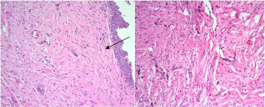
Biomed. Mater. 7 (2012) 065002                                                                                                    W-J Fu et al

its first description in 1967 by Zimskind, the double-J ureteral           2.2. Animals
stent has been an indispensable tool in surgical procedures
                                                                           A total of 18 beagles 12 months old and ranging from 9 to
[2]. Double-J ureteral stents have commonly been placed for
                                                                           11.5 kg were used. Eight were male and 10 were female.
upper urinary tract drainage, for various purposes ranging
                                                                           The animals were anaesthetized with 3% pentobarbital sodium
from urolithiasis to reconstruction for more than four decades.
                                                                           (25 mg kg−1). A firearm fragment injury (combination of blast
However, the conventional double-J stent is associated with
                                                                           injury and burn) was generated by a self-designed explosion
several problems such as irritation, bleeding, pain, reflux,
                                                                           device, which was shown to be reproducible and effective in
infection, migration, calcification formation, obstruction,
                                                                           our previous study [7]. The dogs were randomized into two
periodical exchange and reduction of life quality [3].
                                                                           groups of 9, with both ureters injured by the explosion device.
      Recently, biodegradable polymers have begun to play
                                                                           In group A, a biodegradable ureteral stent was placed into one
an increasingly important role in urology. They have been
                                                                           ureter before ureteroureteral anastomosis was performed and
evaluated as either urethral stent or ureteral stent. The main
                                                                           only end-to-end anastomosis was performed on the other side
advantage of these materials is that stent removal is not
                                                                           as a control. In group B, before ureteroureteral anastomosis
necessary. Olweny et al used a poly-L-lactide-co-glycolide
                                                                           was performed, a biodegradable ureteral stent was placed on
(PLGA) ureteral stent following experimental endopyelotomy
                                                                           one side, while a double-J stent placed on the other side
in pigs and found that the PLGA stent provided effective
                                                                           as a control. The dogs ventilated spontaneously through an
drainage [4]. In a case of UPJ treated by antegrade
                                                                           intubation tube and received fluid replacement treatment.
endopyelotomy, a horn-shaped PLGA stent was used and
successfully functioned as a partial catheter. In addition to
this, the stent removal was avoided [5]. Recently, Kotsar et al            2.3. Surgical procedure
developed a biodegradable braided prostatic stent and the stent            A midline laparotomy was performed on all dogs under general
with dutasteride showed promising results in the treatment of              anesthesia. A mark was made at the ureterovesical junction.
acute urinary retention due to benign prostatic enlargement [6].           The length of the ureter of the beagle is about 15 cm and the
      In this study, we developed a biodegradable ureteral stent           mid-ureter was selected as the location where firearm fragment
and further explored possible advantages of the biodegradable              injury was made. The injured segment of ureter was about
ureteral stent over the traditional double-J stent in the treatment        1 cm in length. In group A, the firearm fragment injuries
of ureteral injury, such as no need for removal, no calcification,         were made to bilateral ureters. The wound was wrapped in
better protection of renal function and the more effective                 wet gauze and left for 30 min to imitate the evacuation time
alleviation of stent-related symptoms. The feasibility and                 needed in battlefield. Then debridement was performed; the
biocompatibility of the biodegradable ureteral stent were                  injured segment of ureter (about 1 cm long) was cut off.
studied in detail using a canine model of ureteral injury.                 The biodegradable ureteral stent was inserted longitudinally
                                                                           into one partial randomly chosen ureter and placed only
2. Materials and methods                                                   at the repaired segment of the ureter, the middle point of
                                                                           the stent was the point at which ureteroureteral anastomosis
The study protocol was approved by the committee of animal                 was performed, and the stent was fixed at the anastomotic
research at the People’s Liberation Army General Hospital and              stoma with absorbable sutures. However, only end-to-end
the procedures were performed in accordance with guidelines                anastomosis was performed using 6-0 absorbable vicryl on the
for the humane handling of animals.                                        opposite ureter. In group B, the equivalent firearm fragment
                                                                           injuries were also made to bilateral ureters at the same position.
2.1. Stents                                                                After debridement and the cutting off of the damaged segment,
                                                                           a biodegradable ureteral stent was implanted using the same
The biodegradable ureteral stents were made from polylactic                method into one randomly chosen ureter and a double-J stent
acid. Poly-L-lactic acid (PLLA) and poly-DL-lactic acid                    on the opposite side before ureteroureteral anastomosis was
(PDLLA) were mixed together in proportion to their mass.                   performed. The biodegradable ureteral stents were fixed to
A 25% barium sulfate additive was applied to the material                  the ureteral walls by means of self-expansion of the stent
to enhance its radio-opacity. The mixture was dissolved in                 material due to absorption of water and absorbable suture at
dichloromethane and cast as films which were evaporated in a               the anastomotic site. The precise implantation of the stents
fume hood. The films were cut into 1 mm wide strips which                  was ensured and documented by radiography postoperatively.
were wound on a 0.8 mm diameter stainless steel wire to form                    Laparotomy wounds were closed in three layers. The
stents 50 mm in length, 0.8 mm in internal diameter and 1.4 mm             abdominal wall was closed with 7-0 silk sutures, and
in external diameter (figure 1). The stents were dried in an oven          then the subcutaneous tissue and the skin were closed
at 65 ◦ C for 2 weeks and then sterilized by 60Co irradiation.             separately with 1-0 silk sutures. The sutures on the
Ultimately partial biodegradable ureteral stents were prepared.            skin were removed after 1 week. The dogs were given
The stents are flexible, and this characteristic will facilitate the       broad-spectrum antibiotics postoperatively for prophylaxis
insertion procedure.                                                       of infection, and wound dressings were replaced every
     Polyurethane BARDEX       R
                                  double-J ureteral stents (4.7Fr          other day. Buprenorphine hydrochloride (0.02 mg kg−1) was
16 cm, Bard, Inc., Tempe, AZ) were used as controls in this                administrated intramuscularly as pain medication. All surgery
study.                                                                     was performed by one experienced surgeon.

                                                                       2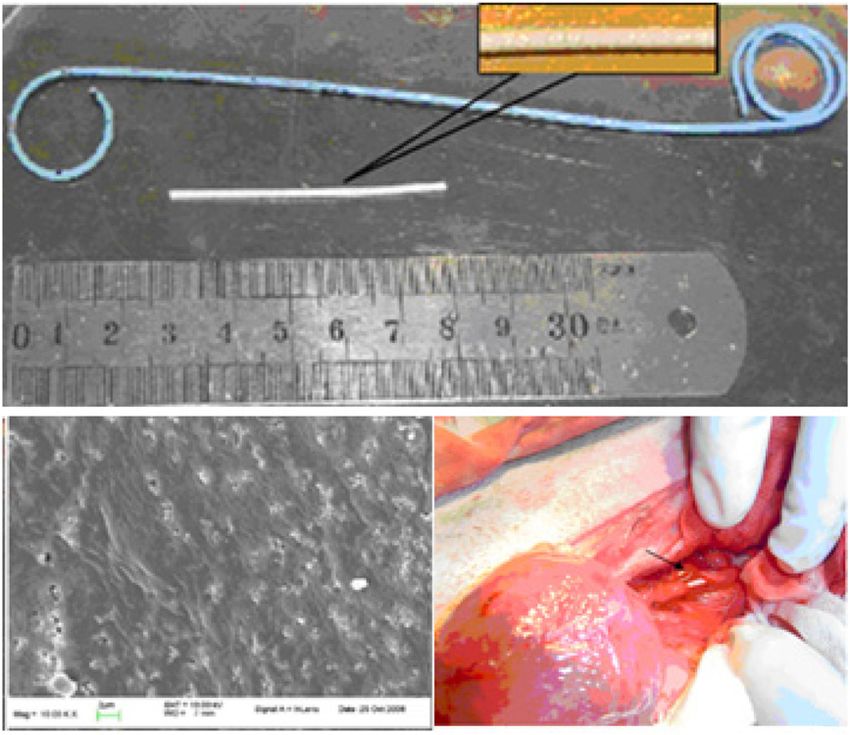
Biomed. Mater. 7 (2012) 065002                                                                                                  W-J Fu et al

                                                                                                            (a)

                                                                  (b)                                        (c)

Figure 1. Biodegradable ureteral stent: (a) double-J stent and biodegradable stent; (b) SEM showed that the surface of the stent was smooth
and particles of barium sulfate evenly distributed (×104); (c) biodegradable ureteral stent was inserted into the ureter.

2.4. Intravenous urography                                               of the fibroplasia and derangement of smooth muscle of
                                                                         ureteral wall: smooth muscle encircled the ureteral lumen,
Intravenous urography (IVU) was performed on all animals at
                                                                         but accompanied by fibrous tissue; fibrous tissue and smooth
1 week preoperatively and 40, 80 and 120 days postoperatively.
                                                                         muscle disarranged, however submucosal loose connective
Iohexol injection (300 mg I/ml, 0.67 ml kg−1) was used as a
                                                                         tissue was still visible; disarrangement of smooth muscle and
contrast medium.
                                                                         fibrous tissue involved full thickness, which were graded from
                                                                         1 to 3; C, transitional epithelial hyperplasia: slight, mild and
2.5. Renogram analysis                                                   severe, which were graded from 1 to 3. This criterion was
99m
    Tc-DTPA renography was carried out to assess renal                   established based on Lumiaho’s study and some modifications
function at the same time points as IVU was performed using              were made according to our study [8]. It reflects the extent of
a gamma camera (Siemens). Following a bolus injection of                 not only the inflammatory reactions, but also the scar formation
120 MBq of 99mTc-DTPA, the total measuring time was 15 min               and fiber disarrangement at the ureteroureteral anastomosis
at the rate of 1 frame s−1 for the first 30 s and at the rate of         site.
1 frame/15 s for the following 14.5 min. The data were saved
on a magnetic disk for further analysis. The ratio of the renal          2.7. Ultrastructural observation and compositional analysis
partial concentration indices (RPCI) and the ratio of the kidney         of the stent surfaces
washout half-time (T1/2) between the kidneys were calculated.
                                                                         At the same time points after the operation, samples
                                                                         of biodegradable ureteral stents and double-J stents were
2.6. Histological examination
                                                                         acquired. Scanning electron microscopy (SEM) was used
Three dogs from each group were randomly selected and                    to further explore the superficial ultrastructure of the
sacrificed with an overdose of narcotic for histological                 biodegradable ureteral stents and double-J stents. The
examination at 40, 80 and 120 days postoperatively. The tissue           elemental compositions of both stents were also analyzed.
at the site of ureteroureteral anastomosis was dissected en
bloc. The excised tissue was fixed in 10% neutral buffered               2.8. Statistical analysis
formalin, processed and paraffin embedded. The tissue was
then sectioned and stained with hematoxylin and eosin                    Results were expressed as mean ± standard deviations. One-
(HE). All histological results were evaluated blind by two               way analysis of variance (ANOVA) was used to evaluate the
experienced pathologists according to the following criteria:            significance of the ratios of RPCI and T1/2 of bilateral kidneys.
A, inflammatory reactions: slight, mild–severe, erosions and             The effect of time in different histologic response groups was
ulcerations, which were graded from 1 to 4; B, extent                    evaluated by the Friedman test. The difference between the

                                                                     3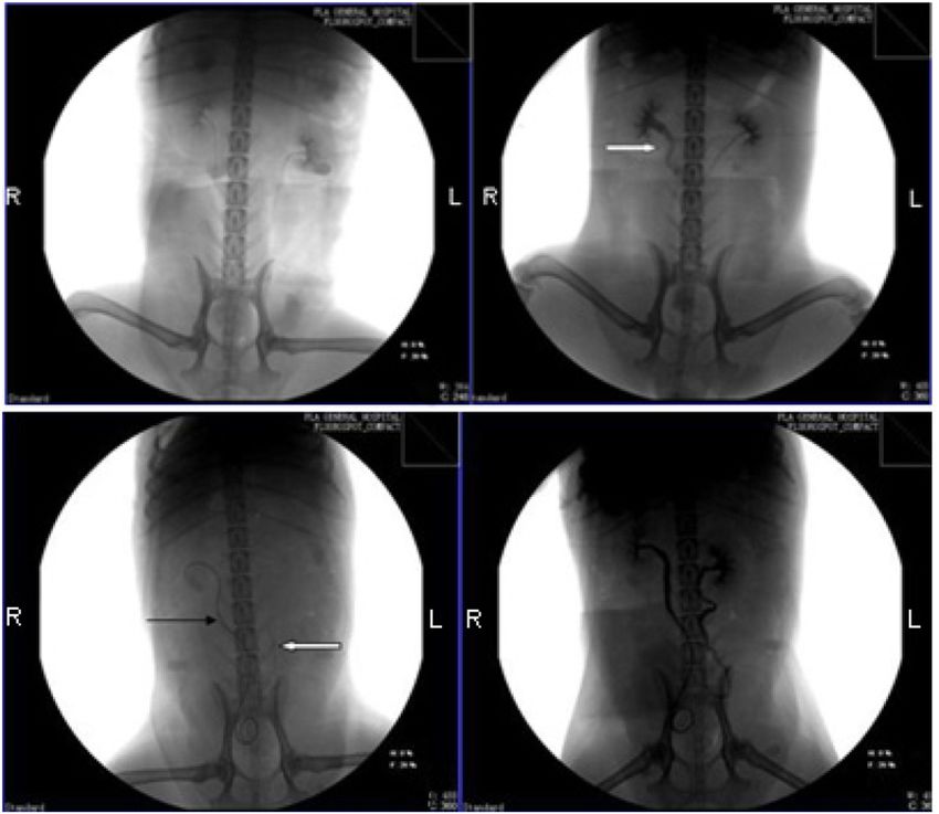
Biomed. Mater. 7 (2012) 065002                                                                                                      W-J Fu et al

                                                                 (a)                                         (b)

                                                                 (c)                                         (d )

Figure 2. IVU image of both groups: (a) normal image at 1 week preoperatively; (b) hydronephrosis and hydroureter were seen on the
stent-free side on right ureter (   ), while no obstruction was noted on the left ureter with the biodegradable stent at 120 days
postoperatively; (c) biodegradable stent on the left side and double-J stent on the right side were both in good position at 80 days; (d) no
signs of hydronephrosis on either stent side were found at 80 days.

two groups was analyzed by the Mann–Whitney test. The P-                   biodegradable stent could be found at this time (figure 2(c)). At
value was set at 0.05 to be statistically significant. Statistical         120 days postoperatively, the biodegradable stents were almost
analysis was carried out using SPSS software (version 17.0,                completely degraded and discharged by urination; there was
SPSS Inc., Chicago, IL).                                                   no sign of ureteral obstruction caused by the degraded stent
                                                                           particles.
3. Results
                                                                           3.2. Quantitative renographic analysis
All biodegradable ureteral stents and double-J stents were
properly implanted into the ureters of the dogs (figure 1). No             The ratios of RPCI and T1/2 of bilateral kidneys were
stent migration or mortality occurred before sacrifice. No signs           used as indicators for quantitative renogram analysis. In
of dysuria, urinary frequency and gross hematuria have been                group B, at 1 week preoperatively and 40, 80 and
observed during follow-up. The double-J stents were removed                120 days postoperatively, the RPCI ratio of bilateral kidneys
through cystotomy when the dogs were sacrificed, while the                 (biodegradable stent side:double-J stent side) showed no
biodegradable stents disappeared gradually due to degradation              statistical difference between groups. The T1/2 ratio of bilateral
in the urine.                                                              kidneys (biodegradable stent side:double-J stent side) also
                                                                           showed no statistical difference between groups (table 1). In
3.1. Intravenous urography                                                 group A, the ratio of RPCI of bilateral kidneys (biodegradable
                                                                           stent side:stent-free side) increased and the T1/2 ratio decreased
Preoperative IVU images showed no signs of hydronephrosis                  with time. The differences were statistically significant
or hydroureter and the ureteral lumens of all dogs were                    (table 2).
smooth (figure 2(a)). Postoperatively, in group A, renal
pelvis and ureter on the biodegradable ureteral stent side                 3.3. Histological findings
appeared normal, without dilation, and ureteral drainage was
patent and clear; however, hydronephrosis and hydroureter                  In the biodegradable ureteral stent group, histological findings
occurred on all stent-free sides at 120 days after surgery                 at 40 days revealed acute inflammatory reactions (graded
(figure 2(b)). In group B, no signs of hydronephrosis were                 at 2.33, table 3) and neutrophilic granulocyte infiltrations.
found on either side at any time point (figure 2(d)). At 80 days           Papillary hyperplasia of the epithelia was obvious at the
postoperatively, intravenous pyelography images showed that                anastomotic stoma of ureter (figure 3(a)). Localized absence of
both stents were in good position and partial degradation of the           epithelium was seen in one case (figure 3(c)). At 80 days, the

                                                                       4
Biomed. Mater. 7 (2012) 065002 W-J Fu et al
(a) (b) (c)
(d) (e) (f)
(g) (h) (i)
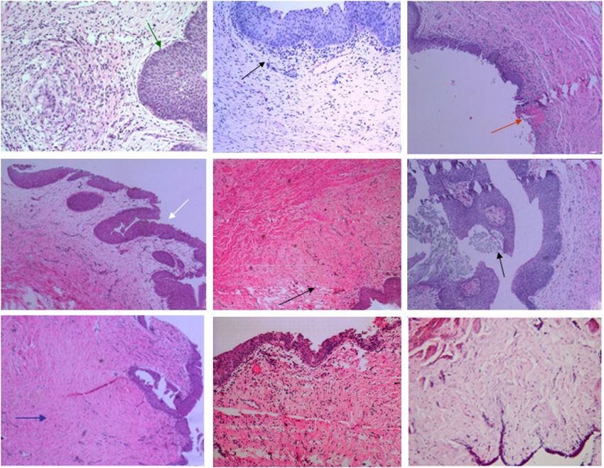
Figure 3. Histological findings of both stent groups: (a) inflammatory reactions and papillary hyperplasia were obvious at 40 days in
biodegradable stent group (HE × 100); (b) inflammatory reactions and epithelial proliferation in the double-J stent group were also obvious
at 40 days (HE × 100); (c) localized absence of epithelium (red arrow) was seen in one case in the biodegradable stent group (HE × 40);
(d) epithelial proliferation in the biodegradable stent group at 80 days was slighter than at 40 days (HE × 40); (e) tissue reactions in the
double-J stent group were similar to that of the biodegradable stent group at 80 days (HE × 40); ( f ) fragments of biodegradable stent were
observed in epithelia (black arrow, HE × 40); (g) fibrosis in the biodegradable stent group was more severe at 120 days (HE × 40); (h)
tissue reactions at 120 days were slighter than that at 80 days in the double-J stent group (HE × 100); (i) normal ureteral mucosa as control
(HE × 40).
Table 1. Bilateral renal function assessed by the RPCI ratio and T1/2 ratio (biodegradable stent side:double-J stent side).
Follow-up (days) 7-pre 40-post 80-post 120-post P-value
RPCI ratio 1.02 ± 0.18 1.09 ± 0.25 1.13 ± 0.17 1.37 ± 0.63 0.305
T1/2 ratio 0.99 ± 0.13 1.14 ± 0.22 1.13 ± 0.13 1.13 ± 0.15 0.263
RPCI = renal partial concentration indices.
T1/2 = the kidney washout half-time.
pre = pre-operation; post = post-operation.
Table 2. Bilateral renal function assessed by the RPCI ratio and T1/2 ratio (biodegradable stent side:stent-free side).
Follow-up (days) 7-pre 40-post 80-post 120-post P-value
RPCI ratio 0.99 ± 0.11 1.24 ± 0.22 1.55 ± 0.27 1.57 ± 0.31 0.000
T1/2 ratio 1.06 ± 0.17 0.86 ± 0.12 0.72 ± 0.06 0.65 ± 0.12 0.000
RPCI = renal partial concentration indices.
T1/2 = the kidney washout half-time.
pre = pre-operation; post = post-operation.
degree of inflammatory reactions (graded at 1.33, table 3) and leukomonocyte infiltration was found (figure 3(d)). Degraded
epithelial hyperplasia (1.67, table 3) were decreased compared fragments of biodegradable stent could be found in the
with tissue inflammatory reactions at 40 days, but submucosal epithelium layer (figure 3( f )). At 120 days, there were no
5Biomed. Mater. 7 (2012) 065002 W-J Fu et al
Table 3. Inflammatory reactions, fibro-hyperplasia and derangement Table 5. Mean quantified histological response to stent materials in
of smooth muscle and epithelial hyperplasia in group B biodegradable stent and double-J stent groups during 120 days.
(biodegradable stent and double-J stent) up to 4 months.
Histological Biodegradable Double-J
Follow-up Day 40 Day 80 Day 120 response stent (n = 9) stent (n = 9) P-value
Biodegradable stent A. Inflammatory reactions 1.67 1.56 0.863
Numbers of ureter n=3 n=3 n=3 B. Fibro-hyperplasia and 1.56 1.78 0.546
A. Inflammatory reactions 2.33 1.33 1.33 derangement of smooth
B. Fibro-hyperplasia and 1.00 1.67 2.00 muscle
derangement of smooth C. Epithelial hyperplasia 1.67 2.10 0.190
muscle Mean(A + B + C) 1.63 1.81 0.400
C. Epithelial hyperplasia 2.00 1.67 1.33
Mean(A + B + C) 1.78 1.56 1.55 P = 0.761 0.01–0.99 = mild changes.
Double-J stent 1.00–1.99 = moderate changes.
Numbers of ureter n=3 n=3 n=3 2.00–3.00 = severe changes.
A. Inflammatory reactions 1.67 1.33 1.67
B. Fibro-hyperplasia and 1.00 2.00 2.33 ureteral stent group was more regularly aligned and paralleled
derangement of smooth
muscle the ureteral lumen (figure 4).
C. Epithelial hyperplasia 2.67 2.00 1.67 In both groups, mean histological reactions showed no
Mean(A + B + C) 1.78 1.78 1.89 P = 0.761 significant changes during the study period (table 3, P >
P-value 0.931 0.436 0.340 0.05). The mean quantified response to stent materials and the
0.01–0.99 = mild changes. operative trauma of the biodegradable stent and double-J stent
1.00–1.99 = moderate changes. were graded at 1.63 and 1.81, respectively, and no significant
2.00–3.00 = severe changes. difference between the two groups was seen (table 5, P >
0.05).
Table 4. Inflammatory reactions, fibro-hyperplasia and derangement
of smooth muscle and epithelial hyperplasia in group A 3.4. Ultrastructural characteristics and elemental
(biodegradable stent and suture alone) up to 4 months. composition of the stent surfaces
Follow-up Day 40 Day 80 Day 120 At 40 days after operation, SEM showed that the surface
Biodegradable stent of the biodegradable ureteral stent was almost integrated
Numbers of ureter n=3 n=3 n=3 (figure 5(a)), but pores were found inside of the stent,
A. Inflammatory reactions 2.00 1.67 1.00 indicating initial degradation (figure 5(b)). However, no signs
B. Fibro-hyperplasia and 1.00 1.67 2.00 of degradation were found in the double-J stent group.
derangement of smooth
muscle Elemental composition analysis revealed C, O, S and Ba
C. Epithelial hyperplasia 2.33 1.67 1.33 elements on the surface of both the biodegradable ureteral stent
Mean(A + B + C) 1.78 1.67 1.44 P = 0.717 and the double-J stent (figure 6). At 80 days, the elemental
Suture alone composition analysis of the surface of the double-J stent
Numbers of ureter n=3 n=3 n=3 confirmed that there existed C, O, S, Ba and Ca elements
A. Inflammatory reactions 1.33 1.33 1.33
B. Fibro-hyperplasia and 1.00 2.33 3.00 (figure 7(a)). However, the surface of the biodegradable
derangement of smooth ureteral stent still only consisted of C, O, S and Ba elements
muscle without Ca (figure 7(b)). At 120 days, the biodegradable
C. Epithelial hyperplasia 1.67 1.67 1.33 stent degraded into small sediment-like particles, due to the
Mean(A + B + C) 1.33 1.78 1.89 P = 0.867 degradable characteristic of the material itself. On SEM, pores
P-value 0.340 0.796 0.190
of varying shapes and sizes and wide cracks were seen on
the surface of discharged degraded small particles of the
biodegradable ureteral stent (figure 8(a)). In the double-J stent
obvious inflammatory reactions, but fibrosis could be observed
group, calcified plaque was observed (figure 8(b)).
at the submucosa of ureteral anastomotic stoma (figure 3(g)).
In the double-J stent group, mean tissue reactions
including inflammatory reactions, fibrous tissue proliferation 4. Discussion
and epithelial hyperplasia at 40, 80 and 120 days were 1.78,
In the treatment of various ureteral injuries, the relief of
1.78 and 1.89, respectively. They were similar to that of obstruction is critical [9]. Ureteral stents have been used
the biodegradable stent group (figures 3(b), (e), (h)). There for various purposes after upper urinary tract surgery or
were no statistically significant differences between the two trauma. The rationale for using ureteral stents is based on
groups at the same time points (table 3, P > 0.05). Tissue the mechanism of bypassing obstructions of the ureter and
reactions in group A were not significantly different between for urinary diversion to ensure flow while causing minimal
the biodegradable stent and suture alone groups (table 4). functional disruption, thus maintaining renal function [10].
Hyperplastic tissue developed both in the biodegradable stent The concept of the ureteral stent was first described in the 19th
and stent-free groups, but fibrous tissue in the biodegradable century [11]. Since then, stents and catheters have been widely
6Biomed. Mater. 7 (2012) 065002 W-J Fu et al
(a) (b)
Figure 4. Histological findings of group A at 120 days: (a) fibrous tissue in the biodegradable stent group aligned more regularly and
parallel to the ureteral lumen (HE × 40); (b) fibrous tissue in the stent-free group was mussy (HE × 100).
(a) (b)
Figure 5. SEM of a biodegradable ureteral stent at 40 days: (a) the surface of the stent was almost integrated (×104); (b) pores, which
indicate that degradation started, can be found in the inside of the stent (×104).
(a) (b)
Figure 6. Results show that there exist C, O, S and Ba elements on the surface of both the biodegradable ureteral stent (a) and the double-J
stent (b) at 40 days.
(a) (b)
Figure 7. Results show that the Ca element exists on the surface of the double-J stent (a) at 80 days; however, no Ca element can be detected
on the surface of biodegradable ureteral stent (b) at the same time.
7Biomed. Mater. 7 (2012) 065002 W-J Fu et al
(a) (b)
Figure 8. SEM at 120 days: (a) pores of varying shapes and sizes and wide cracks can be found on the surface of the discharged degraded
small particle of the biodegradable stent (×6000); (b) a great quantity of calcific plaque can be observed on the surface of the double-J stent
(×6000).
used in urology. Currently, the use of indwelling ureteral stents used to evaluate the renal function when the urinary tract is
is well established. However, the ideal ureteral stent has yet to obstructed. It reflects the effective plasma flux in the kidneys as
be discovered [12]. well as the speed of uptake and the amount of tracer taken in
Numerous investigators have evaluated the ideal size, by the renal tubular epithelium. T1/2 is an indicator of how
material and indwelling time for ureteral stents; it is believed much tracer in urine is washed out from the kidneys and
that the stent serves as a scaffold for the healing ureter to at what speed. It is affected by several factors such as the
drain urine [13]. The ideal stent should be easy to manipulate, body plasma volume. However, system errors are deemed to
have excellent tensile strength, be resistant to encrustation be undiscriminating between kidneys on both sides. For this
and migration, and be biocompatible [14]. Many materials reason, we used the ratios of RPCI and T1/2 of bilateral kidneys
have been tried, including metallic, synthetic, biodegradable to eliminate possible system errors. The RPCI ratio and
and autologous materials. Several metallic stents entered T1/2 ratio of bilateral kidneys (biodegradable stent side:double-
urological practice, but they did not satisfactorily address the J stent side) showed no statistical difference between groups at
issue of frequent stent change, urothelial hyperplastic reaction 1 week preoperatively and 40, 80 and 120 days postoperatively.
or migration [15]. Subsequently, biodegradable materials such However, the RPCI ratio of bilateral kidneys (biodegradable
as poly-lactic acid underwent rapid development as absorbable stent side:stent-free side) increased and T1/2 ratio decreased
fixation materials for orthopedic surgical applications with time (P < 0.01).
and initial urological applications. The development of In this study, the biodegradable ureteral stent was made
biodegradable devices for urologic use started in Finland in from PLLA and PDLLA polymers and these combined
the late 1980s [16]. Nowadays, biodegradable polymers have materials possess good biocompatibility properties with less
an increasingly important role in various medical applications inflammatory reaction, scarring tissue formation and no upper
[17]. For example, Isotalo successfully treated recurrent urinary tract obstruction caused by degraded fragments. The
urethral stricture with a bio-absorbable self-expandable, self- good biocompatibility properties of these materials have
reinforced poly-L-lactic acid urethral stent in combination with been documented; after absorption of the device, the organ
optical urethrotomy [18]. Polylactic acid polymers as a stent preserves its normal function without the need to remove
material possess good biocompatibility, strength and the ability the device [19, 20]. Degradation and biocompatibility are
to degrade in vivo. important characteristics for biodegradable ureteral stents.
Our study demonstrated that a biodegradable ureteral stent The advantages of using biodegradable stents are to preclude
is as reliable in holding the ureter open and allowing drainage a secondary procedure for removal and eliminate the risk of
as the traditional double-J stent in the treatment of ureteral forgotten stents. The forgotten stents can lead to renal failure
injury. The effectiveness of the biodegradable ureteral stent or even death. Although endourology can provide all necessary
was assessed by postoperative intravenous pyelography and solutions for the management of forgotten indwelling stents,
quantitative renogram analysis (RPCI ratio and T1/2 ratio) the best treatment remains prevention [21]. In our study, SEM
of bilateral kidneys. Postoperative intravenous pyelography showed that the degradation of the biodegradable ureteral
showed no signs of hydronephrosis on either stent side at any stent began at 40 days. At 120 days, the stent had almost
time point. Although slight hydroureter due to passive dialation degraded. Some sediment-like particles can be found in
can be found on the IVU image of 80 days in our study, the the urine; this may be the degraded substances of stent
hydroureter on biodegradable stented side was slighter than according to our in vitro observation of the degradation
double-J stented side and vanished spontaneously at 120 days process of the stent. There were also no signs of calcification.
when the biodegradable stent was gone. RPCI is an index In contrast, calcified plaque was found on the surface of
8Biomed. Mater. 7 (2012) 065002 W-J Fu et al
the double-J stent on SEM at 120 days, and the elemental References
composition analysis also confirmed the existence of calcium.
However, there was no obvious sign of calculi formation and [1] Hammontree L N, Wade B K, Passman C M, Prieto J C,
calcifications of the double-J stent did not obstruct urine Burns J R and Kolettis P N 2005 Ureteral injuries: recent
trends in etiologies, treatment, and outcomes J. Pelvic Med.
flow at 120 days; this may be because of a short period Surg. 11 129–36
of follow-up in our study. Previous studies have shown that [2] Zimskind P D, Fetter T R and Wilkerson J L 1967 Clinical use
longer indwelling time of double-J stents was associated with of long-term indwelling silicone rubber ureteral splints
increased prevalence and consequences of all complications, inserted cystoscopically J. Urol. 97 840–4 PMID: 6025928
including stone formation and even death [22, 23]. In the [3] Agrawal S, Brown C T, Bellamy E A and Kulkarni R 2009
The thermo-expandable metallic ureteric stent: an 11-year
present study, no signs of calcification and calcium were seen
follow-up BJU Int. 103 372–6
on the surface of the biodegradable ureteral stent, let alone [4] Olweny E O, Landman J, Andreoni C, Collyer W, Kerbl K,
stone formation. Additionally, the PLLA–PDLLA stent did Onciu M, Välimaa T and Clayman R V 2002 Evaluation
not induce ureteral stricture due to fibrous tissue formation and of the use of a biodegradable ureteral stent after
epithelial hyperplasia. However, a longer period of follow-up retrograde endopyelotomy in a porcine model J. Urol.
is needed. 167 2198–202
[5] Talja M, Multanen M, Välimaa T and Törmälä P 2002
In addition to this, a partial ureteral stent offers various Bioabsorbable SR-PLGA horn stent after antegrade
advantages over traditional long stent design. Upper urinary endopyelotomy: a case report J. Endourol. 16 299–302
tract pressure-flow studies in stented ureters have demonstrated [6] Kotsar A, Isotalo T, Juuti H, Mikkonen J, Leppiniemi J,
that the intravesical pressure can be transmitted directly to the Hänninen V, Kellomäki M, Talja M and Tammela T L 2009
renal pelvis when the pressure in the bladder is increased, such Biodegradable braided poly(lactic-co-glycolic acid) urethral
stent combined with dutasteride in the treatment of acute
as during micturition. A full-length ureteral stent (double-J urinary retention due to benign prostatic enlargement: a
stent) may increase intra-pelvic pressure and cause hydroureter pilot study BJU Int. 103 626–9
and vesicoureteral reflux (VUR) [24]. An advantage of the [7] Li G, Wang Z X, Fu W J, Hong B F, Wang X X, Cao L,
biodegradable stent is that a segmental stent can be placed Xu F Q, Song Q, Cui F Z and Zhang X 2011 Introduction to
only at the repaired segment of ureter, thus could protect biodegradable polylactic acid ureteral stent application for
treatment of ureteral war injury BJU Int. 108 901–6
renal function and avoid VUR. Because no direct connection [8] Lumiaho J, Heino A, Pietiläinen T, Ala-Opas M, Talja M,
between the renal pelvis and the bladder is established, the Välimaa T and Törmälä P 2000 The morphological, in situ
intravesical pressure cannot be transmitted directly to the renal effects of a self-reinforced bioabsorbable polylactide
pelvis when the pressure in the bladder is increased. This (SR-PLA 96) ureteric stent: an experimental study J. Urol.
avoids the reflux of urine into the kidney through the traditional 164 1360–3
[9] López-Huertas H L, Polcari A J, Acosta-Miranda A
double-J stent [25]. On the other hand, the bladder end of
and Turk T M 2010 Metallic ureteral stents: a cost-effective
double-J stent is directly related to bladder irritation [26]. The method of managing benign upper tract obstruction
biodegradable stent will not directly irritate the renal pelvis and J. Endourol. 24 483–5
bladder trigone, and will minimally interrupt normal ureteral [10] Lumiaho J, Heino A, Tunninen V, Ala-Opas M, Talja M,
peristalsis, which may help prevent stent-related symptoms. Välimaa T and Törmälä P 1999 New bioabsorbable
There were no signs of urinary frequency and gross hematuria polylactide ureteral stent in the treatment of ureteral lesions:
an experimental study J. Endourol. 13 107–12
during follow-up. However, there are still several limitations [11] Saltzman B 1988 Ureteral stents. Indications, variations, and
in this study, including further refinement of raw materials and complications Urol. Clin. North. Am. 15 481–91 PMID:
the configuration of the stent to facilitate endoscopic insertion. 3043868
Also a longer follow-up is needed to see if the stricture reforms [12] Beiko D T, Knudsen B E and Denstedt J D 2003 Advances in
once the stent is gone. ureteral stent design J. Endourol. 17 195–9
[13] Kuzaka B, Szymanska K, Borkowski A and Krus S 1996
Restoration of the continuity of dog ureter after resection of
5. Conclusions its 5 cm middle segment Br. J. Urol. 77 342–6
[14] Venkatesan N, Shroff S, Jayachandran K and Doble M 2010
This study demonstrated that a biodegradable ureteral stent Polymers as ureteral stents J. Endourol. 24 191–8
[15] Liatsikos E, Kallidonis P, Stolzenburg J U and Karnabatidis D
could be as effective as the traditional double-J stent for
2009 Ureteral stents: past, present and future Expert Rev.
the treatment of ureteral injury. Moreover, the biodegradable Med. Devices 6 313–24
ureteral stent has several advantages, such as no need for [16] Törmälä P 1992 Biodegradable self-reinforced composite
removal, no calcification, better protection of renal function materials; manufacturing structure and mechanical
by avoiding the reflux of urine and more effective alleviation properties Clin. Mater. 10 29–34
of stent-related symptoms. [17] Ulery B D, Nair L S and Laurencin C T 2011 Biomedical
applications of biodegradable polymers J. Polym. Sci. B:
Polym. Phys. 49 832–64
Acknowledgments [18] Isotalo T, Talja M, Välimaa T, Törmälä P and Tammela T L
2002 A bioabsorbable self-expandable, self-reinforced
This study was supported by National Natural Science poly-L-lactic acid urethral stent for recurrent urethral
strictures: long-term results J. Endourol. 16 759–62
Foundation (no 81070555), Beijing Natural Foundation [19] de Tayrac R, Chentouf S, Garreau H, Braud C, Guiraud I,
(no 2092029) and the Major Project of Clinical High and New Boudeville P and Vert M 2008 In vitro degradation and
Technology of Army hospital. in vivo biocompatibility of poly(lactic acid) mesh for soft
9Biomed. Mater. 7 (2012) 065002 W-J Fu et al
tissue reinforcement in vaginal surgery J. Biomed. Mater. [23] Damiano R, Oliva A, Esposito C, De Sio M, Autorino R
Res. B: Appl. Biomater. 85 529–36 PMID: 18161812 and D’Armiento M 2002 Early and late complications of
[20] Zamiri P, Kuang Y, Sharma U, Ng T F, Busold R H, double pigtail ureteral stent Urol. Int. 69 136–40
Rago A P, Core L A and Palasis M 2010 The [24] Cummings L J, Waters S L, Wattis J A and Graham S J 2004
biocompatibility of rapidly degrading polymeric stents in The effect of ureteric stents on urine flow: reflux J. Math
porcine carotid arteries Biomaterials 31 7847–55 Biol. 49 56–82
[21] Chew B H, Lange D, Paterson R F, Hendlin K, Monga M, [25] Lumiaho J, Heino A, Kauppinen T, Talja M, Alhava E,
Clinkscales K W, Shalaby S W and Hadaschik B A 2010 Välimaa T and Törmälä P 2007 Drainage and antireflux
Next generation biodegradable ureteral stent in a yucatan characteristics of a biodegradable self-reinforced,
pig model J. Urol. 183 765–71 self-expanding x-ray-positive poly-L,D-lactide spiral partial
[22] Ivica S and Dragan S 2009 Long-term indwelling double-J ureteral stent: an experimental study J. Endourol.
stents: bulky kidney and urinary bladder calculosis, 21 1559–64
spontaneous intraperitoneal perforation of the kidney and [26] Miyaoka R and Monga M 2009 Ureteral stent discomfort:
peritonitis as a result of ‘forgotten’ double-J stent etiology and management Indian J. Urol.
Vojnosanit. Pregl. 66 242–4 25 455–60
10You can also read