Viral Shedding in Patients Infected with Pandemic Influenza A (H1N1) Virus in Kenya, 2009
←
→
Page content transcription
If your browser does not render page correctly, please read the page content below
Viral Shedding in Patients Infected with Pandemic
Influenza A (H1N1) Virus in Kenya, 2009
Lilian W. Waiboci1*, Emmaculate Lebo1, John M. Williamson1, William Mwiti2, Gilbert K. Kikwai2, Henry
Njuguna2, Beatrice Olack2, Robert F. Breiman1, M. Kariuki Njenga1, Mark A. Katz1,3
1 US Centers for Disease Control and Prevention-Kenya, Nairobi, Kenya, 2 Kenya Medical Research Institute/US Centers for Disease Control and Prevention-Kenya, Nairobi,
Kenya, 3 Influenza Division, National Center for Immunization and Respiratory Diseases (NCIRD), Centers for Disease Control and Prevention, Atlanta, Georgia, United
States of America

     Abstract
     Background: Understanding shedding patterns of 2009 pandemic influenza A (H1N1) (pH1N1) can inform recommenda-
     tions about infection control measures. We evaluated the duration of pH1N1 virus shedding in patients in Nairobi, Kenya.

     Methods: Nasopharyngeal (NP) and oropharyngeal (OP) specimens were collected from consenting laboratory-confirmed
     pH1N1 cases every 2 days during October 14–November 25, 2009, and tested at the Centers for Diseases Control and
     Prevention-Kenya by real time reverse transcriptase polymerase chain reaction (rRT-PCR). A subset of rRT-PCR-positive
     samples was cultured.

     Results: Of 285 NP/OP specimens from patients with acute respiratory illness, 140 (49%) tested positive for pH1N1 by rRT-
     PCR; 106 (76%) patients consented and were enrolled. The median age was 6 years (Range: 4 months–41 years); only two
     patients, both asthmatic, received oseltamivir. The median duration of pH1N1 detection after illness onset was 8 days (95%
     CI: 7–10 days) for rRT-PCR and 3 days (Range: 0–13 days) for viral isolation. Viable pH1N1 virus was isolated from 132/162
     (81%) of rRT-PCR-positive specimens, which included 118/125 (94%) rRT-PCR-positive specimens collected on day 0–7 after
     symptoms onset. Viral RNA was detectable in 18 (17%) and virus isolated in 7/18 (39%) of specimens collected from patients
     after all their symptoms had resolved.

     Conclusions: In this cohort, pH1N1 was detected by rRT-PCR for a median of 8 days. There was a strong correlation between
     rRT-PCR results and virus isolation in the first week of illness. In some patients, pH1N1 virus was detectable after all their
     symptoms had resolved.

  Citation: Waiboci LW, Lebo E, Williamson JM, Mwiti W, Kikwai GK, et al. (2011) Viral Shedding in Patients Infected with Pandemic Influenza A (H1N1) Virus in
  Kenya, 2009. PLoS ONE 6(6): e20320. doi:10.1371/journal.pone.0020320
  Editor: Patricia V. Aguilar, University of Texas Medical Branch, United States of America
  Received January 22, 2011; Accepted April 21, 2011; Published June 10, 2011
  This is an open-access article, free of all copyright, and may be freely reproduced, distributed, transmitted, modified, built upon, or otherwise used by anyone for
  any lawful purpose. The work is made available under the Creative Commons CC0 public domain dedication.
  Funding: The research was funded by the US Centers for Disease Control and Prevention. The funders had no role in study design, data collection and analysis,
  decision to publish, or preparation of the manuscript.
  Competing Interests: The authors have declared that no competing interests exist.
  * E-mail: lwaiboci@ke.cdc.gov

Introduction                                                                               Much of the information on pH1N1 virus shedding is from
                                                                                        studies conducted in North America, Asia, and Europe
   The 2009 pandemic influenza A (H1N1) (pH1N1) virus has                               [7,8,9,10,11,12]. In many of these studies, real time reverse
been circulating worldwide since the initial cases were detected in                     transcriptase polymerase chain reaction (rRT-PCR) positive
the United States in April 2009 [1]. In order to control the spread                     results were used to identity positive cases and determine the
of infection, public health agencies including the World Health                         duration of virus shedding. A study in Hong Kong found that
Organization (WHO) and the United States Centers for Disease                            pH1N1 virus RNA was shed from the respiratory tract for up to 8
Control and Prevention (CDC) made recommendations on the                                days (median = 4 days) after symptom onset, [10]. Studies carried
duration of isolation of pH1N1 cases. The recommendations were                          out in China and Germany showed that pH1N1-infected patients
based on previous knowledge about duration of shedding of                               shed the virus for a mean of 6 days (Range: 1–17) and 6.6 days
seasonal influenza viruses [2,3,4]. Most patients infected with                         (Standard deviation: 2.6), respectively [10,11]. In addition, a case
seasonal influenza shed the virus for 5–7 days. However, children                       report from the United States reported that pH1N1 virus was
have been shown to shed the virus for up to 21 days and severely                        detected in respiratory samples 5 to 6 weeks after initial diagnosis
immunocompromised individuals up to several months [2,5].                               in 2 severely immunocompromised patients on chemotherapy;
Following the onset of the influenza pandemic in 2009, WHO                              these patients were on oseltamivir treatment and developed
recommended that control precautions such as self-isolation                             resistance to the drug [9].
should be practiced for 7 days from symptom onset or until all                             The first case of pH1N1 in Kenya was detected in June 2009
symptoms resolve [6].                                                                   [13]. At that time, the Kenya Ministry of Public Health and

        PLoS ONE | www.plosone.org                                                  1                                   June 2011 | Volume 6 | Issue 6 | e20320
Influenza A 2009 H1N1 Virus Shedding, Kenya
Sanitation followed WHO guidelines and recommended 7 days of tipped flexible aluminum-shafted NP swab was inserted into the
self-isolation for all pH1N1 patients. In order to assist in providing nose to the nasopharynx, where it was rotated 180 degrees and
evidence-based recommendations for infection control, we tested left in place for 3–5 seconds. The NP swab was inserted into the
serial specimens from laboratory-confirmed pH1N1 patients cryovial containing the OP swab from the same patient. The
attending a clinic in Nairobi to determine the duration of viral specimens were labeled and transported at 4uC to the KEMRI/
shedding, determine the correlation between rRT-PCR-positive CDC-K laboratory where they were tested for influenza A and
results and viral isolation, and evaluate clinical signs and pH1N1 using rRT-PCR within 24 hours.
symptoms associated with shedding. The rRT-PCR testing was performed using the CDC pH1N1
testing protocol [15]. Briefly, total RNA was extracted from
Materials and Methods 100 mL aliquots of each specimen using QIAamp viral RNA
minikit (Qiagen Inc., GmbH, Germany) according to the
Ethics Statement manufacturer’s instructions. One step rRT-PCR was carried out
This study was approved by both the Institutional Review using the AgPath kit (Applied Biosystems, California, USA). For
Board of CDC-Atlanta and the Ethical Review Committee of the influenza A detection, the primers used were 59GAC CRA TCC
Kenya Medical Research Institute (KEMRI). TGT CAC CTC TGA C as the forward, and 59 TG CAG TCC
TCG CTC ACT GGG CAC G as the reverse. The influenza A
Consent detection probe was 59 TGC AGT CCT CGC TCA CTG GGC
Informed written consent was obtained from all participants. ACG. For pH1N1, we used the 59GTG CTA TAA ACA CCA
For children, written consent was obtained from parents or GCC TYC CA as the forward primer, 59 CGG GAT ATT CCT
guardians. TAA TCC TGT RGC as the reverse primer, and 59CA GAA
TAT ACA TCC RGT CAC AAT TGG ARA A as the probe.
Setting and Study Design Specimens were also tested for seasonal influenza A H1 and H3.
We conducted the study in a large informal urban settlement in Following the reverse transcription step, a typical 45 cycle PCR
Nairobi, in an existing population-based surveillance system that reaction was run and fluorescence was read at the annealing/
has been previously described [14]. Consenting participants who extension step. Appropriate negative and positive control speci-
presented to a field clinic, known as Tabitha Clinic, within the mens were run alongside each reaction. The results were recorded
surveillance site with signs and symptoms consistent with as cross-over threshold (CT) values. Any influenza A CT value
influenza-like illness (ILI) or severe acute respiratory illness (SARI) ,40.0 was recorded as positive; CT value 40.0–44 were
had oropharyngeal (OP) and nasopharyngeal (NP) specimens considered indeterminate, and those without a CT reading were
taken. The specimens were tested for influenza by rRT-PCR at the recorded as negative. A specimen was considered to be pH1N1
KEMRI/CDC-K laboratory in Nairobi. positive if both the influenza A and the pH1N1 CT values were
An ILI case was defined as a patient with fever $38uC and ,40.0 as described in the CDC protocol of real time RT-PCR for
cough or sore throat for all ages. For SARI, the definitions varied influenza A(H1N1) [16].
by age group. In children ,5 years old, SARI was defined as Later, in order to evaluate the concordance between rRT-PCR
cough or difficulty breathing along with a danger sign; unable to testing and viral culture for pH1N1, influenza virus isolation in
drink or breast feed, lethargic or unconscious, vomiting every- Mardin-Darby Canine Kidney (MDCK) cells was attempted for
thing, convulsions, nasal flaring, grunts, chest indrawing, stridor in the first rRT-PCR-positive specimen, the last rRT-PCR-positive
a calm child or oxygen saturation #90%. In people $5 years, specimen, and the first rRT-PCR-negative specimen from each
SARI was defined as fever $38uC plus cough or shortness of enrolled patient. Confluent monolayers of MDCK cell line
breath or chest pain or oxygen saturation #90%. growing in T25 cell culture flasks were used. Media were removed
Patients who came to Tabitha Clinic between October 14, from the flasks. Each flask was inoculated with 100 mL of specimen
2009, and November 25, 2009, who had ILI or SARI and NP/OP and inoculum was allowed to adsorb on the cells for 30 min at
specimens that tested positive for pH1N1 were recruited for the 37uC. Following adsorption, 6 mL of Viral Growth Medium
study. Patients who were positive for pH1N1 were contacted at [DMEM supplemented with HEPES buffer, bovine serum
their homes by field workers and requested to return to the clinic albumin, L-Glutamine, trypsin, and antibiotic-antimycotic (Sig-
for follow up every 2 days. During the initial visit and subsequent ma-Aldrich, Inc., St. Louis, MO, USA)] was added and cultures
visits, a trained clinician recorded signs and symptoms and were observed daily for cytopathic effect (CPE) for up to 6 days, at
collected both NP and OP specimens. Patients who had 2 which point all the cultures were harvested. Cultures that showed
consecutive rRT-PCR negative specimens were released from the observable CPE by microscopy were subjected to hemagglutina-
study. A patient was considered to have pH1N1 RNA if a tion assay using guinea pig red blood cells. Supernatants from
specimen collected on that day was positive for pH1N1 by rRT- cultures that showed no CPE were subjected to a second passage,
PCR. Patients were not followed over the weekend. following which cultures with observable CPE were subjected to
hemagglutination assay. Cultures that had no observable CPE
Specimen collection and laboratory testing after the second passage were considered negative by viral
Specimens were obtained according to the following procedure: isolation.
for OP specimens, a sterile nylon-tipped plastic-shafted OP swab
touched the back of the oropharyngeal mucosal membrane for Data collection and statistical methods
3–5 seconds and then was placed into a cryovial containing 1 mL Data were collected in three ways. First, information related to
of viral transport media (VTM). VTM was prepared at the the patient’s initial visit was recorded on computers by clinicians at
KEMRI/ CDC-K laboratory using the standard WHO protocol Tabitha Clinic. Second, during the first follow-up clinic visit and
that includes bovine serum albumin and veal infusion broth subsequent visits, information about signs and symptoms was
supplemented with amphotericin B (www.who.int/csr/resources/ recorded on paper questionnaires and entered into a Microsoft
publications/surveillance/Annex8.pdf). Freshly prepared refrigerated Access database. The signs and symptoms recorded were fever,
VTM was used for up to 3 months. For NP specimens, a polyester- temperature $38uC, cough, sneezing, runny nose, vomiting,
PLoS ONE | www.plosone.org 2 June 2011 | Volume 6 | Issue 6 | e20320Influenza A 2009 H1N1 Virus Shedding, Kenya
diarrhea, chest pain, and difficulty breathing for all patients. In
addition, the following symptoms were recorded for patients $5
years; earache, sore throat, headache, chills, muscle pain, and
abdominal pain. Third, laboratory data were recorded into
Freezerworks software (Dataworks Development, Inc.). Data were
combined into a central database for analysis.
We considered the date of onset of illness to be the date on
which the patient reported the first symptom associated with the
illness, according to the history taken during the patient’s initial
clinic visit. For possible initial symptoms, we considered fever,
cough, and difficulty breathing for patients ,5 years old. In
patients $5 years old, these symptoms and sore throat were
considered as possible initial symptoms. Patients were considered
to have no detectable virus RNA once they had two consecutive
specimens that tested negative for pH1N1 by rRT-PCR. We
defined the last day of virus detection as the midpoint between the
dates of collection of the last rRT-PCR pH1N1-positive specimen
and the first rRT-PCR pH1N1-negative specimen. We were
unable to follow all patients until they had two negative rRT-PCR
tests because some did not return to the clinic for the necessary
follow-up visits. The time to cessation of pH1N1 virus detection Figure 1. Flow chart outlining enrollment in the pH1N1 viral
shedding study undertaken in Kenya from Oct 14–Nov 25,
for these patients was right censored at their last pH1N1 rRT- 2009.
PCR-positive test date. Thus, the duration of pH1N1 virus doi:10.1371/journal.pone.0020320.g001
detection for these patients was only known to occur after their
drop out time and was treated accordingly in the survival analysis (fifty patients had only one rRT-PCR-positive specimen), and100
below. first rRT-PCR-negative specimens from the enrolled patients. No
Comparisons of duration of pH1N1 virus detection between rRT-PCR-negative specimens were obtained from 6 of the
groups, described by the median and mean, were conducted using patients. Of the 162 rRT-PCR-positive specimens, pH1N1 virus
the log-rank test. A multivariable Cox regression model was used was isolated from 132 (81%) specimens. Of rRT-PCR-positive
to determine the association between gender and age with specimens taken on day 0–3 after illness onset, 81/85 (95%) were
duration of pH1N1 virus detection. A Kaplan-Meier plot was
culture-positive, as were 37/40 (93%) taken on day 4–7, 11/20
used to determine duration of pH1N1 virus detection. Odds ratios
(55%) taken on day 8–10, and 3/17 (18%) taken $11 days after
were calculated to determine the odds of isolating pH1N1 virus
illness onset (Figure 2). Viral isolation was successful in 100/106
from rRT-PCR-positive specimens collected over time. All
(94%) of the rRT-PCR positive specimens collected at the initial
analyses were conducted with SAS version 9.1. Findings were
clinic visit. Viral isolation was equally successful in samples with
considered statistically significant if the resulting p-value was
CT values,25 and those with CT values 25–30 (92% vs. 84%
,0.05.
p = 0.25), but more successful for samples with CT values,25
compared to those with CT values 30–39 (92% vs. 63%,
Results p = 0.009). Real time RT-PCR-positive specimens collected from
A total of 481 patients with respiratory illness were seen at
Tabitha Clinic between October 14, 2009 and November 25,
2009; 175 (36%) had ILI and 306 (64%) had SARI. Specimens Table 1. Demographic characteristics of 285 patients with
were collected from 285 (59%) of these patients, and 140 (49%) of acute respiratory illness who presented to Kibera Clinic,
these were positive for pH1N1 by rRT-PCR. Of the 140 pH1N1- Nairobi, Kenya and had specimens collected, Oct–Nov 2009.
positive patients, 106 (76%) consented to participate in the study;
85 (80%) completed the study (Figure 1). Data from all the 106
Characteristics rRT-PCR Results
patients who enrolled in the study were included in the analysis.
The median age of the study population was 6 years (Range: 4 pH1N1-positive pH1N1-negative
months–41 years); 60 (57%) were females (Table 1). The mean Age in years N (%) N (%)
duration between symptom onset and initial clinic visit was 3 days ,5 43 (40) 90(62)
(Range: 0–9 days). Four pH1N1-positive patients had underlying 5–14 41 (39) 32(22)
medical conditions: Out of 16 patients who were tested for HIV, 2
$15 22 (21) 23(16)
were HIV-positive. Two patients had asthma (Table 1) and were
Gender
the only enrolled patients who received oseltamivir treatment.
None of the enrolled patients required hospitalization. Male 46(43) 71(49)
Female 60(57) 74(51)
Pandemic H1N1 virus detection Co-existing medical conditions
From the 106 patients enrolled to the study, 449 specimens were HIV 2(13)* 3(14)
collected including the initial specimen from which pH1N1 was Asthma 2 (2) 0
detected; 200 (44%) were positive for pH1N1 by rRT-PCR. Virus
Treated with oseltamivir 2(2) N/A
isolation was attempted in 262 of the 449 (58%) specimens
collected. The specimens cultured included the 106 initial rRT- *16 people were tested for HIV.
PCR-positive specimens, 56 final rRT-PCR-positive specimens doi:10.1371/journal.pone.0020320.t001
PLoS ONE | www.plosone.org 3 June 2011 | Volume 6 | Issue 6 | e20320Influenza A 2009 H1N1 Virus Shedding, Kenya
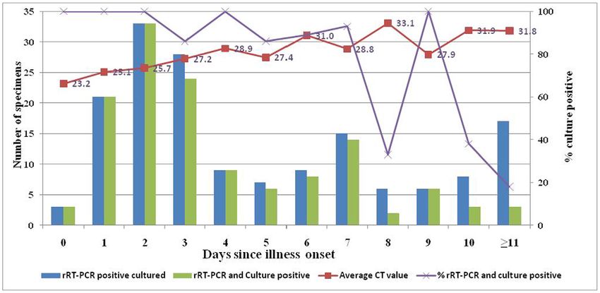
Figure 2. Correlation between rRT-PCR-positive results, cell culture results, and days of illness.
doi:10.1371/journal.pone.0020320.g002
patients on day 0–3 after illness onset were 7.5 times more likely to and symptoms in the patients $5 years old were cough [83/117
be culture-positive than rRT-PCR-positive specimens collected on (71%)], temperature $38uC [63/117 (54%)], reported fever [61/
day 4–7 (CI: 1.4–38.9; P,0.012), 17.6 times more likely to be 117 (52%)], and runny nose [60/117 (51%)] (Table 2).
culture-positive than rRT-PCR-positive specimens collected on Eighteen (17%) pH1N1 enrolled patients who were initially
day 8–10 (CI: 3.2–96.1; p,0.001), and 142.9 times more likely to symptomatic and pH1N1-positive continued to be rRT-PCR-
be culture-positive than specimens collected from patients $11 positive after all of their signs and symptoms had resolved for a
days after illness onset (CI: 2.4–833.3; P,0.001). Of the 100 rRT- median of 9 days (interquartile range 7–10 days) after all signs and
PCR negative specimens, 6(6.4%) were culture-positive. symptoms had resolved. Viable pH1N1 virus was isolated from
The median number of days pH1N1 virus RNA was detectable specimens obtained from 7/18(39%) of these patients.
in patients specimens 8 days (95% CI: 7–10 days) after symptom
onset. There was no statistically significant difference in the
duration of pH1N1 virus detection between children ,5 years old
and those 5 to 14 years or persons $15 years. There was no
difference in the median duration of pH1N1 virus detection
between males and females. In the majority (58%) of patients,
pH1N1 virus RNA was detected for $7 days, and in 16% of
patients for $14 days (Figure 3). In general, the mean CT value
increased with time. One of the HIV-positive patients who was on
antiretroviral therapy and had a CD4 count of 17cells/mm3 had
detectable pH1N1 virus RNA for 4 days after symptom onset. The
other HIV-positive patient, who was not on antiretroviral
treatment and whose CD4 count was unknown, had detectable
pH1N1 RNA for 16 days after symptom onset. These two patients
completed the study with two consecutive pH1N1-negative
specimens.
Association between pH1N1 virus detection and clinical
symptoms
There were 200 patient visits that were associated with pH1N1-
positive rRT-PCR results. We were able to link laboratory results
with clinic visit data on symptoms for 186/200 patients. From the
linkable patient visits, there were 69/186 pH1N1-positive rRT-
PCR results from patients ,5 years old and 117/186 from
patients $5 years old. The most common signs and symptoms in
the pH1N1-positive patients ,5 years old were cough [52/69 Figure 3. Kaplan Meier plot showing the probability of rRT-
(75%)], runny nose [47/69 (68%)], reported fever [44/69 (64%)], PCR-positive pH1N1 test result by day after symptom onset.
and temperature $38uC [40/69 (58%)]. The most common signs doi:10.1371/journal.pone.0020320.g003
PLoS ONE | www.plosone.org 4 June 2011 | Volume 6 | Issue 6 | e20320Influenza A 2009 H1N1 Virus Shedding, Kenya
Table 2. Signs and Symptoms associated with pH1N1 rRT-PCR-positive test results.
Patients ,5 yrs Patients $5 yrs
Days after symptom onset Days after symptom onset
* *
Symptom N 0–3 4–7 8–11 12–20 N 0–3 4–7 8–11 12–20
Total Patient visits 69 36 15 14 4 117 50 29 19 19
Reported Fever (%) 44(64) 35(97) 7(47) 2(14) 0(0) 61(52) 45(90) 16(55) 0(0) 0(0)
Temp ($38) (%) 40(58) 34(94) 6(40) 0(0) 0(0) 63(54) 49(98) 14(48) 0(0) 0(0)
Cough (%) 52(75) 33(92) 12(80) 5(36) 1(25) 83(71) 45(90) 23(79) 9(47) 6(32)
Sneezing (%) 24(35) 19(53) 4(27) 1(7) 0(0) 31(18) 20(40) 9(31) 2(11) 0(0)
Runny nose (%) 47(68) 31(86) 8(53) 6(43) 2(50) 60(51) 32(64) 17(59) 7(37) 4(22)
Vomiting (%) 17(25) 13(36) 3(20) 1(7) 0(0) 9(8) 6(12) 3(10) 0(0) 0(0)
Diarrhea (%) 8(12) 6(17) 2(13) 2(14) 0(0) 5(4) 2(4) 1(3) 1(5) 1(5)
Chest pain (%) 0(0) 0(0) 0(0) 0(0) 0(0) 23(20) 13(26) 7(24) 3(16) 0(0)
Difficulty Breathing (%) 6(9) 4(11) 2(13) 2(14) 0(0) 7(6) 4(8) 1(3) 2(11) 0(0)
Ear problem (%)* 6(5) 5(10) 0(0) 1(5) 0(0)
Sore throat (%)* 34(29) 22(44) 8(28) 3(16) 1(5)
Headache* 48(41) 30(60) 15(52) 2(11) 1(5)
Chills* 22(19) 14(28) 7(24) 1(5) 0(0)
Joint pain* 15(13) 8(16) 6(21) 1(5) 0(0)
Muscle pain* 10(9) 4(8) 5(17) 1(5) 0(00
Abdominal pain* 8(7) 5(10) 2(7) 1(5) 0(0)
*This information was not obtained from patients ,5 years old.
doi:10.1371/journal.pone.0020320.t002
Discussion had resolved. Viable pH1N1 virus, which was potentially
infectious, was isolated from one-third of specimens obtained
This study describes the duration of pH1N1 virus RNA from recovered patients. Therefore, some pH1N1 patients who
detection, the correlation between rRT-PCR-positive results and were no longer symptomatic may still be shedding viable pH1N1
virus isolation, and clinical symptoms associated with pH1N1 virus virus. In addition, half of rRT-PCR-positive samples from patients
detection in patients living in a densely populated community with who were on day 8 to day 10 after symptom onset were culture-
low socioeconomic status in sub-Saharan Africa. Pandemic H1N1 positive. In 2009, in the early stages of the H1N1 pandemic,
RNA was detected from respiratory specimens by rRT-PCR for a WHO recommended that pH1N1 patients remain isolated for 7
median duration of 8 days but up to 17 days after symptoms onset. days or until symptoms resolved [20]. While these guidelines may
This duration of virus RNA detection is similar to the median of 6 be appropriate for the community, in a healthcare setting, where
and 8 days reported in studies from China and Hong Kong, the goal is to prevent as much spread as possible, other measures
respectively [7,10]. In our study, we did not find any differences in need to be considered [21].
the duration of pH1N1 RNA detection among various age groups Of the 2 HIV-positive patients in the study, the patient who was
or between males and females. These results were similar to those not on antiretroviral therapy (ART) had detectable pH1N1 virus
reported by a study in Hong Kong, which reported no correlation RNA for 16 days, much longer than the median duration for the
between influenza viral load and age [17], and one from Canada study population. This is slightly longer than was shown in a study
which showed no differences in shedding between children and of HIV-positive school-aged children carried out in Germany in
adults [8]. In contrast, studies in Hong Kong and China found which the median time from symptoms onset to first negative rRT-
that younger age and male gender were risk factors for prolonged PCR result was 9 days (Range, 5–14 days) and cultures become
pH1N1 virus detection [7,10]. Our study population, was mostly negative after 6 days (Range 3–11 days) [22]. Other studies have
children (79% of the patients were ,14 years old), thus shown that immunosuppressed individuals may shed influenza
associations between age and duration of pH1N1 RNA virus virus longer than the general population [2,9,19]. In a case report
detection were difficult to assess. In addition, our study population from the United States of America, 2 cancer patients who were
included outpatients only, the China and Hong Kong studies severely immunosuppressed were shown to continue having
mentioned above included hospitalized patients [7,10]. detectable pH1N1 virus RNA for 5 and 6 weeks after initial
Cough, fever, and runny nose were the most common clinical diagnosis [9]. In previous studies, severely immunocompromised
symptoms associated with pH1N1 virus RNA detection. This is patients have been shown to shed seasonal influenza for weeks to
similar to studies carried out in Korea [18] and Hong Kong [10]. months [2]. The issue of prolonged shedding in immunocompro-
While sore throat was commonly associated with pH1N1 patients mised patients is especially relevant in sub-Saharan Africa, where
in other studies [1,10,19], in our study, only 29% of the pH1N1- over 22 million people are infected with HIV and only 30% are on
positive patient visits from patients $5 years old were associated ART [23,24]. In Kenya, the HIV prevalence in persons aged 15–
with this symptom. In our study, a substantial proportion of 64 is 7.1% [25], and in the study site, HIV prevalence is 14%
patients continued to shed the virus after respiratory symptoms (KEMRI/CDC-K, unpublished data). In our study, we identified
PLoS ONE | www.plosone.org 5 June 2011 | Volume 6 | Issue 6 | e20320Influenza A 2009 H1N1 Virus Shedding, Kenya
2 HIV-positive patients among 16 tested, one of whom had there was an overlap between the patients who presented with ILI
prolonged shedding. More research should be conducted to better and SARI, thus it was difficult to analyze data for each of the
understand the extent of pH1N1 viral shedding in untreated HIV- syndromes separately. Finally, 20% of patients enrolled in the
infected patients and those on ART therapy. study did not fully complete the study. Therefore, we have little
PCR, which detects viral nucleic acid instead of infectious viral information on these patients regarding shedding. We used right-
particles, is more sensitive than virus culture in detecting influenza censoring in our analysis for these patients. These results were
[26]. However, because it does not detect viable whole virus, comparable to those obtained from the 85 patients who completed
people who have respiratory specimens that are rRT-PCR-positive the study.
may not harbor a sufficient amount of viable virus to infect other In this study, we show that pH1N1 shedding patterns in an
people. The relationship between virus titers and influenza impoverished, densely populated urban community in Nairobi,
transmissibility is not known. However, serial interval studies Kenya, are similar to those described in studies in more affluent
suggest that most seasonal influenza household transmission occurs countries in temperate and subtropical areas of the world. Because
in the first 3 days after the index case’s illness onset [21]. In our of unique co-morbidities in sub-Saharan Africa compared to other
study, we successfully isolated pH1N1 virus from 95% (81/85) of areas of the world [27], more research is needed to characterize
rRT-PCR-positive specimens taken from day 0–3 after symptom shedding dynamics and impact on disease transmission for pH1N1
onset, but only 18% (3/17) rRT-PCR-positive specimens taken infection in African communities.
$11 days after symptom onset were culture-positive. These
findings suggest that if a patient has a respiratory specimen taken Acknowledgments
early in the course of illness that is positive for pH1N1 by rRT-
PCR, that patient is likely shedding live virus. In contrast, a rRT- The authors wish to express gratitude to Leonard Cosmas Otieno, Regina
Waithaka, Fredrick Nindo, Dennis Odhiambo, Beatrice Wambui, Irene
PCR-positive result from a sample taken later in a patient’s course
Omwenga, and Erin Russell for their excellent technical support. CDC
of illness may not mean that the patient is still shedding live virus. Disclaimer: The findings and conclusions in this report are those of the
Our study had some limitations. First, our study population was authors and do not necessarily represent the official position of the US
mainly comprised of persons ,14 years, and the oldest patient was Centers for Diseases Control and Prevention.
41 years old. Thus, we were not able to evaluate viral shedding
patterns in elderly individuals. Second, since few people in our Author Contributions
study had known underlying medical conditions, we were not able
Conceived and designed the experiments: LWW EL MAK MKN HN WM
to determine the impact of co-morbidities, including HIV
RFB BO. Performed the experiments: GKK LWW MKN. Analyzed the
infection, on duration of pH1N1 viral shedding. Some patients data: JMW EL MAK LWW. Contributed reagents/materials/analysis
whose HIV status was unknown may have been HIV-positive; this tools: MAK MKN RFB. Wrote the paper: LWW MAK EL MKN JMW.
could explain the slightly longer duration of rRT-PCR positive Collected clinical specimens and managed clinical data: HN WM.
pH1N1 results in our study compared to other studies. Third, Coordinated field activities: BO WM.
References
1. Novel Swine-Origin Influenza A (H1N1) Virus Investigation Team (2009) 14. Feikin D, Audi A, Olack B, Bigogo GM, Polyak C, et al. (2010) Evaluation of the
Emergence of a novel swine-origin Influenza A (H1N1) virus in humans. The optimal recall period for disease symptoms in home-based morbidity surveillance
New England Journal of Medicine 360: 2605–2615. in rural and urban Kenya. International Journal of Epidemiology 39: 1–9.
2. World Health Organization (2006) Nonpharmaceutical Interventions for pandemic 15. Shinde V, Bridges CB, Uyeki TM, Shu B, Balish A, et al. (2009) Triple-
influenza, international measures. Emerging Infectious Diseases 12: 81–87. reassortant swine influenza A (H1) in humans in the United States, 2005–2009.
3. Centers for Diseases Prevention and Control (2009) Interim Guidance on The New England Journal of Medicine 360: 2616–2625.
Specimen Collection, Processing, and Testing for Patients with Suspected Novel 16. World Health Organization (2009) CDC protocol of realtime RTPCR for
Influenza A (H1N1) Virus infection. May 13 2009 ed. influenza A (H1N1). Atlanta: The WHO Collaborating Center for influenza at
4. Centers for Diseases Control and Prevention (2009) Recommendations for CDC Atlanta.
amount of time persons with influenza-like illness should be away from others. 17. Li IW, Hung IF, To KK, Chan KH, Cheng VC, et al. (2010) The natural viral
Centers for Diseases Control and Prevention. load profile with pandemic 2009 influenza A(H1N1) and effect of oseltamivir
5. Lee N, Chan PK, Hui DS, Rainer TH, Wong E, et al. (2009) Viral loads and treatment. Chest 137: 759–768.
duration of viral shedding in adult patients hospitalized with influenza. Journal 18. Lee CS, Lee JH (2010) Dynamics of clinical symptoms in a case with pandemic
of Infectious Diseases 200: 492–500. influenza A (H1N1). Clinical Microbiology and Infection 16: 389.
6. World Health Organization (2009) Infection prevention and control during 19. Ling LM, Chow AL, Lye DC, Tan AS, Krishnan P, et al. (2010) Effects of early
health care for confirmed or suspected cases of pandemic (H1N1) 2009 virus therapy on viral shedding in 2009 pandemic influenza A (H1N1) virus infection.
infection and influenza-like illness. Clinical Infectious Diseases 50: 963–969.
7. Cao B, Li XW, Mao Y, Wang J, Lu HZ, et al. (2009) Clinical features of the 20. World Health Organization (2009) Global influenza surveillance network:
initial cases of 2009 pandemic influenza A (H1N1) virus infection in China. New Laboratory surveillance and response to pandemic H1N1, 2009. Weekly
England Journal of Medicine 361: 2507–2517. Epidemiological Records 84: 361–365.
8. De Serres G, Rouleau I, Hamelin ME, Quach C, Skowronski D, et al. (2010) 21. Bridges CB, Kuehnert MJ, Hall CB (2003) Transmission of influenza: Implications
Contagious period for pandemic (H1N1) 2009. Emerging Infectious Diseases 16: for control in health care settings. Clinical Infectious Diseases 37: 1094–1101.
783–788. 22. Feiterna-Sperling C, Edelmann A, Nickel R, KlausMagdorf, Bergmann F, et al.
9. Englund J, Zerr D, Pergam S, Kuypers J, Yager J, et al. (2009) Oseltamivir- (2010) Pandemic influenza A (H1N1) outbreak among 15 school-aged HIV-1–
resistant novel influenza A (H1N1) virus infection in two immunosuppressed infected children. Clinical Infectious Diseases 51: e90–e94.
patients, Seattle, Washington, 2009. Morbidity and Mortality Weekly Report 23. UNAIDS (2009) UNAIDS/WHO 2009 AIDS epidemic update. United
58(32): 893–896. Nations. pp 21–36.
10. To KKW, Chan K-H, Li IWS, Tsang T-Y, Tse H, et al. (2010) Viral load in 24. World Health Organization Antiretroviral therapy coverage in sub-Saharan
patients infected with pandemic H1N1 2009 influenza A virus. Journal of Africa, 2003–2007. New York: United Nations.
Medical Virology 82: 1–7. 25. National AIDS/STI Control Programme (Nascop) Kenya (2009) 2007 Kenya
11. Suess T, Buchholz U, Dupke S, Grunow R, Heiden M, et al. (2010) Shedding AIDS indicator survey: Final report. Nairobi, NASCOP. September 2009.
and transmission of novel influenza virus A/H1N1 infection in households— National AIDS/STI Control Programme (NASCOP). pp 15–31.
Germany, 2009. American Journal of Epidemiology 17: 1157–1164. 26. van Elden LJR, van Kraaij MGJ, Nijhuis M, Hendriksen KAW, Dekker AW,
12. Writing Committee of the WHO Consultation on Clinical Aspects of Pandemic et al. (2002) Polymerase chain reaction is more sensitive than viral culture and
(H1N1) 2009 Influenza (2010) Clinical aspects of pandemic influenza A (H1N1) antigen testing for the detection of respiratory viruses in adults with
virus infection. The New England Journal of Medicine 362: 1708–1719. hematological cancer and pneumonia. Clinical Infectious Diseases 34: 177–183.
13. Tabu C, Sharif S, Okoth P, Kioko J, Nzioka C, et al. (2009) Introduction and 27. Hotez PJ, Molyneux DH, Fenwick A, Ottesen E, Sachs SE, et al. (2006)
transmission of 2009 pandemic influenza A (H1N1) virus-Kenya, June–July Incorporating a rapid-impact for neglected tropical diseases with programs for
2009. Morbidity and Mortality Weekly Report 58: 1143–1146. HIV/AIDS, tuberculosis, and malaria. PLoS Medicine 3: 0567–0584.
PLoS ONE | www.plosone.org 6 June 2011 | Volume 6 | Issue 6 | e20320You can also read