Use of a -hCG Discriminatory Zone With Bedside Pelvic Ultrasonography
←
→
Page content transcription
If your browser does not render page correctly, please read the page content below
IMAGING/ORIGINAL RESEARCH
Use of a !-hCG Discriminatory Zone With Bedside
Pelvic Ultrasonography
Ralph Wang, MD, RDMS, Teri A. Reynolds, MD, Hugh H. West, MD, Deepa Ravikumar, BA, Christina Martinez, BA,
Ian McAlpine, BA, Vanessa L. Jacoby, MD, MAS, John C. Stein, MD, MAS
From the Department of Emergency Medicine (Wang, Reynolds, West, Ravikumar, Martinez, McAlpine, Stein) and the Department of Obstetrics,
Gynecology, and Reproductive Sciences (Jacoby), University of California, San Francisco, San Francisco, CA.
Study objective: We seek to assess the performance of the ! human chorionic gonadotropin (!-hCG)
“discriminatory zone” when using bedside pelvic ultrasonography in the evaluation of symptomatic pregnant
emergency department (ED) patients.
Methods: This was a cross-sectional study of bedside pelvic ultrasonography performed on consecutive
pregnant patients in the first trimester who presented to the ED with abdominal pain or vaginal bleeding.
Patients received pelvic ultrasonography, serum !-hCG testing, and blinded formal radiologic ultrasonography. All
patients were followed for 8 weeks to determine outcomes. The sensitivity and specificity of a discriminatory
!-hCG level of 3,000 mIU/mL for the diagnosis of ectopic pregnancy were calculated for patients without an
intrauterine pregnancy visualized by bedside ultrasonography.
Results: Thirty-six faculty physicians performed bedside pelvic ultrasonography on 256 patients. There were 161
cases with a confirmed visualizable intrauterine pregnancy and 29 ectopic pregnancies. Bedside ultrasonography
identified 115 intrauterine pregnancies. The range of !-hCG for cases of confirmed visualizable intrauterine
pregnancy with a nondiagnostic bedside ultrasonography was 15 mIU/mL to 123,368 mIU/mL (median 6,633;
interquartile range 1,551 to 32,699). For patients with nondiagnostic bedside ultrasonography, using a
discriminatory !-hCG level of 3,000 mIU/mL to further assess for ectopic pregnancy showed sensitivity of 35%
(95% confidence interval [CI] 18% to 54%) and specificity of 58% (95% CI 48% to 67%). Finally, the overall
sensitivity of bedside pelvic ultrasonography for the detection of intrauterine pregnancy was 71% (95% CI 63% to
78%), and the specificity was 99% (95% CI 94% to 100%).
Conclusion: When bedside pelvic ultrasonography does not demonstrate an intrauterine pregnancy, serum !-
hCG level is not helpful in differentiating intrauterine from ectopic pregnancy in symptomatic ED patients. [Ann
Emerg Med. 2011;58:12-20.]
Please see page 13 for the Editor’s Capsule Summary of this article.
Provide feedback on this article at the journal’s Web site, www.annemergmed.com.
A podcast for this article is available at www.annemergmed.com.
0196-0644/$-see front matter
Copyright © 2011 by the American College of Emergency Physicians.
doi:10.1016/j.annemergmed.2010.12.023
INTRODUCTION Currently, the diagnostic tests most commonly used to
Background evaluate patients with possible ectopic pregnancy include pelvic
Ectopic pregnancy, a common and potentially fatal ultrasonography and quantitative serum ! human chorionic
condition, must be considered in all first-trimester patients gonadotropin (!-hCG). Pelvic ultrasonography may reveal a
presenting to the emergency department (ED) with normal or abnormal intrauterine pregnancy, ectopic pregnancy,
abdominal pain, vaginal bleeding, or syncope. Ectopic molar pregnancy, or none of the above (also known as an
pregnancy has an overall prevalence of approximately 2% in “indeterminate result”). The indeterminate result occurs in
all pregnancies. Several studies suggest that the prevalence of approximately 10% to 30% of symptomatic first-trimester
ectopic pregnancy ranges from 3% to 13% in symptomatic patients undergoing radiologic ultrasonography and may
first-trimester ED patients.1-5 Although early detection represent early intrauterine pregnancy, ectopic pregnancy, or
during the last 2 decades has greatly improved outcomes, embryonic demise.7 In such cases, the patient’s indeterminate
ectopic pregnancy remains a leading cause of maternal findings are often interpreted in the context of the
morbidity and mortality.6 “discriminatory zone,” the level of !-hCG above which the
12 Annals of Emergency Medicine Volume , . : July Wang et al Use of !-hCG Discriminatory Zone With Bedside Pelvic Ultrasonography
hCG has any utility in the setting of bedside ultrasonography
Editor’s Capsule Summary
performed on symptomatic patients presenting to the ED.
What is already known on this topic Although multiple authors have suggested the use of a cutoff at
There is a common belief that intrauterine 2,000 mIU/mL to help determine the disposition of ED
patients with an indeterminate bedside pelvic ultrasonography
pregnancies of sufficient size to produce a ! human
result,4,18-20 we are unaware of any published data about the
chorionic gonadotropin (!-hCG) level greater than
clinical performance of this !-hCG discriminatory zone cutoff
3,000 mIU/mL can be observed on emergency used after bedside pelvic ultrasonography.
physician–performed pelvic ultrasonography.
What question this study addressed Goals of This Investigation
Whether emergency physicians can identify The purpose of our study was to assess the clinical utility of
intrauterine pregnancy in patients whose !-hCG the traditional !-hCG discriminatory zone in differentiating
ectopic from normal pregnancy after indeterminate bedside
level exceeds the threshold.
pelvic ultrasonography in symptomatic pregnant patients
What this study adds to our knowledge presenting to the ED. We hypothesized that the !-hCG
Collectively, the 36 emergency physicians in this discriminatory zone cutoff of 3,000 mIU/mL would not
study failed to identify an intrauterine pregnancy in demonstrate test characteristics suitable for differentiating
ectopic from normal pregnancy in these patients. Further, we
29 of 111 patients who had an intrauterine
sought to evaluate the !-hCG discriminatory zone for bedside
pregnancy and a !-hCG level greater than 3,000 pelvic ultrasonography and to explore whether a revised zone
mIU/mL. may be more helpful in clinical decisionmaking.
How this is relevant to clinical practice
In this study, when an emergency MATERIALS AND METHODS
physician–performed pelvic ultrasonography result Study Design
was indeterminate for intrauterine pregnancy, serum This was a cross-sectional study of emergency physician–
performed bedside pelvic ultrasonography in first-trimester
!-hCG level alone did not differentiate intrauterine
pregnant patients presenting to the ED with symptoms of
and ectopic pregnancy. abdominal pain or vaginal bleeding.
sensitivity of ultrasonography for intrauterine pregnancy Setting
detection approaches 100%. Using the discriminatory zone The study was conducted at the University of California, San
combined with the clinical history, providers attempt to Francisco, an urban tertiary care university teaching hospital
differentiate between normal intrauterine pregnancy and with an annual ED volume of approximately 40,000 patients
abnormal pregnancy, including ectopic pregnancy. Finding a per year, and was approved by the Committee on Human
serum !-hCG level greater than the discriminatory zone, in Research (institutional review board).
combination with an indeterminate ultrasonography, has been
shown to be associated with ectopic pregnancy.8,9 Typical Selection of Participants
transvaginal discriminatory zone values in the literature range We attempted to enroll consecutive patients meeting the
from 1,500 to 3,000 mIU/mL and have largely been developed following inclusion criteria: adult women of reproductive age
according to radiology-performed ultrasonography in who presented to the ED with a complaint of vaginal bleeding,
outpatients,10 with intrauterine pregnancy defined as the abdominal pain, or syncope and had a positive serum or urine
visualization of a gestational sac.11-13 pregnancy test result. Exclusion criteria were previous pelvic
ultrasonography during the same pregnancy, with available
Importance result, or clinical instability as assessed by the attending
In recent years, bedside pelvic ultrasonography has shown physician. Research assistants identified potential study
significant promise in the evaluation of pregnant patients candidates by using the ED electronic tracking system or by
presenting to the ED, allowing for quick and accurate detection activation by pager from a clinician during weekdays from 7 AM
of intrauterine pregnancy in 50% to 70% of cases, with a to 11 PM. Physicians identified patients after hours.
decrease in resource utilization and length of stay.14-17 Bedside pelvic ultrasonography was performed and
However, both the equipment and the examination protocol of interpreted by attending faculty credentialed in the use of pelvic
bedside ultrasonography usually differ substantially from that ultrasonography according to American College of Emergency
used in radiology-performed ultrasonography, and the distinct Physicians’ (ACEP’s) guidelines. The full details of our
test performance characteristics of bedside ultrasonography have credentialing policy have been reported previously.21 No
not been well described. Specifically, it is unclear whether !- additional training was conducted for this study.
Volume , . : July Annals of Emergency Medicine 13Use of !-hCG Discriminatory Zone With Bedside Pelvic Ultrasonography Wang et al
Data Collection and Processing physician interpreted that no intrauterine pregnancy was
Patient consent was obtained by a research assistant or visualized or if the interpretation was reported as an inadequate
physician. Enrolled patients received a serum CBC count, study. The bedside ultrasonographic reporting form did not
serum !-hCG measurement, Rh screen (for symptoms of include a specific category for ectopic pregnancy, and no cases
bleeding), and bedside pelvic ultrasonography. A bedside pelvic of ectopic pregnancy were positively identified as such on the
ultrasonography protocol was instituted such that emergency text portion of the bedside ultrasonographic report. For the
physician faculty performed an initial transabdominal pelvic purposes of this study, radiology examinations were also
scan, using either a SonoSite Micromaxx or M-Turbo (SonoSite interpreted as positive for intrauterine pregnancy if an
Inc., Bothell, WA) with a 5-2 MHz curved probe. Transverse intrauterine gestational sac with yolk sac or a fetal pole was
and longitudinal views were obtained and stored. If no obvious visualized. The research assistant used a standard abstraction
intrauterine pregnancy was observed, a transvaginal scan would form to record the results from the radiology interpretation. To
then be performed using either machine with an 8-5 MHz assess interrater reliability between the research assistant and
intracavitary probe. senior author, a 10% random sample of cases was assessed and
Operators evaluated the uterus to identify presence or showed an actual agreement of 96%, " 0.92 (95% confidence
absence of intrauterine pregnancy (as defined by the intrauterine interval [CI] 0.78 to 1.00).
presence of gestational sac containing at least yolk sac or fetal We created a subset of patients with a confirmed intrauterine
pole), the pelvis for evidence of free fluid, and the adnexa for pregnancy to assess what proportion of intrauterine pregnancies
obvious masses. Physicians documented their interpretation was actually detected by the emergency physician and then
before receiving any subsequent radiology report. For constructed the discriminatory zone for emergency physician
documentation purposes, physicians could interpret their scans ultrasonography. Patients met our definition of a confirmed
in the following 3 ways: intrauterine pregnancy identified, no intrauterine pregnancy for this study only by meeting one of the
intrauterine pregnancy identified, or inadequate study. They following criteria: (1) patients who had a gestational sac with
were also allowed to enter additional free text. Physicians were yolk sac or a fetal pole visualized by radiologist at initial or
generally not aware of the results of the !-hCG level at their subsequent ultrasonography, or (2) confirmed pregnancy at
interpretation because they performed the ultrasonography 8-week follow-up clinic visit or telephone call. This definition of
before it returned, but there was no procedure in place to confirmed intrauterine pregnancy is not intended to indicate
purposefully blind them from the result. Patients subsequently that the intrauterine pregnancy should have been observed at
received pelvic ultrasonography performed by the radiology bedside ultrasonography, only that these patients were
department (blinded to the results of the bedside subsequently proven to have an intrauterine pregnancy that
ultrasonography) during their ED visit. could have been observed if our technology were perfect (as
Research assistants contacted and performed a standardized opposed to an intrauterine pregnancy that had already
interview with all enrolled patients at 8 weeks. The research aborted at presentation, which, regardless of technology or
assistants were blinded to the patients’ ultrasonography results serum !-hCG level, would never be visualized). Only
(ED and radiology). Patients’ clinical records were also reviewed patients classified as having a confirmed intrauterine
for demographic and clinical history information (age, race, pregnancy by either of these criteria were included in the
ethnicity, presenting complaint, and risk factors for ectopic subset of patients used to evaluate the discriminatory zone
pregnancy), as well as formal ultrasonographic interpretation, for bedside ultrasonography.
consultant notes, subsequent clinic notes about the status of the
pregnancy, and pathology reports. One research assistant (I.M.) Primary Data Analysis
who had been trained by one of the authors (J.C.S.) and had All data were entered into a Microsoft Excel spreadsheet
evaluated 10 practice charts with him at the initiation of the (Version 12.2.8, Microsoft, Redmond, WA). To assess
project performed the chart review. A standard abstraction form physician compliance with the bedside ultrasonography
for all patients was used to obtain the chart review data, and the protocol, we analyzed a 10% random sample of the enrolled
abstractor followed a defined data dictionary for this purpose. patients. One of the authors (R.W.) reviewed the physician note
Only physician data were reviewed. For any ambiguous entries, and bedside ultrasonographic images. To evaluate how our test
the abstractor reviewed the case with one of the authors (J.C.S.); performance compared with that in similar previous reports of
at 2 separate times during data collection, that author reviewed bedside pelvic ultrasonography by emergency physicians, we
10 cases with the abstractor to ensure quality of data. The analyzed sensitivity and specificity for the identification of
abstractor was not blinded to the study goals and objectives. intrauterine pregnancy in our entire data set. To evaluate the
test characteristics of the currently defined !-hCG
Outcome Measures discriminatory zone of 3,000 mIU/mL for the identification of
The bedside ultrasonographic study result was interpreted as ectopic pregnancy after use of bedside ultrasonography, we
positive for intrauterine pregnancy if an intrauterine gestational analyzed sensitivity and specificity. To assess the discriminatory
sac with yolk sac or a fetal pole was visualized.2 It was zone for bedside pelvic ultrasonography, we evaluated only
interpreted as negative for intrauterine pregnancy if the those patients from the data set who had a confirmed
14 Annals of Emergency Medicine Volume , . : July Wang et al Use of !-hCG Discriminatory Zone With Bedside Pelvic Ultrasonography
Figure 1. Enrollment process and outcomes of all patient participants. IUP, Intrauterine pregnancy; D&C, dilation &
curettage.
intrauterine pregnancy.9 We present this data in aggregate and approached (were “missed”) by research assistant or physician
by physician to better represent individual variation in ability to and were identified only by review of daily discharges from the
identify an intrauterine pregnancy. To test whether increased ED. There were 6 patients who refused to participate and 8 who
experience with ultrasonography may impart a different withdrew before actually receiving ultrasonography. Twenty
discriminatory zone among emergency physicians, we planned patients were subsequently excluded because they did not
an a priori analysis of 2 physician subgroups: (1) faculty who undergo serum !-hCG measurements in the ED. Seventeen
enrolled more than 10 patients in the cohort, according to a patients were excluded because of lack of follow-up (7 of these
previous ED study of deep venous thrombosis22; and (2) faculty patients had no intrauterine pregnancy observed by bedside or
who were trained in the use of ultrasonography during radiologic ultrasonography and were never treated or contacted
residency. Summary statistics, diagnostic test characteristics, and again, and 10 of these patients had intrauterine pregnancy
95% CIs were performed with Stata (version 10; StataCorp,
observed by the bedside ultrasonography, with no subsequent
College Station, TX).
formal radiologic ultrasonography or clinical follow-up). Thus,
RESULTS 256 patients remained for preliminary analysis. To ensure that
Of 356 adult women of reproductive age who presented to physicians followed the bedside ultrasonography protocol
the ED with a complaint of vaginal bleeding, abdominal pain, (transabdominal first, and if no intrauterine pregnancy was
or syncope and had a positive serum or urine pregnancy test observed, then transvaginal sonography), a review of a 10%
result, 293 were enrolled in the study between January 2007 random subset showed 92% compliance (95% CI 75% to
and June 2009 (Figure 1). We attempted to screen consecutive 98%). Thus, there were 2 of 25 patients for whom the attending
patients, but 49 of the 356 patients (14%) were never physician did not visualize an intrauterine pregnancy on
Volume , . : July Annals of Emergency Medicine 15Use of !-hCG Discriminatory Zone With Bedside Pelvic Ultrasonography Wang et al
Table 1. Baseline characteristics of the study populations: (1) Table 2. Diagnostic accuracy of emergency
256 patients who were enrolled, received a !-hCG test, and physician–performed ultrasonography for the detection of
had clinical follow-up available, and (2) the 36 physicians who intrauterine pregnancy.*
performed the studies. Outcome
Patient Characteristics (n!256) No. (%)/Median (IQR)
EPPU Diagnosis IUP No IUP Total
Age, y, mean (SD) 28.4 (7)
IUP 114 1 115
Race, No. (%)
No IUP 47 94 141
Asian 49 (19)
Total 161 95 256
Black 68 (27)
White 77 (30) EPPU, Emergency physician-performed ultrasonography; IUP, intrauterine preg-
Other 51 (20) nancy.
Unknown 11 (4) *Sensitivity!71% (95% CI 63% to 78%); specificity!99% (95% CI 94% to
100%); positive predictive value!99% (95% CI 95% to 100%); negative predic-
Ethnicity, No. (%)
tive value!67% (95% CI 58% to 74%).
Hispanic 36 (14)
Non-Hispanic 203 (79)
Unknown 17 (7) Table 3. Diagnostic performance of a discriminatory zone of
Presenting complaints, No. (%) 3,000 mIU/mL for patients with no intrauterine pregnancy by
Abdominal pain/cramping 209 (82) emergency physician–performed pelvic ultrasonography for the
Vaginal bleeding/spotting 167 (65) detection of ectopic pregnancy.*
Risk factors for ectopic pregnancy, No. (%)
Previous ectopic pregnancy 9 (3.5) "-hCG Level, mIU/mL Ectopic No Ectopic
History of sexually transmitted disease 9 (3.5) Positive (#3,000) 10 47 57
History of intrauterine device 4 (1.6) Negative ("3,000) 19 65 84
!-hCG at enrollment, mIU/mL, median 10,676 (1,724–51,681) Total 29 112 141
(interquartile range)
Physician characteristics (n!36) *Prevalence of ectopic pregnancy with no IUP on EPPU!20.6% (95% CI 14.2%
Residency ultrasonographic training, No. (%) 23 (64) to 28.2%); sensitivity!35% (95% CI 18% to 54%); specificity!58% (95% CI 48%
to 67%); positive predictive value!18% (95% CI 9% to 30%); negative predictive
Years of ultrasonographic experience, 3 (3–4)
value!77% (95% CI 67% to 86%).
excluding residency, median (IQR)
Number of ultrasonographic studies 5 (2–9)
performed, median (IQR)
characteristics for bedside ultrasonographic identification of
intrauterine pregnancy (Table 2) showed a sensitivity of 71%
(95% CI 63% to 78%) and specificity of 99% (95% CI 94% to
transabdominal views and who did not proceed to transvaginal 100%). Positive predictive value was 99% (95% CI 95% to
sonography. 100%) and negative predictive value was 67% (95% CI 58% to
Table 1 reports the characteristics of the physician study 74%).
population, as well as the pregnant women. Thirty-six Because it is often quoted that in the setting of an
emergency physicians performed bedside ultrasonographic indeterminate ultrasonography, finding a serum !-hCG level
examinations. Thirteen physicians had not received any greater than the discriminatory zone is associated with ectopic
ultrasonographic training during their residency. Physicians had pregnancy, we calculated test characteristics for indeterminate
a median of 3 years of ultrasonographic experience at the onset bedside pelvic ultrasonography.8,9 Table 3 shows the diagnostic
of the study, exclusive of residency training (interquartile range test performance using a !-hCG cutoff of 3,000 mIU/mL for
[IQR] 3 to 4). One faculty physician was fellowship trained in the diagnosis of ectopic pregnancy in the subset of patients who
emergency ultrasonography. Physicians enrolled a median of 5 did not have an intrauterine pregnancy identified by bedside
patients in this study (IQR 2 to 9). pelvic ultrasonography. Sensitivity was 35% (95% CI 18% to
Of the study population of 256 patients, 161 were found to 54%), and specificity was 58% (95% CI 48% to 67%). Positive
show a confirmed intrauterine pregnancy, leaving 95 who were likelihood ratio was 0.82 (95% CI 0.48 to 1.40), and negative
never proven to have an intrauterine pregnancy that could have likelihood ratio was 1.13 (95% CI 0.83 to 1.50).
been visible at the ED visit. These 95 patients showed eventual To evaluate whether there may be a different discriminatory
clinical outcomes as follows: 54 with spontaneous abortion, 10 zone that applies to bedside ultrasonography, we set up an
with dilation and curettage (with no report of an intrauterine analysis similar to the one originally described by Kadar et al.9
pregnancy), 1 molar pregnancy, and 1 indeterminate (patient From our total 256 patients, 161 (63%) were ultimately
still reported irregular bleeding at 8 weeks but also reported classified after follow-up as having an intrauterine pregnancy
never requiring a subsequent ED or clinic visit; she was that was visualizable. Table 4 shows this group of patients and
subsequently lost to follow-up). ED bedside ultrasonography compares those who were diagnosed with intrauterine
identified 115 intrauterine pregnancies, and all were true pregnancy by bedside pelvic ultrasonography with those who
intrauterine pregnancies except for 1 case that proved were not, stratified by serum !-hCG levels. Although there was
subsequently to be a molar pregnancy. Our overall test no cutoff at which 100% of the intrauterine pregnancies were
16 Annals of Emergency Medicine Volume , . : July Wang et al Use of !-hCG Discriminatory Zone With Bedside Pelvic Ultrasonography
Table 4. Discriminatory zone evaluation: Comparison of the random sample of cases, this protocol was followed more than
number of visualizable intrauterine pregnancies (n!161) that 90% of the time. However, it is possible that if we excluded
were identified by emergency physician–performed pelvic cases in which only transabdominal sonography was performed,
ultrasonography with those that were not identified at varying
our results would differ.
serum !-hCG values.
Additionally, among the key factors that determine the
Serum "-hCG IUP Observed No IUP Observed discriminatory zone are operator training and the equipment
Level, mIU/mL in ED* in ED Total Cases
and scanning protocols used. Certainly, our results reflect our
"3,000 3 18 21 site-specific operator training. However, because we used the
3,000–4,999 5 1 6
ACEP emergency ultrasonography guidelines as the basis for our
5,000–9,999 7 8 15
10,000–24,999 12 8 20 training and all faculty ultrasonographers met these minimum
25,000–49,999 30 6 36 criteria, our study conditions should be reproducible. Further,
50,000–100,000 35 4 39 our faculty has a wide variation in experience and duration of
#100,000 22 2 24 ultrasonographic use, which is likely similar to that of many
Total 114 47 161
EDs nationally. Several of our faculty are particularly interested
*Defined as yolk sac or fetal parts within gestational sac. in ultrasonography, many were trained during residency (23/
36), and a smaller proportion trained after residency and may
have relatively limited experience. Thus, although the particular
identified, using a !-hCG level of more than 25,000 mIU/mL training and level of experience probably contribute to the
identified 87 of 99 (88%), and using a level of more than discriminatory zone for our physicians, it seems likely that our
50,000 mIU/mL identified 57 of 63 (90%). The range of !- current expertise mix is similar to that of many other clinical
hCG for cases of confirmed intrauterine pregnancy with environments, though distinct from the radiology environment.
nondiagnostic bedside ultrasonography was 15 mIU/mL to In addition, as mentioned above, other key factors in
123,368 mIU/mL (median 6,633; IQR 1,551 to 32,699). determining the discriminatory zone include the equipment and
We conducted a similar analysis for our 2 a priori physician scanning protocols used, and these may be particular to bedside
subgroups (those enrolling more than 10 patients and those ultrasonography and the emergency setting. The technology
with residency training in ultrasonography) but were unable to used for formal radiologic ultrasonography (here, an Acuson
identify any important differences when results were compared Sequoia) is widely considered to be more sensitive than the
with the full group analysis. For the subgroup of physicians who technology used at the bedside in the ED. It is possible that, had
performed more than 10 ultrasonography tests in this study the improved technology been available in the ED, the
(n!4), above a !-hCG value of 100,000 mIU/mL, 100% (5/5)
sensitivity of bedside ultrasonography would have been
of the intrauterine pregnancies were identified, and above a !-
enhanced. However, although this may limit the generalizability
hCG value of 25,000 mIU/mL, 89% (32/36) of the intrauterine
of our results, we sought precisely to evaluate the diagnostic
pregnancies were identified. For the group of physicians who
performance of bedside ultrasonography in the clinical setting.
trained in ultrasonography during residency (n!22), above a
Beyond equipment factors, the definition of intrauterine
!-hCG value of 100,000 mIU/mL, 100% (12/12) of the
pregnancy used in our protocol required the visualization of a
intrauterine pregnancies were identified, and above a !-hCG
yolk sac or fetal pole, in addition to a gestational sac (a
value of 25,000, 88% (57/64) of the intrauterine pregnancies
conservative definition of intrauterine pregnancy designed to
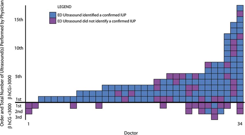
were identified. Figure 2 displays the identification of confirmed
avoid mistaken identification of a pseudosac as a gestational
intrauterine pregnancy by individual physician.
sac). Although this would not change the performance of
LIMITATIONS bedside ultrasonography relative to radiologic ultrasonography
One potential limitation to our study was that 18% of within this study (because radiologic examinations were also
eligible patients were not enrolled. This may have introduced interpreted by the same criteria), this definition would shift the
spectrum bias because it is possible that there were differences discriminatory zone relative to published studies that allowed
between this group of patients and the enrolled group. determination of intrauterine pregnancy by gestational sac
Another potential limitation is that we did not specifically alone.7,23-25
require physicians to document whether they performed Finally, our inclusion criteria included the presence of pain
transvaginal ultrasonography in addition to transabdominal or bleeding, and a symptomatic cohort may be expected to have
ultrasonography. All physicians were trained in both a higher prevalence of abnormal pregnancies (in which the
transabdominal and transvaginal ultrasonography, and all had relationship between size/visibility and !-hCG level may be
met ACEP guidelines for credentialing. Our ED bedside clinical disrupted) than an asymptomatic clinical cohort. Our particular
protocol, as well as study protocol, was to perform aim was to evaluate the utility of the discriminatory zone in this
transabdominal ultrasonography, and if no identifiable symptomatic population, and our results are not necessarily
intrauterine pregnancy was found, to then proceed to directly comparable to a discriminatory zone derived in
transvaginal ultrasonography. We have demonstrated that in a asymptomatic or mixed populations.
Volume , . : July Annals of Emergency Medicine 17Use of !-hCG Discriminatory Zone With Bedside Pelvic Ultrasonography Wang et al
Figure 2. Accuracy of bedside ultrasound examinations for each provider, stratified by discriminatory zone.
DISCUSSION In evaluating our data, we first intended to establish its
The correlation of ultrasonographic findings to !-hCG generalizability. We compared the test characteristics of ED
measurements has become a standard part of clinical practice in bedside pelvic ultrasonography in the identification of
symptomatic first-trimester patients.6 Although several authors intrauterine pregnancy with those of previous studies. We found
have suggested extending this approach to symptomatic ED the data in our study to be similar to reported values from a
patients with an initial indeterminate bedside ultrasonographic recent review.26 Our sensitivity of 71% (95% CI 63% to 78%)
result, this concept has not been rigorously evaluated.4,18-20 In our compares with 67% (95% CI 59% to 75%) reported by Wong
study, we found that the test characteristics for a !-hCG cutoff of et al,27 79% (95% CI 73% to 84%) reported by Mateer et al,4
3,000 mIU/mL are not acceptable for use in clinical practice when and 91% (95% CI 83% to 95%) reported by Durham et al.28
attempting to identify ectopic pregnancy in symptomatic patients Our specificity was 99% (95% CI 94% to 100%) and compares
with an indeterminate result on bedside pelvic ultrasonography with 92% (95% CI 65% to 99%) reported by Wong et al,27
(sensitivity of 35% and specificity of 58%). 100% (95% CI 96% to 100%) reported by Mateer et al,4 and
In the seminal work by Kadar et al,9 the initial 100% (95% CI 88% to 100%) reported by Durham et al.28
discriminatory zone was defined with radiology-performed The higher sensitivity in the studies by Durham et al28 and
transabdominal ultrasonography in a mixed cohort of Mateer et al4 may be accounted for by the fact that both studies
outpatients. Intrauterine pregnancy, defined as the visualization used a small, selected group of operators to perform the
of a gestational sac, was not detected in patients with a !-hCG ultrasonographic examinations in a consecutive sample. Because
level less than 6,000 mIU/mL. At a level greater than 6,500 we have used standardized training and credentialing following
mIU/mL, gestational sacs were reliably visualized, resulting in a the ACEP guidelines and because our test characteristics are
narrow discriminatory zone of 6,000 to 6,500. This finding similar to those of previous reports, we believe that the
subsequently was proven to help further differentiate pregnant subsequent results of our study are likely to be generalizable to
patients at risk for ectopic pregnancy who had no intrauterine those of other groups of ACEP-credentialed physicians.
pregnancy observed on their pelvic ultrasonographic result; The primary goal of this investigation was to assess the test
those with a low !-hCG level had a reasonable chance of an performance of a !-hCG cutoff of 3,000 mIU/mL in reference
early intrauterine pregnancy, whereas patients with a !-hCG to patients who do not receive a diagnosis of intrauterine
more than 6,500 mIU/mL had an increased likelihood of pregnancy on bedside pelvic ultrasonography. The low
ectopic pregnancy.8 With the advent of transvaginal sensitivity (35%) of this !-hCG cutoff derives from the fact that
ultrasonography and high-resolution technology in the among patients with ectopic pregnancy who have a
following decades, the discriminatory zone values were nondiagnostic bedside pelvic ultrasonographic result, the
redefined, resulting in the current accepted lower range of 1,500 proportion with a “positive” !-hCG test result (greater than the
to 3,000 mIU/mL.2,11-13,24 It has been shown that the 3,000 mIU/mL discriminatory zone) is only 35%. The majority
sensitivity for ectopic pregnancy in this situation ranges from of patients with ectopic pregnancy who have an indeterminate
73% to 93% and is dependent on equipment, gestational age, bedside pelvic ultrasonographic result have a !-hCG level lower
and operator skill.6 than 3,000 mIU/mL. Overall, this implies that in the setting of
18 Annals of Emergency Medicine Volume , . : July Wang et al Use of !-hCG Discriminatory Zone With Bedside Pelvic Ultrasonography
nondiagnostic ED ultrasonography, using a !-hCG cutoff of way diminishes the valuable contribution of bedside pelvic
3,000 mIU/mL will not aid with the exclusion of ectopic ultrasonography to the evaluation of women at risk for ectopic
pregnancy; rather, it will miss 65% of the cases. pregnancy in the ED. This diagnostic modality remains an
In our study, of the 256 total patients, 141 had efficient means of assessing patients for ectopic pregnancy and
nondiagnostic bedside pelvic ultrasonography. There were a was again shown to be highly accurate in our study when an
total of 29 cases of ectopic pregnancy. Thus, the prevalence of intrauterine pregnancy was identified. There were no patients
ectopic pregnancy when no intrauterine pregnancy is observed who received a diagnosis of intrauterine pregnancy by bedside
on initial bedside ultrasonography in our study was 21% (95% ultrasonography who were subsequently found to have ectopic
CI 14% to 28%). If one uses a !-hCG cutoff of 3,000 mIU/ pregnancy (although one was later found to have a molar
mL, patients with results above this level will have a posttest pregnancy). Among all patients with complete follow-up, 45%
prevalence of ectopic pregnancy of 18%, and patients with (115/256) received a diagnosis of intrauterine pregnancy by the
results below this level will have a posttest prevalence of ectopic emergency physician and thus would have required no further
pregnancy of 23%. Therefore, it does not seem appropriate to emergency evaluation or consultant ultrasonography. However,
use the traditional !-hCG discriminatory zone in clinical although no heterotopic pregnancies were identified in our
algorithms involving bedside ultrasonography in symptomatic study, in patients who have concerning symptoms the
ED patients because the test result does not significantly alter identification of an intrauterine pregnancy does not preclude
the probability of disease. this diagnosis. Appropriate further evaluation should be
We turned to the subset of patients who had a confirmed obtained if the clinical situation warrants.
intrauterine pregnancy to evaluate whether a different This re-evaluation of the discriminatory zone with respect to the
discriminatory zone might be helpful when bedside specific clinical practice of bedside pelvic ultrasonography in
ultrasonography is used. Intrauterine pregnancy was visualized symptomatic ED patients should alert practitioners to avoid the
by bedside ultrasonography at a !-hCG as low as 1,440 mIU/ inappropriate interpretation of serum !-hCG level in patients with
mL. However, it was not until levels reached 25,000 mIU/mL nondiagnostic results. Although ED bedside pelvic
or higher that we were able to identify more than 80% of cases ultrasonographic examinations can reliably exclude ectopic
(Table 4). This results in a wide zone for identifying pregnancy when they demonstrate a clear intrauterine pregnancy,
intrauterine pregnancy with bedside ultrasonography, unlike the the use of the traditional discriminatory zone does not appear to be
narrow discriminatory zone reported by radiologists using helpful in the further differentiation of ectopic pregnancy from
intrauterine pregnancy when the ultrasonography result does not
different imaging protocols and more sensitive equipment. In
demonstrate a clear intrauterine pregnancy.
evaluation of the data by individual physicians (Figure 2), 47 of
161 (29%) of confirmed intrauterine pregnancies were not
diagnosed. The median !-hCG of the 47 missed intrauterine Supervising editor: Alan E. Jones, MD
pregnancies was 6,633 mIU/mL (IQR 1,551 to 32,699). Even By Annals policy, submissions authored by faculty in the
when we evaluated subgroups that were more likely to have department of the editor in chief (Dr. Callaham) are handled
increased experience or training, we found discriminatory zones entirely by other senior editors, and Dr. Callaham plays no
that were comparable to those of the overall physician group. role in their decisionmaking nor is informed of any details
For patients who do not have an identified intrauterine during the process.
pregnancy on initial bedside ultrasonographic examination and Author contributions: RW and JCS developed the study
who are subsequently found to have serum !-hCG levels greater concept and design, conducted study revisions and response
than the traditional discriminatory zone of 3,000 mIU/mL, to the reviews, supervised the study, had full access to all the
there is still a high likelihood of intrauterine pregnancy. Thus, data in the study, and take responsibility for the integrity of
emergency physicians should not prematurely counsel patients the data and the accuracy of the data analysis. DR, CM, and
to expect an adverse outcome if their emergency physician– IM collected data. RW, TAR, DR, CM, IM, VLJ, and JCS drafted
performed ultrasonographic examination does not reveal an the article. RW, TAR, VLJ, and JCS were responsible for critical
revision of the article for important intellectual content. RW
intrauterine pregnancy, regardless of the !-hCG level.
and JCS conducted statistical analysis. RW, TAR, HHW, VLJ,
As described above, given the myriad factors that may affect and JCS interpreted the data. RW takes responsibility for the
the discriminatory zone, including characteristics of operators, paper as a whole.
equipment, protocols, and patient populations, it is not
surprising that the discriminatory zone for bedside pelvic Funding and support: By Annals policy, all authors are required
to disclose any and all commercial, financial, and other
ultrasonography is notably wider than that for formal radiology-
relationships in any way related to the subject of this article
performed studies. ED bedside studies are limited by the use of as per ICMJE conflict of interest guidelines (see
portable miniaturized ultrasonographic technology, by operator www.icmje.org). This study was funded by Agency for
training and experience, and by imaging protocols that are Healthcare Research and Quality grant 1K08HS015569. The
necessarily constrained by the demands of ED setting. However, Agency for Healthcare Research had no role in design and
the finding of a significantly wider discriminatory zone in no conduct of the study; the collection, management, analysis,
Volume , . : July Annals of Emergency Medicine 19Use of !-hCG Discriminatory Zone With Bedside Pelvic Ultrasonography Wang et al
and interpretation of the data; or the preparation, review, or 13. Peisner DB, Timor-Tritsch IE. The discriminatory zone of beta-hCG
approval of the article. for vaginal probes. J Clin Ultrasound. 1990;18:280-285.
14. Blaivas M, Sierzenski P, Plecque D, et al. Do emergency
Publication dates: Received for publication March 31, 2010. physicians save time when locating a live intrauterine pregnancy
Revisions received August 20, 2010, and October 26, 2010. with bedside ultrasonography? Acad Emerg Med. 2000;7:988-
Accepted for publication November 15, 2010. Available online 993.
February 18, 2011. 15. Shih CH. Effect of emergency physician-performed pelvic
sonography on length of stay in the emergency department. Ann
Address for correspondence: Ralph Wang, MD, RDMS, Emerg Med. 1997;29:348-351; discussion 52.
Department of Emergency Medicine, 505 Parnassus Ave, Box 16. Burgher SW, Tandy TK, Dawdy MR. Transvaginal ultrasonography
208, University of California, San Francisco, San Francisco, by emergency physicians decreases patient time in the
CA; 415-353-8309, fax 415-353-1792; E-mail emergency department. Acad Emerg Med. 1998;5:802-807.
ralphc.wang@ucsfmedctr.org. 17. Tayal VS, Cohen H, Norton HJ. Outcome of patients with an
indeterminate emergency department first-trimester pelvic
ultrasound to rule out ectopic pregnancy. Acad Emerg Med.
REFERENCES 2004;11:912-917.
1. Kohn MA, Kerr K, Malkevich D, et al. Beta-human chorionic 18. Burgher SW, Tandy TK, Dawdy MR. Transvaginal ultrasonography
gonadotropin levels and the likelihood of ectopic pregnancy in by emergency physicians decreases patient time in the
emergency department patients with abdominal pain or vaginal emergency department. Acad Emerg Med. 1998;5:802-807.
bleeding. Acad Emerg Med. 2003;10:119-126. 19. Valley VT, Mateer JR, Aiman EJ, et al. Serum progesterone and
2. Kaplan BC, Dart RG, Moskos M, et al. Ectopic pregnancy: endovaginal sonography by emergency physicians in the
prospective study with improved diagnostic accuracy. Ann Emerg evaluation of ectopic pregnancy. Acad Emerg Med. 1998;5:309-
Med. 1996;28:10-17. 313.
3. Tayal V. Outcome of patients with an indeterminate emergency 20. Durston W. Ultrasound availability in the evaluation of ectopic
department first-trimester pelvic ultrasound to rule out ectopic pregnancy in the ED: comparison of quality and cost-effectiveness
pregnancy. Acad Emerg Med. 2004;11:912-917. with different approaches. Am J Emerg Med. 2000;18:408-417.
4. Mateer JR, Valley VT, Aiman EJ, et al. Outcome analysis of a 21. Stein JC, Nobay F. Emergency department ultrasound
protocol including bedside endovaginal sonography in patients at credentialing: a sample policy and procedure. J Emerg Med.
risk for ectopic pregnancy. Ann Emerg Med. 1996;27:283-289. 2009;37:153-159.
5. Moore C, Todd W, O’Brien E, et al. Free fluid in Morison’s pouch 22. Kline JA, O’Malley PM, Tayal VS, et al. Emergency
on bedside ultrasound predicts need for operative intervention in clinician–performed compression ultrasonography for deep
suspected ectopic pregnancy. Acad Emerg Med. 2007;14:755- venous thrombosis of the lower extremity. Ann Emerg Med. 2008;
758. 52:437-445.
6. Barnhart KT. Clinical practice. Ectopic pregnancy. N Engl J Med. 23. Cacciatore B, Stenman UH, Ylöstalo P. Comparison of abdominal
2009;361:379-387. and vaginal sonography in suspected ectopic pregnancy. Obstet
7. Condous G, Kirk E, Lu C, et al. Diagnostic accuracy of varying Gynecol. 1989;73:770-774.
discriminatory zones for the prediction of ectopic pregnancy in 24. Dart RG, Kaplan B, Cox C. Transvaginal ultrasound in patients
women with a pregnancy of unknown location. Ultrasound Obst with low beta-human chorionic gonadotropin values: how often is
Gynecol. 2005;26:770-775. the study diagnostic? Ann Emerg Med. 1997;30:135-140.
8. Kadar N, Taylor KJ, Rosenfield AT, et al. Combined use of serum 25. Mol BW, Hajenius PJ, Engelsbel S, et al. Serum human chorionic
HCG and sonography in the diagnosis of ectopic pregnancy. AJR gonadotropin measurement in the diagnosis of ectopic pregnancy
Am J Roentgenol. 1983;141:609-615. when transvaginal sonography is inconclusive. Fertil Steril. 1998;
9. Kadar N, DeVore G, Romero R. Discriminatory hCG zone: its use 70:972-981.
in the sonographic evaluation for ectopic pregnancy. Obstet 26. McRae A, Murray H, Edmonds M. Diagnostic accuracy and clinical
Gynecol. 1981;58:156-161. utility of emergency department targeted ultrasonography in the
10. Barnhart KT, Simhan H, Kamelle SA. Diagnostic accuracy of evaluation of first-trimester pelvic pain and bleeding: a systematic
ultrasound above and below the beta-hCG discriminatory zone. review. CJEM. 2009;11:355-364.
Obstet Gynecol. 1999;94:583-587. 27. Wong TW, Lau CC, Yeung A, et al. Efficacy of transabdominal
11. Nyberg DA, Filly RA, Mahony BS, et al. Early gestation: correlation ultrasound examination in the diagnosis of early pregnancy
of HCG levels and sonographic identification. AJR Am J complications in an emergency department. J Accid Emerg Med.
Roentgenol. 1985;144:951-954. 1998;15:155-158.
12. Bree RL, Edwards M, Bohm-Velez M, et al. Transvaginal 28. Durham B, Lane B, Burbridge L, et al. Pelvic ultrasound
sonography in the evaluation of normal early pregnancy: performed by emergency physicians for the detection of ectopic
correlation with HCG level. AJR Am J Roentgenol. 1989;153: pregnancy in complicated first-trimester pregnancies. Ann Emerg
75-79. Med. 1997;29:338-347.
Did you know?
Annals accepts audio and video files as ancillaries to the main article.
Visit http://www.annemergmed.com/content/instauth/ for more details!
20 Annals of Emergency Medicine Volume , . : July You can also read