Case Report ST-Segment Elevation Myocardial Infarction from Septic Emboli Secondary to Infective Endocarditis by Abiotrophia Defectiva
←
→
Page content transcription
If your browser does not render page correctly, please read the page content below
Hindawi
Case Reports in Cardiology
Volume 2020, Article ID 8811034, 6 pages
https://doi.org/10.1155/2020/8811034
Case Report
ST-Segment Elevation Myocardial Infarction from Septic Emboli
Secondary to Infective Endocarditis by Abiotrophia Defectiva
Bashar Khiatah ,1 Sam Jazayeri ,1 John Wilde ,1 Mathew Westfall ,1
Thomas Q. Kong Jr.,2 and Amanda Frugoli 3
1
Department of Internal Medicine, Community Memorial Hospital, 147 N Brent St, Ventura, CA 93003, USA
2
Ventura Cardiology Consultants, 100 North Brent Street, Suite 301, Ventura, CA 93003, USA
3
Department of GME Internal Medicine, GME Internal Medicine of Community Memorial Hospital of Ventura, 147 N Brent St,
Ventura, CA 93003, USA
Correspondence should be addressed to Bashar Khiatah; bkhiatah@cmhshealth.org
Received 13 May 2020; Revised 5 July 2020; Accepted 8 July 2020; Published 13 July 2020
Academic Editor: Ertugrul Ercan
Copyright © 2020 Bashar Khiatah et al. This is an open access article distributed under the Creative Commons Attribution License,
which permits unrestricted use, distribution, and reproduction in any medium, provided the original work is properly cited.
This article showcases a young patient who presented with STEMI secondary to septic emboli due to endocarditis with Abiotrophia
Defectiva in the setting of a congenital bicuspid aortic valve. We aim to discuss current considerations for STEMI in young
individuals including embolism due to IE, especially in patients with known or suspected congenital heart valve disease.
1. Introduction 2. Clinical Case
Bicuspid aortic valve “BAV” is the most common congenital A 22-year-old healthy male presented to the hospital with
cardiac defect and is the most common valvular malforma- complaints of sudden onset substernal chest pain and short-
tion. Several studies have shown that bicuspid aortic valve ness of breath. Shortly after arrival, ventricular fibrillation
(BAV) has a higher incidence of IE than the general popula- occurred and he was defibrillated immediately. ECG
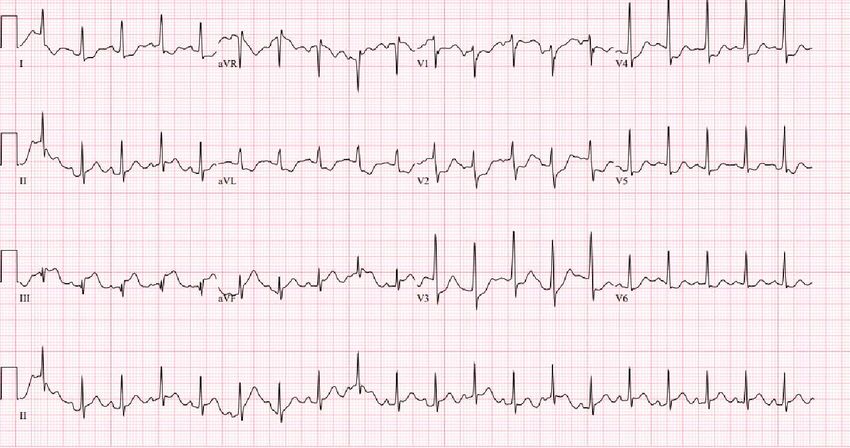
tion; thus, it is currently considered intermediate-risk cardiac (Figure 1) showed ST-segment elevation in the inferior leads,
conditions for IE. Infective endocarditis “IE” prevalence has and he was transferred for an urgent angiogram which dem-
been increasing drastically due to expansion of at-risk persons onstrated total occlusion of the proximal RCA for which a
including a generally aging population, an increase in intrave- bare metal stent was placed that successfully restored blood
nous drug users (IVDU), increased need for hemodialysis, and flow (Figures 2(a) and 2(b)).
an increase in cardiac devices. IE can occur on any single valve
or, in rarer cases, may include all valves. It has a wide range of 2.1. Differential Diagnosis. STEMI among the young is often
complications that can occur, including prolonged bacteremia, divided into the following categories including atheromatous
valvular regurgitation, or dehiscence leading to pulmonary CAD, nonatheromatous CAD (e.g., Kawasaki), sympathomi-
edema and heart failure, interruption in the cardiac electrical metic abuse, spontaneous coronary artery or aortic dissec-
system, development of metastatic infection focus, and cardio- tion, hypercoagulable states, and cardioembolism.
embolic events causing MI or stroke. These are some of the
most common findings but others can develop such as 2.2. Investigation. ECG showed ST-elevation MI in the distri-
mycotic aneurysm, perivalvular abscess, and free wall rupture. bution area of the RCA with elevated troponin I. He was found
In this case report, we present a unique case of a healthy to have an elevated leukocyte count, creatinine (minor), and
young male sustaining sudden onset STEMI secondary to BNP levels with mild normocytic anemia. Other basic labs
infectious endocarditis from Abiotrophia Defectiva in the such as TSH, lipid panel, and urine drug screen were within
setting of a congenital bicuspid aortic valve. normal limits. Further history investigation after stabilizing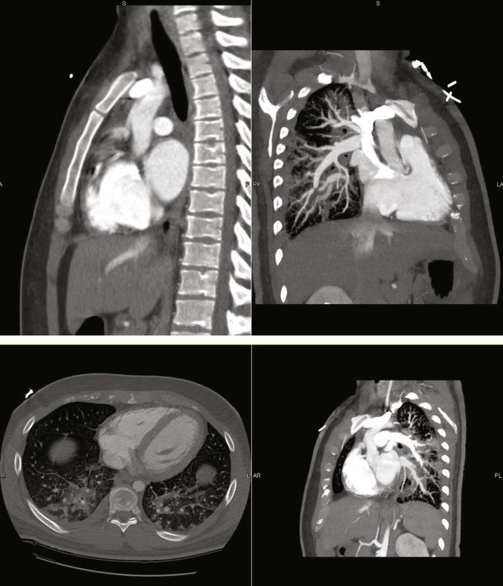
2                                                                                                         Case Reports in Cardiology

             Figure 1: Sinus tachycardia with fusion complexes, ST elevation in lead II, III, AVF, ST depression in V2-V4.

                                     (a)                                                      (b)

                        Figure 2: (a) Occlusive septic embolism of the proximal RCA. (b) RCA post stenting.

the patient revealed weight loss, occasional hemoptysis, and         These tests included JAK2, APAS, Factor V Leiden, prothrom-
night sweats for the last month for which he was prescribed          bin mutation, and DIC panel. Infectious workup for HIV,
antibiotics for presumed community-acquired pneumonia.               syphilis, and tuberculosis testing was also negative.
He denied any drug abuse, recent invasive surgery, or recent
trauma. His family history was negative for early MI or famil-       2.3. Management. He was treated with empiric antibiotics
ial hyperlipidemia. A 2D transthoracic echocardiogram dem-           pending cultures and experienced refractory heart failure
onstrated left atrial enlargement, a bicuspid aortic valve with      due to severe aortic insufficiency and mitral regurgitation.
a large mobile echodensity measuring 0:9 × 1:7 cm, severe            He subsequently underwent successful aortic and mitral
aortic insufficiency, and a malcoaptation of the anterior mitral       valve replacement with bioprosthetic valves and was dis-
valve leaflet with moderate to severe eccentric mitral insuffi-         charged with an extended course of IV antimicrobials.
ciency and turbulent flow in the left inferior pulmonary vein
(Figures 3(a)–3(d)). CTA (Figure 4) was negative for aortic          3. Discussion
aneurysm or dissection but revealed perihilar pulmonary infil-
trates with pulmonary edema, mediastinal adenopathy, and             ST-segment elevation myocardial infarction in very young
splenomegaly. Blood cultures grew Abiotrophia defectiva              patients (≤35 years) is most likely related to illicit or pre-
from two different bottles. Workup for hypercoagulable disor-         scribed drug use or to hypercoagulable status such as anti-
ders was performed and found to be within normal limits.             phospholipid, nephrotic syndrome, and factor V Leiden.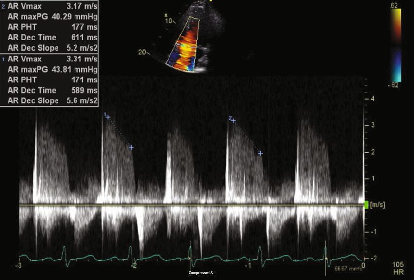
Case Reports in Cardiology                          3

                                     (a)

                                     (b)

                                     (c)

                             Figure 3: Continued.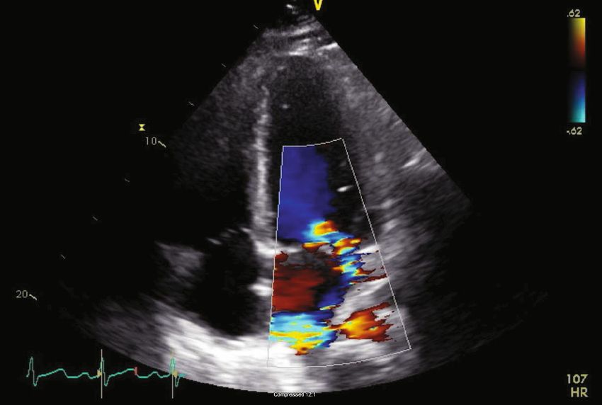
4                                                                                                                Case Reports in Cardiology

                                                                       (d)

Figure 3: (a) Bicuspid aortic valve. (b) Vegetation on the Aortic valve. (c) Moderate to severe eccentric mitral regurgitation. (d) Severe Aortic
valve insufficiency.

                                                                             with ST-segment elevation myocardial infarction is therefore
                                                                             not well defined [1, 2].
                                                                                  The incidence of IE in the United States is 15 per 100,000
                                                                             population; however, it is much higher in those with congen-
                                                                             ital bicuspid aortic heart valves (about one percent of the
                                                                             population) [3]. The incidence of IE among this group is 14
                                                                             per 10,000 patient-years, which is almost 11 times higher
                                                                             than the general population [4, 5]. Only 0.4% of IE cases
                                                                             are caused by Abiotrophia defectiva, which is a nutritionally
                                                                             variant streptococcus and part of normal oral, urogenital,
                                                                             and intestinal flora [6]. Despite the low incidence rate of IE
                                                                             by Abiotrophia defectiva, it has been reported in the litera-
                                                                             ture in multiple case reports and series demonstrating that
                                                                             the incidence may be uptrending [6]. Notably, 10.5% of all
                                                                             patients with IE by AD had congenital heart disease includ-
                                                                             ing bicuspid aortic valve [4]. Among all the patients with
                                                                             IE, only 3% have the complication of acute coronary syn-
                                                                             drome [7]. The pathophysiology of ACS in IE could be
                                                                             caused by obstructive coronary embolism, pseudoaneurysm
                                                                             formation, large vegetation blocking the coronary ostia, or
                                                                             compression of the coronary artery by an abscess [7]. It has
                                                                             been reported that a septic embolism of the coronary artery
                                                                             causing a cardioembolic myocardial infarction is a very rare
                                                                             complication of bacterial IE, accounting for 10 mm, and previ-
                                                                             ous embolic events [8]. It has been reported that the embolic
                                                                             events were more frequent at early stages [9].
Smoking, dyslipidemia, and family history remain the most                         While guidelines for treating infectious endocarditis
significant traditional risk factors for atheromatous CAD.                    remain the same [8], the best practices for management of
Spontaneous coronary artery dissection, vasculitis, and septic               STEMI in IE are not known, and the data is controversial
emboli from an infected valve are also causes of nonathero-                  due to the rarity of cases. Although thrombolytic agents have
matous STEMI. Physicians providing care for such patients                    been used successfully in a few cases, the data militate against
are less likely to consider nontraditional cardiac etiologies                the use of such agents due to increased risk of severe intracra-
for myocardial ischemia which may result in delaying diag-                   nial hemorrhage, which is thought to be attributed to the
nosis. The pattern of care and outcomes of the very young                    high prevalence of silent cerebral infarctions and mycotic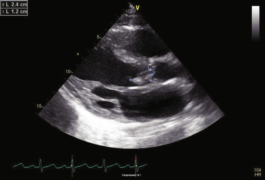
Case Reports in Cardiology                                                                                                               5

aneurysms [10]. Despite limited evidence on the efficacy of          should be considered and further evaluation for undiag-
PCI, there appears to be a trend in using recanalization as a      nosed congenital heart defect should be pursued.
treatment for STEMI in IE. It has been reported, however,
that balloon inflation at the site of the occlusion might cause     Abbreviations
a displacement of the vegetation and increase the risk of fur-
ther embolic phenomena and coronary artery mycotic aneu-           STEMI:    ST-elevation myocardial infarction
rysms [11]. Recently, Nazir S et al. have reported the success     ROSC:     Return of spontaneous circulation
rate of each intervention as follows: 56% in balloon angio-        CAD:      Coronary artery disease
plasty, 68% in aspiration thrombectomy, and 81% in coro-           ECG:      Electrocardiogram
nary stenting [10].                                                RCA:      Right coronary artery
    Surgery remains indicated in patients who have large veg-      DIC:      Diffuse intravascular coagulation
etations associated with severe valvular disease, those pre-       CTA:      Computed tomography angiography
senting with moderate to severe HF, or in patients with            ACS:      Acute coronary syndrome
uncontrolled infection. Surgical timing [i.e., urgent versus       PCI:      Percutaneous coronary intervention
early (within 48 hours) versus delayed] should be based on         IE:       Infective endocarditis.
individual risk-benefit analysis, with early surgery being
strongly indicated when benefits exceed operative risks. Sur-
gery is usually delayed in patients with intracerebral hemor-
                                                                   Additional Points
rhage and patients with large cerebral infarction as surgery       Learning Objectives. (i) Review the differential diagnosis for
may pose a significant risk of neurological deterioration           STEMI in very young patients. (ii) Treatment options for
and preoperative cerebral bleeding [12].                           STEMI secondary to IE.
    The choice of replacement with mechanical or tissue
prosthesis valves in IE of a native aortic valve remains based
on the patient’s factors, especially given similar survival and    Conflicts of Interest
endocarditis recurrence rate in both bioprosthetic and             The authors confirm that there are no conflicts of interest
mechanical valves [13]. It has been reported that there is a       regarding this publication.
national trend in using bioprosthetic rather than mechanical
valves for aortic valve replacement in IE patients [14]. This is
juxtaposed to IE of a native mitral valve, when repair is not an   References
option, replacement with a prosthetic valve is the only choice
                                                                    [1] S. Bangalore, G. C. Fonarow, E. D. Peterson et al., “Age and
as there are no established alternatives yet. [13]                      gender differences in quality of care and outcomes for patients
    In our case, the patient presented with a STEMI requiring           with ST-segment elevation myocardial infarction,” The Amer-
urgent angiography and PCI due to occlusive septic embo-                ican Journal of Medicine, vol. 125, no. 10, pp. 1000–1009, 2012.
lism of the proximal RCA. Early mitral and aortic valve             [2] M. Egred, G. Viswanathan, and G. K. Davis, “Myocardial
replacement were indicated due to the large size of the vege-           infarction in young adults,” Postgraduate Medical Journal,
tation, severe mitral regurgitation, severe aortic insufficiency,         vol. 81, no. 962, pp. 741–745, 2005.
and refractory heart failure in an otherwise young, healthy         [3] S. Pant, N. J. Patel, A. Deshmukh et al., “Trends in infective
male with a congenital bicuspid aortic valve.                           endocarditis incidence, microbiology, and valve replacement
                                                                        in the United States from 2000 to 2011,” Journal of the Amer-
                                                                        ican College of Cardiology, vol. 65, no. 19, pp. 2070–2076, 2015.
4. Follow-Up                                                        [4] H. I. Michelena, O. Katan, R. M. Suri, L. M. Baddour, and
                                                                        M. Enriquez-Sarano, “Incidence of infective endocarditis in
One month postdischarge and after completing a course of                patients with bicuspid aortic valves in the community,” Mayo
six weeks antibiotics regimen, the patient was recovering               Clinic Proceedings, vol. 91, no. 1, pp. 122-123, 2016.
well.                                                               [5] J. I. E. Hoffman and S. Kaplan, “The incidence of congenital
                                                                        heart disease,” Journal of the American College of Cardiology,
                                                                        vol. 39, no. 12, pp. 1890–1900, 2002.
5. Conclusion                                                       [6] A. Téllez, J. Ambrosioni, J. Llopis et al., “Epidemiology, clinical
                                                                        features, and outcome of infective endocarditis due to Abiotro-
Increasing the awareness of the differential diagnosis for
                                                                        phia species and Granulicatella species: report of 76 cases,
STEMI in young patients, especially given the increased
                                                                        2000-2015,” Clinical Infectious Diseases, vol. 66, no. 1,
incidence of septic coronary artery emboli from infective               pp. 104–111, 2018.
endocarditis, and determining the most appropriate treat-           [7] M. C. Manzano, I. Vilacosta, J. A. San Román et al., “Acute
ment (thrombolysis vs PCI vs surgery), which itself                     Coronary Syndrome in Infective Endocarditis,” Revista Espa-
remains a subject of debate, may help to clarify the stan-              ñola de Cardiología, vol. 60, no. 1, pp. 24–31, 2007.
dard of care in such patients. Furthermore, given the dis-          [8] G. Habib, P. Lancellotti, M. J. Antunes et al., “2015 ESC guide-
parity of evidence and practice, further studies to                     lines for the management of infective endocarditis: the task
substantiate the use of PCI in STEMI patients during IE                 force for the management of infective endocarditis of the
is warranted. Lastly, the consideration of normal flora as               European Society of Cardiology (ESC). Endorsed by: European
a cause of infective endocarditis in a healthy young patient            Association for Cardio-Thoracic Surgery (EACTS), the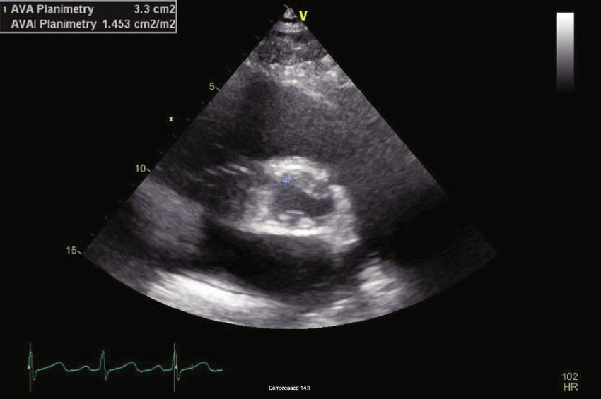
6                                                                            Case Reports in Cardiology

       European Association of Nuclear Medicine (EANM),” Euro-
       pean Heart Journal, vol. 36, no. 44, pp. 3075–3128, 2015.
 [9]   J. Fabri Jr., V. S. Issa, P. M. A. Pomerantzeff, M. Grinberg, A. C.
       P. Barretto, and A. J. Mansur, “Time-related distribution, risk
       factors and prognostic influence of embolism in patients with
       left-sided infective endocarditis,” International Journal of Car-
       diology, vol. 110, no. 3, pp. 334–339, 2006.
[10]   S. Nazir, E. Elgin, R. Loynd, M. Zaman, and A. Donato, “ST-
       elevation myocardial infarction associated with infective endo-
       carditis,” The American Journal of Cardiology, vol. 123, no. 8,
       pp. 1239–1243, 2019.
[11]   F. Khan, R. Khakoo, and C. Failinger, “Managing embolic
       myocardial infarction in infective endocarditis: current
       options,” The Journal of Infection, vol. 51, no. 3, pp. e101–
       e105, 2005.
[12]   D. Kang, “Timing of surgery in infective endocarditisHeart,”
       Heart, vol. 101, pp. 1786–1791, 2015.
[13]   G. B. Pettersson, J. S. Coselli, G. B. Pettersson et al., “2016 The
       American Association for Thoracic Surgery (AATS) consensus
       guidelines: Surgical treatment of infective endocarditis: Execu-
       tive summary,” The Journal of Thoracic and Cardiovascular
       Surgery, vol. 153, no. 6, pp. 1241–1258.e29, 2017.
[14]   E. B. Savage, P. Saha-Chaudhuri, C. R. Asher, J. M. Brennan,
       and J. S. Gammie, “Outcomes and prosthesis choice for active
       aortic valve infective endocarditis: analysis of the Society of
       Thoracic Surgeons adult cardiac surgery database,” The Annals
       of Thoracic Surgery, vol. 98, no. 3, pp. 806–814, 2014.
You can also read