Guide to inflammatory bowel disease - bowel diseae - Southern.IML Pathology
←
→
Page content transcription
If your browser does not render page correctly, please read the page content below

Guide to Guide to inflammatory
         inflammatory
                 bowel disease
         bowel diseae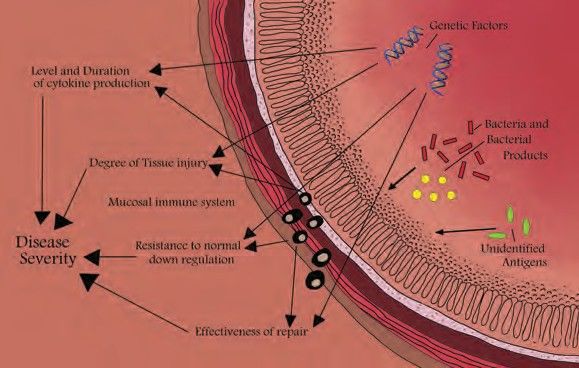
Inflammatory bowel disease (IBD) Symptoms of IBD
The term ‘inflammatory bowel disease’ (IBD) refers to two separate While there may be some overlap in the symptoms of UC and CD,
conditions that both cause chronic relapsing inflammation of the the diseases typically have a slightly different presentation. UC
intestinal tract. These conditions are ulcerative colitis (UC) and presents as a relapsing disorder marked by episodes of bloody
Crohn’s disease (CD). Although they produce differing patterns of mucoid diarrhoea accompanied by crampy abdominal pain; the
inflammation, there is some overlap in the clinical symptoms and the pain may persist for days, weeks or months: then subside, only to
serious consequences of these conditions. recur after an asymptomatic interval. In the worst cases, the initial
disease may produce such serious bleeding and fluid loss that it
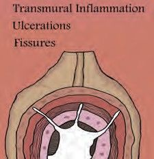
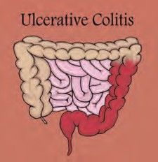
In the case of ulcerative colitis, the inflammatory process affects the may constitute an emergency. The onset of UC peaks between 20
colon. The inflammation is typically continuous, extending a variable and 25 years but the condition may arise in both younger and
distance from the rectum and is typically confined to the mucosal considerably older individuals. In the case of CD, the clinical
lining of the colon. This leads to broad areas of superficial ulceration. manifestations are more variable. The disease usually begins with
On rare occasions, UC may present as a more fulminant severe intermittent attacks of relatively mild diarrhoea, fever and
colitis, with more extensive inflammation and ulceration in association abdominal pain, spaced by asymptomatic periods lasting from
with systemic illness. Unlike UC, Crohn’s disease may affect any weeks to many months. In those with disease affecting the colon,
portion of the gastrointestinal tract from the mouth to the anus. The overt bleeding or undetected bleeding leading to anaemia may
disease is segmental and produces discrete, separate areas of occur. In about one-fifth of patients, the onset is more abrupt, with
inflammation. The inflammation in CD extends through the full acute pain in the lower right quadrant, fever and diarrhoea. Most
thickness of the intestinal wall, leading to deep fissuring ulcers, patients present with CD in the second decade of life. However, a
fibrous thickening of the bowel wall and narrowing of the lumen with second smaller peak occurs in adulthood. CD is more common in
stricture formation. Complications may also include the formation of Western developed countries. In both conditions, the onset of
fistula tracts, which may extend into other segments of the intestinal symptoms may be preceded by a period of physical or emotional
tract or other organs such as the urinary bladder. The majority of stress.
patients with CD will also have anal complications, including fissures
and fistulas.
Causes of IBD
Numerous familial studies1,2 have demonstrated a genetic
predisposition to IBD, with first-degree relatives exhibiting an
increased prevalence. In the case of CD, relatives have a 12–15
times greater risk of contracting the disease. Conversely, 15% of
IBD patients have affected first-degree relatives. These studies are
supported by gene linkage studies1,3,4,5 which have shown linkage
to gene loci on chromosomes 3, 5, 7, 12 and 16. However, a recent
article in the journal Nature6 has reported 71 genetic associations in
IBD, breaking the record for the largest number of associations for
any common disease. At several of these loci, causative genes
have now been identified. At present a gene called the CARD15
gene on chromosome 16 is the most understood susceptibility
gene, explaining around 20% of the genetic predisposition to CD.
As noted above, IBD shows considerable genetic variability. This
partly explains the variability in clinical disease presentation in IBD,
as differing mutations lead to differences in the nature of the IBD.
As an example, the CARD15 gene mutations are associated with a
small bowel disease location and a modestly earlier age of onset
The clinical variability may also relate to interactions with various
external factors including diet, infections, smoking and other
environmental factors. These external factors appear to interact with
the intestinal immune system, which plays a critical role in the
development and progression of the disease. Normally, the intestine
is in a steady state of physiologic inflammation and represents a
Figure 1: Patterns of disease and inflammation in Crohn’s disease
dynamic balance between factors that activate the immune system
and ulcerative colitis.
and host defences that maintain the integrity of the mucosa and
down-regulate inflammation. In IBD host mucosal immunity is
Over many years, both UC and CD lead to pre-malignant changes stimulated in response to substances in the bowel, particularly
(dysplasia) in the epithelial cells lining the GI tract called dysplasia. If bacterial products and secreted toxins, and then fails to switch off.
left untreated, this will inevitably lead to cancer. The risk of cancer is This leads to activation of inflammatory cells whose products cause
greatest in UC patients whose entire colon is affected; with a tissue injury. Interestingly, the second half of the 20th century saw an
20–30 fold increased risk of cancer after 10 years. Therefore regular increase in the incidence of IBD which closely mirrors the reported
colonoscopies are required to monitor for this complication. increase in allergic and autoimmune conditions. This has led some
Both conditions may also be associated with extra-intestinal authors to suggest that modern living conditions may contribute to
manifestations. In the case of Crohn’s disease, approximately 25% defective maturation in the normal regulation of the immune system
of patients have at least one extra intestinal manifestation. These by specific regulatory T lymphocytes. In summary, IBD results from
include arthritis, sacroiliitis, ankylosing spondylitis, skin lesions and an inappropriate response of the mucosal immune system to
liver diseases such as primary sclerosing cholangitis. antigenic stimuli, including the normal enteric flora, in a genetically
susceptible individual.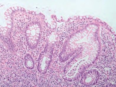
A recent meta-analysis reported the pooled sensitivity and pooled
                                                                         specificity of calprotectin in detecting inflammatory bowel disease in
                                                                         adults was 93% and 96% respectively. Studies assessing faecal
                                                                         calprotectin levels in IBD patients have found sensitivities and
                                                                         specificities from 90 to 95% for the identification of organic disease7.
                                                                         Calprotectin is particularly suitable for analysis as its level is relatively is
                                                                         uniform within the stool; therefore, a small sample is representative. In
                                                                         addition it is not degraded rapidly, with no change after storage at room
                                                                         temperature for up to 7 days. One recent review of the literature
                                                                         suggests that screening with faecal calprotectin could significantly
                                                                         reduce the number of adults requiring endoscopy.

Figure 2: IBD likely results from a combination of genetic
prediposition, cellular alterations and upregulated immunity.

Diagnosing IBD
There are a number of diseases that have similar presenting
symptoms to IBD, including infections, coeliac disease and
tumours. In addition there are conditions that produce GI
symptoms but that have no identifiable cause. These fall into the
category of functional disorders, of which irritable bowel
syndrome (IBS) is the most common example. Until recently the
only way to reliably differentiate organic disease from IBS was by            Figure 3: Endoscopic appearance of ulcerative colitis.
performing a colonoscopy and upper GI endoscopy. During this
procedure a camera is inserted into the colon, enabling the                                                                         (a)
gastroenterologist to visualise the mucosal lining and take
biopsies of any lesions seen. These are then processed in the
pathology lab and examined under the light microscope for any
abnormalities. The biopsies may be clearly diagnostic or
alternatively may show nonspecific inflammation, in which case
further investigations such as a stool culture may be required. In
the case of CD involving parts of the small bowel not accessible
to the endoscope, radiological investigation and possibly
‘capsule endoscopy’ may be required. The latter investigation
involves swallowing a capsule that contains a small camera.

While endoscopic assessment and microsopic examination of
biopsies represents the gold standard in diagnosing IBD,
                                                                                                                                    (b)
endoscopy is associated with unpleasant bowel preparation,
some discomfort and a small risk of complications. In the primary
care setting, in a majority of patients with non-acute abdominal
complaints, no organic pathology will be found with endoscopy.
Ideally, these patients should initially be assessed by a non-
invasive test that be identifies those patients who require a
subsequent colonoscopy. Blood tests for systemic markers of
inflammation, such as CRP and ESR, may indicate an
inflammatory process. However, the tests are not specific for
intestinal inflammatory disease and tend to correlate poorly with
symptoms and disease activity. Attention has therefore turned to
investigation of faeces to look for markers of inflammation. Of the
various faecal markers available, faecal calprotectin has been the
most widely adopted test.                                                Figure 4: (a) Normal colonic mucosa showing regular crypt
                                                                         architecture and no acute inflammation; (b) colonic biopsy showing
Calprotectin is a cell protein derived predominantly from neutrophils,   ulcerative colitis. There is architectural distortion with crypt
one of the types of white blood cells responsible for inflammation.      branching, an increase in inflammatory cells within the mucosa and
Calprotectin is found in various body fluids in proportion to the        a collection of neutrophils within a crypt.
degree of inflammation and has antibacterial and antifungal
properties.

                       For a complete list of our collection rooms please visit our website:
                                           www.southernpath.com.au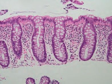
Faecal calprotectin in clinical use
       Faecal calprotectin can be useful in the following situations:                                   In addition to the diagnostic setting, recent studies have highlighted
                                                                                                        the usefulness of faecal calprotectin in monitoring the response of
       •    Differentiation of irritable bowel syndrome (IBS) and inflammatory                          patients with IBD to treatment, the aim of which is the suppression
            bowel disease (IBD), or other organic GI disorder
                                                                                                        of the inflammatory response. Even in cases of successful
       •    Assessment of disease activity in IBD
                                                                                                        symptomatic treatment, subclinical inflammation may persist,
       •    Monitoring of response to treatment in IBD
                                                                                                        contributing to the risk of relapse. Studies have demonstrated that
       While faecal calprotectin is indicative of organic disease, it cannot                            high faecal calprotectin identifies those patients with IBD in clinical
       differentiate the type of disease. In addition to IBD, increased faecal                          remission who art at risk of early relapse because of persistent mild
       calprotectin concentrations have been reported in bacterial                                      inflammation. The predictive value appears to be stronger for
       gastroenteritis, colorectal cancer and diverticular disease.                                     ulcerative colitis (UC) than for Crohn’s disease (CD).
       Therefore, patients with markedly elevated calprotectin levels will
       require further investigation, including endoscopy. Patients may also                            Critically, there are a number of clinical settings where immediate
       have variable calprotectin concentrations. Therefore, patients with                              colonoscopy/endoscopy is indicated and faecal calprotectin testing
       slightly raised calprotectin levels who present to the GP for the first                          is inappropriate. These have been referred to as ‘red flag’ indicators
       time may require a repeat sample 1–2 weeks later. The regular use                                and include age over 50 years, a positive family history of bowel
       of non-steroidal anti-inflammatory drugs (NSAIDs) may also cause                                 cancer, unexplained weight loss, rectal bleeding, a prolonged
       inflammation of the intestine and it has been recommended that                                   change in bowel habit in an older patient, anaemia and a palpable
       patients stop NSAIDs use several weeks before measuring faecal                                   abdominal or rectal mass. Should any of these indicators be
       calprotectin. Below is one suggested clinical algorithm for the use of                           present, an immediate referral to a gastroenterologist is required.
       faecal calprotectin measurements in the differential diagnosis of                                However, in cases where ‘red flag’ indicators are not present, a
       irritable bowel syndrome and inflammatory bowel disease.                                         faecal calprotectin measurement has the potential for clinicians to
                                                                                                        assess the need for further investigation and prevent unnecessary
                                                                                                        invasive testing.

                                                                           Faecal Calprotectin (FCP)

                                                                                 FCP can be negative in small
                                                                                   bowel Crohn’s disease

                                                    Calprotectin                        Calprotectin                          Calprotectin
                                                    < 50mg/kg                          50–150 mg/kg                           >150 mg/kg

                                                                                  Exclude other possible                 Organic disease
                                                    IBS likely                       cause of GI tract                  including IBD and
                                                                                        inflammation                  colorectal cancer likely.
                                                                                 (e.g. infection, NSAIDS).            Proceed to colonscopy
                                                                                    Repeat calprotectin

                                                            Calprotectin normal                      Calprotectin > 50 mg/kg
                                                                 IBS likely                           Suggest colonscopy

                             Figure 5: An algorithm for the use of faecal calprotectin measurements in the differential diagnosis of irritable bowel syndrome
                             and inflammatory bowel disease8.

      References
      1. ‘The Genetic Jigsaw of inflammatory Bowel Disease’, Watts D A and Satsangi J. Gut 2002, 50 (Suppl 3):iii3-iii36.
      2. ‘Crohn’s Disease severity in Familial and Sporadic cases’. Carbonnel, F, Macaigne G, Beaugerie L, Gendre JP and Cosnes J. Gut, 1999,44:91-95.
      3. The role of Genetics in Inflammatory Bowel Disease’. Current Drug targets. 2008 May; 9(5):361-
      4. ‘New IBD Genes’. McGovern D, Ahmad T. Gut 2005;54:1060-61.
      5. ‘Genome wide scan in a Flemish inflammatory bowel disease population: support for the IBD4 locsu, population heterogeneity, and epistasis.’ Verweire S et al. Gut 2004;53:980-986.
      6. Host–microbe interactions have shaped the genetic architecture of inflammatory bowel disease Luke Jostins, Stephan Ripke, Rinse K. Weersma, Richard H. Duerr, Dermot P. McGovern,
         et al. Nature 31 Oct 2012. 491,: 119-124
      7. Faecal calprotectin for screening of patients with suspected inflammatory bowel disease: diagnostic meta-analysis’. Van Rheenen P, Van de Vijver E, Fidler V. BMJ 2010:341:c3369.
      8. Faecal markers of gastrointestinal inflammation’ Sherwood, R. J. Clin Pathol 2012; 65:981-985.

This test is non rebatable, there will be an out of pocket cost of $85.00 Correct at time of printing May 2015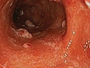


You can also read