Coexistence of bilateral macular edema and pale optic disc in the patient with Cohen syndrome - De Gruyter
←
→
Page content transcription
If your browser does not render page correctly, please read the page content below
Open Medicine 2021; 16: 156–160
Case Report
Klaudia Rakusiewicz*, Krystyna Kanigowska, Wojciech Hautz, Dorota Wicher,
Marlena Młynek, Marta Wyszyńska, Anna Rogowska, Joanna Jędrzejczak-Młodziejewska,
Małgorzata Danowska, Agnieszka Czeszyk
Coexistence of bilateral macular edema and pale
optic disc in the patient with Cohen syndrome
https://doi.org/10.1515/med-2021-0208 Results ‒ In the ophthalmologic examination, the girl
received July 2, 2020; accepted December 14, 2020 had bilateral astigmatism accompanied by myopia and
Abstract a marked reduction in central corneal thickness. Fundus
Background ‒ Cohen syndrome (Q87.8;ORPHA:193; examination showed pale optic nerve discs and “salt and
OMIM#216550) is an autosomal recessive inherited genetic pepper” retinopathy. Bilateral cystic macular edema was
disorder caused by mutation in the VPS13B/COH1 gene. It revealed in handheld optical coherence tomography.
is characterized by variable clinical symptoms such as Electroretinography showed a reduced response ampli-
deformity of the head, face, hands and feet, eye abnorm- tude of cones and rods.
alities, abdominal obesity, neutropenia and nonprogres- Conclusion ‒ In a patient with high myopia, macular
sive intellectual disability. The typical lesions in the eye- edema, pale optic disc and facial dysmorphism, Cohen
ball in Cohen syndrome include high myopia, retinal syndrome should be considered in the differential diag-
dystrophy, strabismus, maculopathy and lens subluxa- nosis. The severity of individual clinical features in
tion. The present study describes the coexistence of bilat- patients with Cohen syndrome varies. It can be assumed
eral macular edema with pale optic disc in a patient with a that the type of mutation affects the occurrence and
homozygous deletion in the VPS13B/COH1 gene. severity of individual symptoms.
Material and methods ‒ A 6-year-old Caucasian girl Keywords: Cohen syndrome, macular edema, pale optic
with facial dysmorphism, microcephaly, prominent upper disc, facial dysmorphism, CGH test
incisors, narrow hands with slender fingers, congenital
heart defect and ophthalmic symptoms was subjected to
genetic testing. The genetic evaluation revealed a homo-
zygous deletion on the long arm of chromosome 8 encom- 1 Introduction
passing 20–25 exons of the VPS13 gene, as confirmed by
Cohen syndrome. She underwent a full ophthalmological Cohen syndrome (Q87.8;ORPHA:193; OMIM#216550) is a
examination with the assessment of slit lamp examination rare genetic disorder that is inherited in an autosomal
of anterior segment and fundoscopy, refraction error, bio- recessive manner [1–6]. The diagnosis is mainly based
metry, central corneal thickness and additionally electro- on the clinical picture, but no unequivocal diagnostic
retinography, optical coherence tomography and fundus criteria have been established so far [5,7]. A genetic test
photography. that reveals mutations in the VPS13B gene, also known as
COH1, confirms the diagnosis of Cohen syndrome [1–5].
The gene is located on the long arm of chromosome
8q22.2, and the gene protein product plays a role in pro-
tein sorting, vesicle-mediated protein transport, glycosy-
* Corresponding author: Klaudia Rakusiewicz, Department of
Pediatric Ophthalmology, Children’s Memorial Health Institute, lation and lysosomal function [1,3]. As a consequence,
Warsaw, Poland, e-mail: k.rakusiewicz@ipczd.pl mutation in the gene leads to the production of defective,
Krystyna Kanigowska, Wojciech Hautz, Marta Wyszyńska, Anna faulty, inefficient proteins. Depending on the function of
Rogowska, Joanna Jędrzejczak-Młodziejewska, Małgorzata a particular protein, it affects many systems and organs.
Danowska, Agnieszka Czeszyk: Department of Pediatric
In 1973, Cohen et al. [2] described the syndrome by
Ophthalmology, Children’s Memorial Health Institute, Warsaw,
Poland
presenting three patients with similar, representative facial
Dorota Wicher, Marlena Młynek: Department of Medical Genetics, features with concomitant obesity, diminished muscle tone
Children’s Memorial Health Institute, Warsaw, Poland (hypotonia) and nonprogressive intellectual disability.
Open Access. © 2021 Klaudia Rakusiewicz et al., published by De Gruyter. This work is licensed under the Creative Commons Attribution 4.0
International License.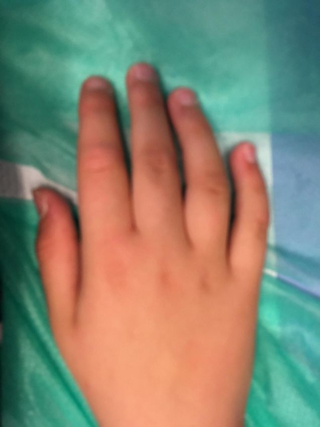
Coexistence of bilateral macular edema and pale optic disc in the patient with Cohen syndrome 157
In 1987, Norio et al. [8] examined six patients in the in after mid-childhood) and intermittent chronic neutro-
Finnish population with similar clinical manifestations penia associated with compromised immunity [1,2,4].
and additionally observed ocular symptoms such as myopia Abnormalities of the eyes described in patients
and retinal dystrophy. with Cohen syndrome include progressive myopia and
The frequency of the disorder in the general popula- “salt and pepper” retinopathy [1,2,4]. Other ophthalmic
tion is unknown. Cohen syndrome occurs more frequently symptoms consist of strabismus, iris coloboma, choroidal
in the Finnish people and in the Amish families [2]. coloboma, posterior subcapsular cataract, astigmatism,
Based on the literature, it appears that the intensity microcornea, microphthalmia, maculopathy, ptosis, optic
of typical clinical features in patients varies greatly [1,5]. atrophy, exophthalmos and lens subluxation [1,2]. Although
Identification of new mutations in VPS13B also presents the symptoms of visual disturbances are serious, they
extensive heterogeneity, which may explain clinical varia- usually do not lead to blindness. According to some
bility in Cohen syndrome [4]. However, there is no observed authors, optimal vision is preserved up to the fourth
consistent correlation between specific mutations and the decade of life [1,9].
severity of specific clinical features [4,5].
Cohen syndrome is characterized by low birth weight,
delay in reaching normal milestones in infancy and dimin-
ished muscle tone [1,2,4,5]. The affected individuals 2 Case study
usually have a distinct appearance such as short stature,
small, narrow hands and feet, microcephaly, a prominent In this case, a 6-year-old Caucasian girl is presented. She
nasal bridge, almond-shaped palpebral fissures, long has been diagnosed with Cohen syndrome and is under
eyelashes, thick eyebrows and hair. Most children with multi-specialized care at the Children’s Memorial Health
Cohen syndrome are described as sociable, open minded Institute. Informed consent has been obtained from par-
with a cheerful disposition [1,2,4,5].Typical features also ents of the patient presented in this study. In the array
include mental impairment, abdominal obesity (appearing CGH testing, the presence of 218 Kb homozygous deletion
Figure 1: (a) and (b) Results of a CGH analysis pointing a partial loss of both copies of genetic material at the VPS13B gene:
8q22.2(100290888_100508951)x0 (Agilent Technologies SurePrint G3 ISCA V2 CGH 8x60K [hg19]).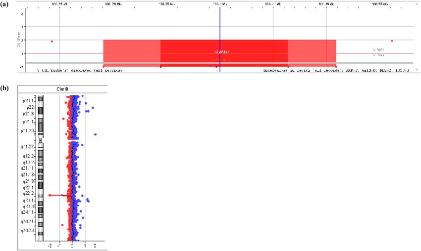
158 Klaudia Rakusiewicz et al.
in the long arm of chromosome 8 (region q22.2) was
detected in the girl [Figure 1]. The identified deletion
encompassed the 20–25 exons of the OMIM VPS13B
gene, so these findings confirmed the diagnosis of Cohen
syndrome.
The child was born by spontaneous delivery, at the
38th week of pregnancy with low birth weight −2,300 g.
An echocardiographic examination was performed on the
second day of life due to abnormal heart murmur. The
examination showed atrial and ventricular septal defect
and aortic coarctation. Some distinct features that draw
attention to the girl are microcephaly, micrognation,
facial dysmorphism: a prominent nasal bridge, thick
hair and eyebrow, abnormalities of the palpebral fissures
(downslanting and almond-shaped palpebral fissures)
[Figure 2], prominent upper incisors and small and short
hands [Figure 3]. During the first weeks of life, the patient
developed larynx flaccidity and chronic neutropenia
causing compromised immunity. At the ophthalmology
department, the girl underwent a full ophthalmological Figure 3: Small and short hands in girl with Cohen syndrome.
examination, which was performed under short, inhala-
tion anesthesia due to the patient’s failure of cooperation.
Informed consent: Voluntary consent was obtained from
Visual acuity was impossible to assess because of difficult
the child’s legal guardian to take and publish photos.
contact with the child. Eyeball movement was normal,
and no nystagmus was found. Refraction after accommo-
dation paralysis revealed high myopic astigmatism: right
eye −0.5 Dsph to −6.25 Dcyl ax 178 and left eye −3.25 Dsph 3 Discussion
to −4.5 Dcyl ax 180. The anterior segment was within
normal limits in the slit lamp examination. Ophthalmo- In Cohen syndrome, myopia is refractive in type due to
scopic examination of both eyes demonstrated pale optic high corneal and lenticular power, not the axial length of
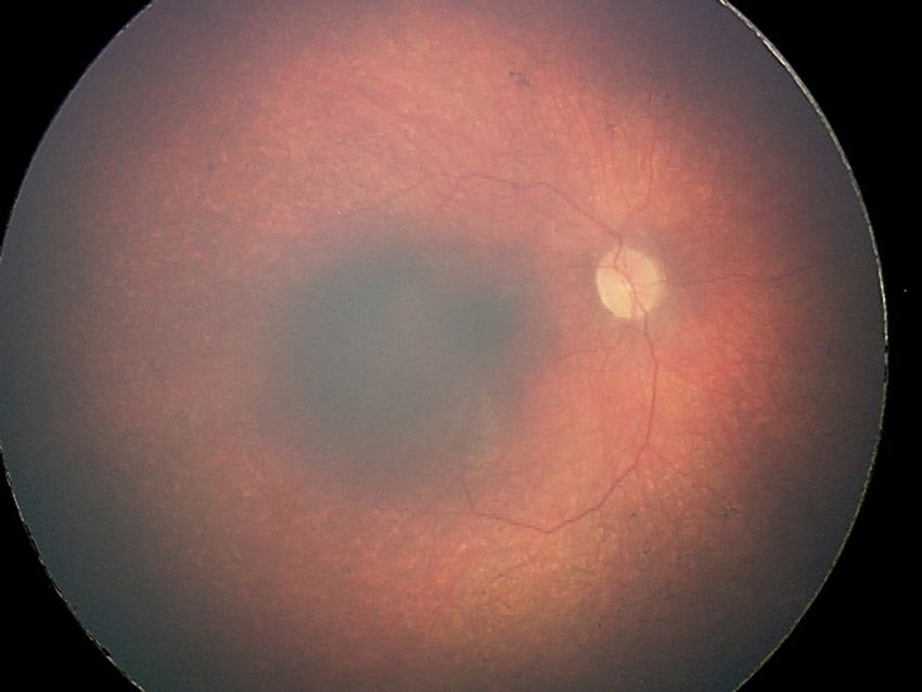
discs and “salt and pepper” retinopathy, accompanied by eyeball. This is most likely due to dysgenesis, corneal and
arterial stenosis [Figure 4]. The intraocular pressure in ciliary body atrophy [1,4,10]. Kivitie-Kallio et al. [6]
the right eye was 12 mm Hg and in the left was 10 mm Hg.
The axial length of both eyeballs was similar, i.e., in the
right eye was 20.63 mm and in the left was 19.40 mm. The
central thickness of cornea in the right eye was 413 µm and
in the left was 439 µm. Handheld optical coherence tomo-
graphy revealed macular edema in both eyes [Figure 5].
Electroretinography showed a reduced response amplitude
of cones and rods [Figure 6].
Figure 2: The girl with Cohen syndrome – note thick hair and eye- Figure 4: Right fundus photography of a girl with Cohen syn-
brow, abnormalities of the palpebral fissures (downslanting and drome – note eyes pale optic discs and “salt and pepper”
almond-shaped palpebral fissures). retinopathy.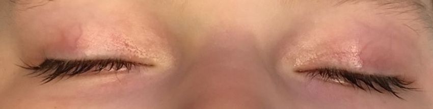
Coexistence of bilateral macular edema and pale optic disc in the patient with Cohen syndrome 159
pigment granularity and “salt and pepper” retinopathy
with narrowed vessels. At an advanced stage, retino-
pathy with bone spicule formed a classic picture of reti-
nitis pigmentosa.
Kivitie-Kallio et al. [6] in a study of 22 subjects, the
majority observed typical lesions for retinal retinopathy
accompanied by pale optic disc in all the patients. Taban
et al. [5] based on their own analysis detected pigment
granularity and pale optic disc in all the patients. Chandler
et al. [11] in 11 of 22 examined patients observed advanced,
severe retinopathy with narrow vessels, bone spicule and
pale optic disc.
Our patient is noticed by the pale, atrophic optic disc,
which is accompanied by lesions in the retina, such as
pigment granularity and narrow arterial vessels, while no
typical bone spicule are found. Severe retinopathy with
bone spicule has been reported in older people, so in our
Figure 5: (a) and (b) Results of optical coherence tomography – 6-year-old patient it may not be present yet.
macular edema in both eyes. Regarding lesions located in the macula, Beck et al.
[9] presented a case of nonleaking cystoid macular
analyzed 22 patients with Cohen syndrome and reported edema in an 11-year-old patient with Cohen syndrome.
high myopia and large astigmatism in the 0.5–6.0 Dcyl Uyhazi et al. [3] focused on the analysis of the initial
range in all the subjects. modification that occurs in the structure of the retina in
Chandler et al. [11] examined 22 patients with geneti- the course of this syndrome. The authors described the
cally confirmed disease and found a refractive error in the case of a 13-month-old girl in whom OCT revealed loss
range from −0.25 to −18 Dsph. Myopia was documented in of the interdigitation signal between the photoreceptor
68% of patients before 5 years of age [11]. The authors outer segments and the apical retinal pigment epithe-
agree that myopia and astigmatism progress with the lium. Loss of only the photoreceptor outer segments
patient’s age [4,10,12]. Our patient was diagnosed with was also noted, suggesting that these are the first visible
myopia as early as at 3 years of age, which was accom- symptoms of retina that later lead to macular edema and
panied by high astigmatism and thinning of the central retinal dystrophy characteristic of Cohen syndrome.
thickness of the cornea. Mutation of the VPS13B gene causes an incorrect
In the literature, the most commonly reported retinal function of the Golgi apparatus membrane protein, which
lesions quintessential of Cohen syndrome are bull’s affects, among others, the function of retinal photorecep-
eye maculopathy, chorioretinopathy, dystrophy with tors [3,9,13]. It is assumed that the edema of the macula
Figure 6: Electroretinography result.160 Klaudia Rakusiewicz et al.
does not occur due to fluid accumulation, or an increase References
in vascular permeability, but only because of the impaired
adhesion and splitting of several retinal layers [3,9,13]. The [1] Rodrigues JM, Fernandes HD, Caruthers C, Braddock SR,
macular edema in hereditary retinal dystrophies is caused Knutsen AP. Cohen syndrome: review of the literature. Cureus.
2018;10(9):e3330.
by a similar mechanism resulting from mutations in indi-
[2] Cohen MM, Hall BD, Smith DW, Graham CB, Lampert KJ. A new
vidual genes [13].
syndrome with hypotonia, obesity, mental deficiency and
Bilateral macular edema was confirmed in the patient facial, oral, ocular and limb anomalies. J Pediatr.
in handheld OCT. Macular lesions in Cohen syndrome have 1973;83:280–4.
been reported in the literature, but only in two cases, a [3] Uyhazi KE, Binenbaum G, Carducci N, Zackai EH, Aleman TS.
characteristic picture of OCT macular edema has been docu- Early photoreceptor outer segment loss and retinoschisis
in Cohen syndrome. Ophthalmic Genet. 2018;39(3):
mented [3,9]. It can be assumed that this was due to the lack
399–404.
of access to this study technique in the past. [4] Hennies HC, Rauch A, Seifert W, Schumi C, Moser E, Al-Taji E,
It cannot be excluded that the coexistence of the et al. Allelic heterogeneity in the COH1 gene explains clinical
described ophthalmologic lesions may result from a homo- variability in Cohen syndrome. Am J Hum Genet.
zygous deletion in our patient. Hennies et al. [4] examined 2004;75(1):138–45.
the inheritance of Cohen syndrome in 20 patients. The [5] Taban M, Memoracion-Peralta DS, Wang H, Al-Gazali LI,
Traboulsi EI. Cohen syndrome: report of nine cases and review
authors confirmed the homozygous mutation in seven
of the literature, with emphasis on ophthalmic features. J
patients from the consanguineous parents and two patients AAPOS. 2007;11(5):431–7.
from parents without known consanguinity – both from [6] Kivitie-Kallio S, Summanen P, Raitta C, Norio R.
Poland. Heterozygous mutation has been documented in Ophthalmologic findings in Cohen syndrome. A long-term
all other patients from unrelated parents. follow-up. Ophthalmology. 2000;107(9):1737–45.
[7] Kivitie-Kallio S, Noria R. Cohen syndrome: essential features,
natural history, and heterogeneity. Am J Med Genet.
2001;102(2):125–35.
4 Conclusion [8] Norio R, Raitta C, Lindahl E. Further delineation of the Cohen
syndrome; report on chorioretinal dystrophy, leukopenia and
consanguinity. Clin Genet. 1984;25:1–14.
It can be assumed that the type of mutation may affect the [9] Beck KD, Wong RW, Gibson JB, Harper CA. Nonleaking cystoid
severity and diversity of ophthalmic features in Cohen macular edema in Cohen syndrome. J AAPOS.
syndrome. In a child with coexistence of high myopia 2019;23(1):38–9.
and astigmatism, retinal dystrophy, pale optic disc and [10] Summanen P, Kivitie-Kallio S, Norio R, Raitta C, Kivelä T.
Mechanisms of myopia in Cohen syndrome mapped to chro-
other abnormalities and facial dysmorphics, ophthalmol-
mosome 8q22. Invest Ophthalmol Vis Sci.
ogists should consider Cohen syndrome. Finding charac-
2002;43(5):1686–93.
teristic lesions in the eye in a group of children with [11] Chandler KE, Biswas S, Lloyd IC, Parry N, Clayton-Smith J,
suspected Cohen syndrome has a significant impact on Black GC. The ophthalmic findings in Cohen syndrome. Br
making the correct diagnosis. Early diagnosis gives the J Ophthalmol. 2002;86(12):1395–8.
possibility of appropriate visual rehabilitation and is cru- [12] Duplomb L, Duvet S, Picot D, Jego G, El Chehadeh-Djebbar S,
Marle N, et al. Cohen syndrome is associated with major gly-
cial in the further development of the child.
cosylation defects. Hum Mol Genet. 2014;23:2391–9.
[13] Lingao M, Ganesh A, Karthikeyan A, Al Zuhaibi S, Al-Hosni A, Al
Conflict of interest: No potential conflict of interest was Khayat A, et al. Macular cystoid spaces in patients with retinal
reported by the authors. dystrophy. Ophthalmic Genet. 2016;37(4):377–83.You can also read