Rare presentation of subcutaneous sarcoid granulomas: A case report and review of the literature - KEI ...
←
→
Page content transcription
If your browser does not render page correctly, please read the page content below
Matson A. et al. Medical Research Archives, vol. 6, issue 1, January 2018 issue Page 1 of 7 CASE REPORTS Rare presentation of subcutaneous sarcoid granulomas: A case report and review of the literature First Authors: Andrea Matson*, D.O. PGY-3, Kelly Shortridge, D.O. PGY-2, Ashley Brown, D.O. PGY-1 Authors’ affiliation: Mountain Vista Medical Center, Department of Surgery, 10238 East Hampton Avenue Suite 301C, Mesa, AZ 85209, Phone: 541-660-8747, Fax: 480-993-3422 Email: Andrea_Matson@iasishealthcare.com * Corresponding author Second author: Raul Lopez, M.D. Author’s affiliation: Mountain Vista Medical Center, Department of Surgery, Mesa, Arizona 10238 East Hampton Avenue Suite 301C Mesa, AZ 85209 Phone: 480-257-2670 Fax: 480-993-3422 Email: Raul_Lopez@iasishealthcare.com The authors have no conflicts of interest to report Abstract Few cases of Darier-Roussy type sarcoidosis have ever been presented in the literature due to the rarity of this condition. Sarcoidosis occurs in only 10-20 per 100,000 in the population and can be present throughout various organs and sites of the body. Skin manifestation as a broad category represent just 25-35% of these cases and even more rare is the Darier-Roussey type which has not had prevalence quantified. We report a new case of Darrier-Roussey sarcoidosis in a 59-year old Caucasian female with flesh colored, firm subcutaneous nodules on her bilateral forearms and hips that developed over 6 months time. Histopathology with special stains identified these nodules as noncaseating sarcoid granulomas. She was also found to have bilateral hilar lymphadenopathy. This is a literature review and case report of subcutaneous sarcoidosis in an adult patient with diagnosis confirmed by clinical exam, imaging, and histopathology who had successful treatment outcome with oral prednisone. Keywords: sarcoidosis; Darier-Roussy type; nodules; naked granuloma Copyright 2018 KEI Journals. All Rights Reserved http://journals.ke-i.org/index.php/mra
Matson A. et al.       Medical Research Archives, vol. 6, issue 1, January 2018 issue       Page 2 of 7

INTRODUCTION                                             (90%), there are many different forms of
                                                         cutaneous sarcoidosis [1,11]. It is the
Sarcoidosis is a well-known multi-system                 second most common manifestation at 25-
disorder most recognized by non-caseating                30% [4,11]. Among the rarest is a form of
granulomas throughout various organs and                 skin manifestation known as Darrier-Roussy
sites in the body [1,4,5]. Its etiology                  which manifests as subcutaneous nodules
remains a mystery but research has shown a               palpable just under the skin (See Table 1 for
combination of genetic (HLA genes) and                   a complete list of skin manifestations) [1,2].
environmental factors are at play [6]. This              Due to the rarity of this form of sarcoid in an
disease occurs in 10-20 per 100,000 in the               already infrequent disease only a handful of
population. Although lungs are the most                  case presentations have been reported some
common site of presentation in this disease              dating back to the 1930’s.

                   Table 1. Various cutaneous presentations of sarcoidosis [4,8,12]

 Type                                       Skin manifestation
 Papular sarcoid                            Presents with papules on the skin
 Lichenoid sarcoid                          Presents as plaques
 Nasal ala papules                          Papules around nasal ala with scaling
 Sarcoidal alopecia                         Scarring and scaling areas with associated hair loss
 Darier-Roussy type                         Subcutaneous nodules most commonly extremities
 Ichthyosiform sarcoid                      Dry thickened scaling skin
 Ulcerative sarcoidosis                     Ulcerative skin lesions
 Scar associated sarcoid                    Granuloma formation/ fibrosis at scar sites
 Tattoo-associated sarcoid                  Similar to scar associated sarcoid
 Lupus pernio                               Extensive violaceous plaques with scales in perinasal
                                            region and mid face
 Erythema Nodosum                           Acute nodular erythematous eruptions

In 1904, Darier and Roussy reported the first            with sparse lymphocytes is known as a
case of subcutaneous sarcoidosis identifying             naked granuloma and is a key feature of
it as a disease with numerous subcutaneous               sarcoid lesions [1]. Gram stains on biopsy
nodules on trunk and extremities [4, 10].                samples to rule out infectious organisms,
Because sarcoidosis is a diagnosis of                    imaging to rule out involvement of other
exclusion many other causes of granuloma                 organs, extensive history and physical exam,
formation must be ruled out with a litany of             chest x-ray, pulmonary function tests, eye
tests and studies involving multiple                     exams, CBC, CMP, EKG, UA, Tb skin test
specialties (as in this case). A punch biopsy            or interferon-y release assay, Thyroid
should be used and will show noncaseating                testing, and Vitamin D25, and D1,25 are
granulomas       with     central   organized            included in the workup. Also useful is the
collections of epithelial macrophages and                serum ACE which is elevated in 40-80% of
multinucleated giant cells surrounded by                 patients with sarcoidosis [1].
sparse lymphocytes [1]. This presentation

 Copyright 2018 KEI Journals. All Rights Reserved                  http://journals.ke-i.org/index.php/mra
Matson A. et al. Medical Research Archives, vol. 6, issue 1, January 2018 issue Page 3 of 7
CASE REPORT nonpathologically enlarged mediastinal
lymph nodes.
This is the case of a 59-year old female who
came to our clinic in March of 2017 with a On 4/25/17 the patient underwent repeat
5-week history of bilateral indurated biopsies of the arm lesions and these were
cutaneous lesions. They were firmly fixed tested for AFP and GMS stains both of
to the skin, mobile, and nontender ranging in which returned negative along with negative
size from 1-6cm. She also reported lesions repeat anaerobic and aerobic cultures. The
on her bilateral upper thighs. She had no pathology again showed non-necrotizing
history of any trauma to the regions, skin granuloma. A quantiferon gold level was
cancer, or skin infections. Her past medical negative ruling out a mycobacterial
history was notable for myocardial infection. ACE levels are elevated in
infarction, hypertension, hyperlipidemia, approximately half of all sarcoidosis patients
hypothyroid, pulmonary embolism, and according to one source. However, in our
gout. She had complained of a chronic dry patient it was found to be within normal
cough that had occurred after moving to limits [1]. The test results were discussed
Arizona from California and denied history with the patient’s pulmonologist who
of smoking. Also noteworthy was a change decided to do a bronchoscopy and right
in her medications upon arriving in Arizona. lower lobe lavage on 5/1/17. Lavage fluid
She had discontinued steroid injections for was sent to pathology and returned with no
her hips upon arriving in Arizona (see Table sign of infection or malignancy. It was
2 for a full list of medications). The determined after ruling out all other
decision was made to remove a lesion from possibilities of infectious, malignant, and
each arm and send it to pathology. She had foreign body reactions that the patient most
operative resection of the arm lesions on likely had an undiagnosed sarcoidosis
3/30/17. She had no complications or related illness. The patient was started on
recurrence and path showed non-necrotizing high dose steroids 3 weeks after her forearm
granulomata negative for neoplasia. Gram biopsies to allow appropriate time for
stains and anaerobic and aerobic cultures surgical wound healing. The steroids were
were negative. She was also sent to a then tapered to the lowest effective dose to
pulmonologist for appropriate work up of suppress the cough and skin nodules.
her cough. On pulmonary function studies Shortly after beginning the steroids the
the patient had a mild restrictive lung defect nodules completely resolved. It was
with normal diffusion capacity. Suspecting hypothesized that the nodules and cough
sarcoidosis, further testing was requested by were being suppressed while the patient was
the pulmonologist including, repeat biopsies in California due to the steroid injections she
of the lesions with special stains, and a CT had been receiving for her hips.
of the chest revealing multiple bilateral
Copyright 2018 KEI Journals. All Rights Reserved http://journals.ke-i.org/index.php/mraMatson A. et al. Medical Research Archives, vol. 6, issue 1, January 2018 issue Page 4 of 7
Table 2. Complete list of patient medications and doses.
Medication Dose Frequency
Fenofibrate 54mg tablet PO QD
Furosemide 20mg tablet PO QD
Levothyroxine 50mcg tablet PO QD
Prednisone* 30mg QD
20mg tablet PO 8/16-10/14,
10mg tab PO 7/3-8/13
Klor-Con Sprinkle 10mEq capsule PO QD
Metformin 500mg tablet PO QD
Atorvastatin 20mg tablet PO QD
Allopurinol 100mg tablet PO QD
Valsartan 320mg tablet PO QD
Clonidine HCl 0.1 mg tablet PO QD
Nifedipine ER 60 mg tablet PO QD
Xarelto 20 mg tablet PO QD
Breo Ellipta 100 mcg-25 mcg/ dose 1Puff QD
powder
*New medications given after the patient developed her nodules
DISCUSSION and cough [1]. On imaging, bilateral hilar
adenopathy is often noticed as in this patient
Sarcoidosis is a diagnosis of exclusion [13]. Other similar granulomatous skin
typically presenting in patients between 20 diseases have characteristic findings that
and 60 years of age [1]. It most frequently were not seen in our patients work up (see
involves the lungs (over 90%) commonly Table 2).
presenting with upper respiratory symptoms
Table 3. Histopathology was used to differentiate and confirm the diagnosis of subcutaneous
sarcoidosis based on presence or absence of the above findings. [3]
Skin manifestation Differentiating findings
Sarcoidosis Noncaseating granulomas
Granuloma annulare Mucin and collagen surrounding granulomas
Necrobiosis lipoidica Granulomas with collagen between
histiocytes
Necrobiotic xanthogranuloma Xanthogranulomas, cholesterol clefting
Foreign body Multinucleated giant cells, polarization
Infections Postitive stains for microorganisms
Crohn’s disease Dermal granulomatous infiltrate composed
of epithelioid histiocytes
Siliconosis Fibrotic nodules with onion skinned
arrangement of collagen fibers
Copyright 2018 KEI Journals. All Rights Reserved http://journals.ke-i.org/index.php/mraMatson A. et al. Medical Research Archives, vol. 6, issue 1, January 2018 issue Page 5 of 7
In the case report by Plana et al, a 40-year To confirm the diagnosis of sarcoidosis,
old female with common variable immune supporting physical exam findings,
deficiency (CVID) there was the similarity histopathology, and imaging must be
of sarcoidal granulomas of the skin. present. On balance, diseases with similar
However in CVID there will frequently be presentation must have been effectively
synovial membrane involvement, history of ruled out. All criteria listed were met in our
recurrent infections, cytopenias, and patient. Therefore, we can say with certainty
autoimmune conditions [6]. Our patient had that she had systemic sarcoidosis as
a normal cbc, and no history of recurrent evidenced by the hilar adenopathy seen on
infections making this diagnosis unlikely. CT of her lungs, as well as subcutaneous
sarcoidosis discovered by biopsy results and
Vedove and colleagues—In contrast— special staining performed on the specimen.
present two confirmed cases of subcutaneous
sarcoidosis which manifest with many As for the trigerring event leading to the
similarities to this case presentation, sarcoidal granuloma formation, historically
following a similar work-up and treatment research has shown that a variety of genetic
protocol to ours with successful results[1]. mutations in HLA type genes combined with
environmental exposures lead to the
In the first case reported an 81-year old male formation of sarcoid type granulomas [7].
presented with nontender firm nodules of Frequently infectious organisms, scars,
bilateral forearms and thighs that appeared polarized material such as tattoos, or foreign
in 6 months time [1]. Biopsies, and special bodies have been required to trigger the
stains confirmed noncaseating granulomas inflammatory cascade leading to granuloma
of sarcoid origin [1]. He did also have an formation. In many cases the cause of
elevation in ACE which is common granuloma formation remains unknown.
with sarcoidosis but not required for Upon questioning our patient thoroughly
diagnosis [1]. He responded very well to there was no obvious identifiable exposure
prednisone [1]. that led to the formation of the sarcoid
In the second case a 69-year old woman granulomas seen on her forearms and hips
presented with multiple large erythematous [7]. Current treatments in the literature
nodules appearing over 2 months time. Her reflect a constantly evolving approach. One
biopsy results also confirmed noncaseating approach that has remained constant and
granulomas with negative staining and with the best results is corticosteroids either
cultures ruling out foreign body particles, systemic or locally injected [9]. Topical or
fungi, or infectious organisms. This patient intralesional steroids have been shown to
also had bilateral hilar adenopathy on CT of have good outcomes as well in isolated cases
the chest consistent with sarcoidosis[1]. without systemic symptoms [9].
During a review of 85 cases of subcutaneous
sarcoidosis the majority presented with REFERENCES
cutaneous nodules as their initial
symptom[1]. Sixty-seven cases out of 85 1. Wanat, K. A., & Rosenbach, M. (2014,
presented with symmetric subcutaneous May 13). A Practical Approach to
nodules on the extremities and the majority Cutaneous Sarcoidosis. American
of them were subsequently found to have Journal of Clinical Dermatology, 283-
enlarged hilar lymph nodes.[1] 297.
Copyright 2018 KEI Journals. All Rights Reserved http://journals.ke-i.org/index.php/mraMatson A. et al. Medical Research Archives, vol. 6, issue 1, January 2018 issue Page 6 of 7
2. Rafiei, P., & Vijayakumar, V. (2011, disease. Journal of Cutaneous
June). Be Aware of Disseminated Pathology, 43, 475-477.
Sarcoidosis With Diffuse
Subcutaneous Nodules on F-18 FDG 8. Noiles, K., Beleznay, K., Crawford, R.
PET. Clinical Nuclear I., & Au, S. (2013, December).
Medicine, 36(6), 42-44. Sarcoidosis Can Present with
Necrotizing Granulomas
3. Wanat, K., & Rosenbach, M. (2015). Histologically: Two Cases of
Cutaneous Sarcoidosis. Clinical Chest Ulcerated Sarcoidosis and Review of
Medicine, 36, 685-702. the Literature. Journal of Cutaneous
Medicine and Surgery, 17(6), 377-383.
4. Kempf, W., Zollinger, T., Sachs, M.,
Ullmer, E., Cathomas, G., & 9. Marcoval, J., Mana, J., & Rubio, M.
Dirnhofer, S. (2014). Granulomas are a (2011). Specific cutaneous lesions in
source of interleukin-33 expression in patients with systemic sarcoidosis:
pulmonary and extrapulmonary relationship to severity and chronicity
sarcoidosis☆. Human Pathology, 45, of disease. Clinical Dermatology, 36,
2202-2210. 739-744.
5. Grunewald, J., Spagnolo, P., 10. Vedove, C. D., Colato, C., &
Wahlstrom, J., & Eklund, A. (2015, Girolomoni, G. (2011, April 2).
March 20). Immunogenetics of Subcutaneous sarcoidosis: report of
Disease-Causing Inflammation in two cases and review of the
Sarcoidosis. Clinical Review Allergy literature. Clinical Rheumatology, 30,
Immunology, 49, 19-35. 1123-1128.
6. Pla, A. P., Bassas-Vila, J., Roure, S., 11. Ismail, A., Beckum, K., & McKay, K.
& Ferrandiz, C. (2015). Necrotizing (2014). Transepithelial elimination in
and sarcoidal granulomas in the skin sarcoidosis: a frequent
and synovial membrane, associated finding. Journal of Cutaneous
with common variable Pathology, 41, 22-27.
immunodeficiency. Clinical
Dermatology, 40, 379-382. 12. Baughman, R. P., & Lower, E. E.
(2015, May 20). Treatment of
7. Janegova, A., Janega, P., Kovac, O., Sarcoidosis. Clinical Review Allergy
Dragun, J., & Zakutansky, A. (2016). Immunology, 49, 79-92.
Plantar subcutaneous sarcoidosis – a
rare form of skin sarcoidosis: unusual 13. Miida, H., & Ito, M. (2010).
plantar aponeurosis location of Tuberculoid granulomas in cutaneous
sarcoidosis as primary manifestation of sarcoidosis: a study of 49
asymptomatic systemic cases. Journal of Cutaneous
Pathology, 37, 504-506.
Copyright 2018 KEI Journals. All Rights Reserved http://journals.ke-i.org/index.php/mraMatson A. et al. Medical Research Archives, vol. 6, issue 1, January 2018 issue Page 7 of 7
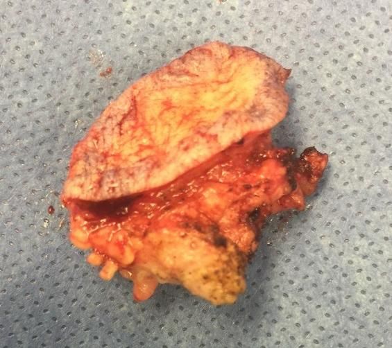
Figure 1. Right sided 1cm nodule removed from forearm.
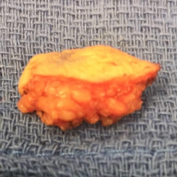
Figure 2. Left sided 2cm nodule removed from forearm.
Copyright 2018 KEI Journals. All Rights Reserved http://journals.ke-i.org/index.php/mraYou can also read