Vitamin C Administration Attenuated Artemether-Induced Hepatic Injury in Rats - SciELO
←
→
Page content transcription
If your browser does not render page correctly, please read the page content below
Int. J. Morphol.,
38(1):48-55, 2020.

               Vitamin C Administration Attenuated Artemether-
                       Induced Hepatic Injury in Rats

                                   La Administración de Vitamina C Atenúa la Lesión
                                       Hepática Inducida por Artemeter en Ratas

                          Refaat A. Eid1; Mohamed Samir Ahmed Zaki2,3; Mansour A. Alghamdi2;
               Abulqasim Mohammed Sideeg2; Kamal, Z. M. Ali2; Mohamed Andarawi1 & Mohamed A. Haidara4

EID, R. A.; ZAKI, M. S. A.; ALGHAMDI, M. A.; SIDEEG, A. M.; ALI, K. Z. M.; ANDARAWI, M. & HAIDARA, M. A. Vitamin
C administration attenuated Artemether-induced hepatic injury in rats. Int. J. Morphol., 38(1):48-55, 2020.

         SUMMARY: This research was designed to investigate the potential protective effect of vitamin C supplementation against
hepatocyte ultrastructural alterations induced by artemether (antimalarial drug) administration. Twenty-four adult male albino rats were
used in this study and were divided into four groups (n=6). Group I served as a control and rats in group II administrated artemether (4
mg/kg B.W) orally for three consecutive days. Group III administered artemether plus a low dose of vitamin C (2.86 mg/kg/l water)
while group IV received artemether plusa high dose of vitamin C (8.56 mg/kg). At the end of the experimental period (14 days), the
harvested liver tissues were examined by transmission electron microscopy (TEM), and blood samples were assayed for biomarkers of
liver injury and oxidative stress. Artemether significantly (p30 %). Artemether is characterized                  erythrocytes has been reported to depend upon the presence
by its novel structure which is very effective against                    or absence of glutathione. In the presence of glutathione,
multidrug-resistant Plasmodium falciparum malaria                         ascorbic acid has synergistic antioxidant activity against
(Meshnick, 2002).                                                         haem-mediated cell toxicity (Li et al., 2006). In glutathione
                                                                          deficient red cells, as often happens in parasitized RBCs
       Oxidative stress occurs when the generation of                     due to oxidative stress, ascorbic acid can react with iron or
reactive oxygen species in the body exceeds the ability of                iron-containing compounds to generate hydrogen peroxide
the body to neutralize and eliminate them (Li et al., 2018).              or hydroxyl radical and accentuate the haemolytic
The susceptibility of liver tissues to this stress due to                 mechanisms in malaria (Li et al., 2006).
exposure to drugs is a function of overall balance between
the degree of oxidative stress and the antioxidant capacity                     Therefore, the aim of the present work is to study the
(Khan et al., 2005).                                                      artemether induced liver toxicity on the liver and to evaluate

1
  Pathology department, College of Medicine, King Khalid University, Abha 61421, Saudi Arabia.
2
  Anatomy department, College of Medicine, King Khalid University, Abha 61421, Saudi Arabia.
3
  Histology department, College of Medicine, Zagazig University, Egypt.
4
  Physiology department, Kasr al-Aini Faculty of Medicine, Cairo University, Cairo, Egypt.

48
EID, R. A.; ZAKI, M. S. A.; ALGHAMDI, M. A.; SIDEEG, A. M.; ALI, K. Z. M.; ANDARAWI, M. & HAIDARA, M. A. Vitamin C administration attenuated Artemether-induced hepatic injury
in rats. Int. J. Morphol., 38(1):48-55, 2020.
the possible protective effect of vitamin C against this Determination of serum levels of superoxide dismutase
toxicity in rats. (SOD) and Glutathione peroxidase (GPx). After two weeks,
animals were sacrificed, and serum levels of (SOD) and
(GPx) were measured using commercial kits supplied by
MATERIAL AND METHOD SPINREACT, Spain, according to the manufacturer’s
instructions.
Animals: All experimental procedures were approved by Light Microscopy (LM): The fixed liver specimens
the medical research ethical committee at King Khalid (formalin fixed tissues) were trimmed, washed, dehydrated
University and according to the Guide for the Care and Use in ascending grades of ethyl alcohol, cleared in methyl
of Laboratory Animals published by the US National benzoate and embedded in paraffin after having completed
Institutes of Health. (NIH publication No. 85-23, revised the routine follow-up steps. Sections at 3-5 m sections were
1996). Sprague–Dawley rats (n=24) weighing 150- obtained from liver using rotary microtome and stained by
250 g were used in this study. All rats were bred and housed hematoxylin and eosin (H&E) stain for light microscopically
in the research centre of King Khalid University, college of investigation to Bancroft & Gamble (2008).
medicine (Abha, Saudi Arabia), at a temperature of 23 ±1
°C and a 12 h light: 12 h dark cycle. Rats had free access to Transmission Electron Microscopy (TEM): Small pieces
tap water and fed standard laboratory chow during the of liver tissues were removed and immediately fixed in 2.5
acclimatization period. % glutaraldehyde for 24 hours and washed with phosphate
buffer (0.1 M, PH 7.4). Post for 1-2 hours. The samples
Experimental design: After one-week adaptation. All rats washed in phosphate buffer to remove excess fixative,
were fed a standard laboratory diet. The rats were randomly dehydrated through ascending grades of ethanol followed
divided into four groups (n=6 rats each). Animals in the first by clearing in propylene oxide. The specimens were
group (Control) were fed with standard laboratory chow for embedded in Araldite 502, to form gelatin capsules.
two weeks. Animals in the second group (Artemether), rats Polymerization was obtained by placing the capsules at 60
o
were given artemether supplementation (4 mg/kg B.W / daily C. Semi-thin sections (~1 mm thick) were stained with
by oral gavage) for three consecutive days and continue on toluidine blue for orientation and observation. Ultrafiltration
a standard diet for two weeks. The third group, rats was made in 1 % osmium tetroxide buffered to PH 7.4 with
administered artemether, 4 mg/kg B.W /day by oral gavage 0.1 M phosphate buffer at 4 oC -thin sections (100 nm) were
for three consecutive days, plus a low dose of vitamin C, prepared using ultra-microtome and picked up on uncoated
2.86 mg/kg /l water for two weeks. Animals in the fourth rats copper grids. Following double staining with uranyl acetate
administered artemether, 4 mg/kg B.W /day by oral gavage and lead citrate, three to five random micrographs for each
for three consecutive days, plus a high dose of vitamin C, section were examined and photographed using a JEM-1011
8.56 mg/kg /l water for two weeks. transmission electron microscope, JEOL Ltd., Musashino,
Akishima, Tokyo, Japan, at 80 Kv (Haidara et al., 2018).
Biochemical measurements
Statistical analysis. The data were expressed as mean ± stan-
Blood samples: At the end of the experimental period, blood dard deviation (SD). Data were processed and analyzed using
samples were collected by cardiac puncture under the SPSS version 10.0 (SPSS, Inc., Chicago, Ill., USA). One-
anaesthesia (sodium thiopentone at 40 mg/kg body weight) way ANOVA was done followed by Tukey’s post hoc test.
after an overnight fast of 12 hours. These blood samples Pearson correlation statistical analysis was done for the
were collected without anticoagulant, left for 10 min, then detection of a probable significance between two different
centrifuged for 10 min at 4000 r/min to obtain serum, which parameters. Results were considered significant if p ≤ 0.05.
was stored at –20 °C until further biochemical analysis for
determination of serum liver enzymes, oxidative stress
biomarkers. RESULTS
Determination of serum levels of ALT, AST: After two
weeks, animals were sacrificed, and liver function was Vitamin C reduces biomarkers of liver injury and
evaluated by assessing serum ALT and AST levels using an oxidative stress induced artemether. To determine whether
enzymatic kit (Randox Laboratories, Crumlin, UK) vitamin C can inhibit artemether-induced up-regulation of
according to the manufacturer's instructions. liver injury enzymes (ALT and AST), and biomarkers of
oxidative stress (SOD and GPx) and inflammation in our
49EID, R. A.; ZAKI, M. S. A.; ALGHAMDI, M. A.; SIDEEG, A. M.; ALI, K. Z. M.; ANDARAWI, M. & HAIDARA, M. A. Vitamin C administration attenuated Artemether-induced hepatic
injury in rats. Int. J. Morphol., 38(1):48-55, 2020.
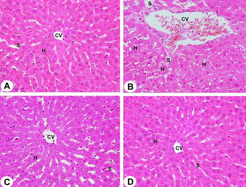
animal model of artemether –induced liver injury, we Histopathological findings (LM): H&E stained sections of
measured the blood levels of ALT, AST, SOD and GPx in liver of the control group revealed normal characteristic of
all rat groups. Artemether caused the augmentation of ALT hepatic architecture; the hepatic lobules appeared to be made
(Fig. 1A), AST (Fig. 1B), which were significantly (pEID, R. A.; ZAKI, M. S. A.; ALGHAMDI, M. A.; SIDEEG, A. M.; ALI, K. Z. M.; ANDARAWI, M. & HAIDARA, M. A. Vitamin C administration attenuated Artemether-induced hepatic injury
in rats. Int. J. Morphol., 38(1):48-55, 2020.
Fig. 2. LMs X400 of harvested tissues obtained from the liver of the control group (A) compared to the Artemether-treated group (B),
Artemether-treated + 2.86 mg/kg Vit-C group (C), and Artemether-treated + 8.56 mg/kg Vit-C group (D) rats are visualized using light
microscopy. Abbreviations: H, hepatocytes; CV, central vein; S, blood sinusoid.
cisternae of rough endoplasmic reticulum appeared dilated DISCUSSION
and fragmented. The bile canaliculi with junctional complex
were distorted. (Fig. 5B). Moreover, the blood sinusoids were
damaged with abnormal microvilli (Figs. 3B and 4B). The main objective of our study was to investigate
the potential protective effect of vitamin C to the hepatocyte
Electron microscopic examination of the third and ultrastructure against artemether -induced liver injury in a
fourth group revealed minor changes in most liver cells. The rat model of the disease using TEM. In addition, a
hepatocytes have normally-shaped mitochondria, the comparison was also made between the pathological and
cisternae of the rough endoplasmic reticulum were normally biochemical changes occurred in response to the disease
distributed, and intact bile canaliculi with junctional complex and its potential treating drug, vitamin C. The main findings
are seen. Some vacuoles are still present. Normally of our study were that (i) Artemether induced hepatic
distributed and intact blood sinusoids with normal microvilli profound damage to hepatocyte ultrastructure; (ii) low dose
were seen (Figs. 3C, 4C and 5C). vitamin C substantially but not completely slowed down
the progression of the disease in rats; and (iii) high dose
Examination of the liver sections of the fourth group by vitamin C significantly reduced certain biomarkers of liver
electron microscopy, the histological architecture of the injury. These conclusions are supported by the data
hepatic lobules exhibited a more or less normal appearance indicating that Artemether markedly increased liver injury
(Figs. 3D, 4D and 5D). enzymes (ALT and AST), and reduced anti-oxidative stress
51EID, R. A.; ZAKI, M. S. A.; ALGHAMDI, M. A.; SIDEEG, A. M.; ALI, K. Z. M.; ANDARAWI, M. & HAIDARA, M. A. Vitamin C administration attenuated Artemether-induced hepatic injury
in rats. Int. J. Morphol., 38(1):48-55, 2020.
Fig. 3. TEMs X5000 (5 µm) of harvested tissues obtained from the liver of the control group (A) compared to the Artemether-treated
group (B), Artemether-treated + 2.86 mg/kg Vit-C group (C), and Artemether-treated + 8.56 mg/kg Vit-C group (D) rats are visualized
using transmission electron microscopy. Note that large black arrows point to intercellular spaces between hepatocytes. Abbreviations:
N, nucleus; m, mitochondria; RER, rough endoplasmic reticulum; Ly, lysosomes.
biomarker (GPx and SOD), which were significantly degenerative cells, which is in accordance with a previous
improved with vitamin C treatment (Fig. 1 ). Also, vitamin study (Wood, 1965).
C partially prevented damages occurred to liver cells after
two weeks in rats fed on Artemether (Figs. 2-5). The results of the TEM examination of hepatocytes
support that there was a diversity of cellular damage.
Many xenobiotics, drugs and chemicals cause Spaces of Disse were seen around hepatocytes expanded
diverse forms of liver injury (Sturgill & Lambert, 1997), and filled with long fragmented microvilli which cause
and this may result in distortion in liver histology. Earlier the distinct clarification of the cell's borders under the light
reports have also shown that artemether treatment results microscopy examination. It also showed cytoplasmic
in congestion of hepatic sinusoids in healthy rats (Izunya vacuoles and sinusoids were filled with hypertrophy of
et al., 2010). Kupffer cells which is in accordance with Bjørndal et al.
(2018) who reported that the neutral fat accumulates in
Our results showed that treatment with artemether the liver frequently, is due to inhibition of aerobic oxidation.
has a toxic effect on liver cells, associated changes in Furthermore, artemether has been shown to induce transient
52EID, R. A.; ZAKI, M. S. A.; ALGHAMDI, M. A.; SIDEEG, A. M.; ALI, K. Z. M.; ANDARAWI, M. & HAIDARA, M. A. Vitamin C administration attenuated Artemether-induced hepatic injury
in rats. Int. J. Morphol., 38(1):48-55, 2020.
Fig. 4. Higher magnification of TEMs X10000 (2 µm) of the Vit-C protects against endoplasmic reticulum; ne, nuclear envelop; Chr,
chromatin; Ly, lysosomes; Bc, bile canaliculiArtemether-induced hepatocyte ultrastructural damage in rats, at the end of the experiment,
after two weeks. (A) Control group. (B), Artemether group. (C), Artemether +2.86 mg/kg Vit-C group, and Artemether +8.56 mg/kg Vit-
C group (D). Note that large black arrows point to intercellular spaces between hepatocytes. Abbreviations: N, nucleus; nu, nucleolus; L,
lipid droplets; V, vacuoles; m, mitochondria; RER, rough.
and moderate elevations in liver transaminases (Nwanjo in body fluids, scavenging reactive oxygen and nitrogen
et al., 2007). species (Elzoghby et al., 2015).
Pro-oxidant chemicals which stimulate the In conclusion, the overall results showed that
oxidation effort either by synthesis or by inhibiting vitamin C ameliorates the hepatotoxic effect of artemether,
antioxidant may cause damage to cells and tissues (Kumar which was possibly mediated via free radical scavenging
& Muralidhara, 2007) due to the formation of the and inhibition of free radical generation.
superoxidation, which leads to rupture of the plasma
membrane and organelles.
ACKNOWLEDGMENTS.The authors extend their
Vitamin C has a reducing potential that reacts with appreciation to the Deanship of Scientific Research at King
most of the important radicals and oxidants (Magdy et al., Khalid University for funding this work through research
2015) where it acts as a powerful hydrosoluble antioxidant groups program under grant number G.R.P.186 -39.
53EID, R. A.; ZAKI, M. S. A.; ALGHAMDI, M. A.; SIDEEG, A. M.; ALI, K. Z. M.; ANDARAWI, M. & HAIDARA, M. A. Vitamin C administration attenuated Artemether-induced hepatic injury
in rats. Int. J. Morphol., 38(1):48-55, 2020.
Fig. 5. TEMs X5000 (5 µm) (Blood sinusoids) of the Vit-C protects against Artemether-induced blood sinusoid and hepatocyte ultrastructural
damage in rats, at the end of the experiment, after two weeks. (A) Control group. (B), Artemether group. (C), Artemether +2.86 mg/kg
Vit-C group, and Artemether +8.56 mg/kg Vit-C group (D). Note that large black arrows point to intercellular spaces between hepatocytes.
Abbreviations: H, hepatocytes; N, nucleus; m, mitochondria; RER, rough endoplasmic reticulum; Ly, lysosomes; MF, myelin figures; S,
blood sinusoids; R, erythrocytes; V, vacuole; Mo, monocytes; mv, short microvilli.
EID, R. A.; ZAKI, M. S. A.; ALGHAMDI, M. A.; SIDEEG, A. grupo IV recibió arteméter más una dosis alta de vitamina C
M.; ALI, K. Z. M.; ANDARAWI, M. & HAIDARA, M. A. La (8,56 mg / kg). Al final del período experimental (14 días), los
administración de vitamina C atenúa la lesión hepática inducida tejidos hepáticos recolectados se examinaron por microscopía
por Artemeter en ratas. Int. J. Morphol., 38(1):48-55, 2020. electrónica de transmisión (MET), y las muestras de sangre se
analizaron en busca de biomarcadores de daño hepático y estrés
RESUMEN: Esta investigación fue diseñada para in- oxidativo. El arteméter aumentó significativamente (pEID, R. A.; ZAKI, M. S. A.; ALGHAMDI, M. A.; SIDEEG, A. M.; ALI, K. Z. M.; ANDARAWI, M. & HAIDARA, M. A. Vitamin C administration attenuated Artemether-induced hepatic injury
in rats. Int. J. Morphol., 38(1):48-55, 2020.
la vitamina C es un agente protector parcial contra la lesión he- Corresponding author:
pática inducida por arteméter. Dr. Refaat A. Eid
Department of Pathology
PALABRAS CLAVE: Arteméter; Ratas; Vitamina C; College of Medicine
Ultraestructura de hepatocitos; Lesión hepática de King Khalid University
biomarcadores; Estrés oxidativo. Abha
SAUDI ARABIA
REFERENCES
Email: refaat_eid@yahoo.com
Bancroft, J. D. & Gamble, M. Theory and Practice of Histological
Received: 02-07-2019
Techniques. 6th ed. New York, Churchill Livingstone, 2008. pp.440-
50.
Accepted: 29-07-2019
Bhatt, S.; Weiss, D. J.; Cameron, E.; Bisanzio, D.; Mappin, B.; Dalrymple,
U.; Battle, K.; Moyes, C. L.; Henry, A.; Eckhoff, P. A.; et al. The effect
of malaria control on Plasmodium falciparum in Africa between 2000
and 2015. Nature, 526(7572):207-11, 2015.
Bjørndal, B.; Alterås, E. K.; Lindquist, C.; Svardal, A.; Skorve, J. & Berge,
R. K. Associations between fatty acid oxidation, hepatic mitochondrial
function, and plasma acylcarnitine levels in mice. Nutr. Metab. (Lond.),
15:10, 2018.
Elzoghby, R. R.; Hamoda, A. F.; Abdel-Fatah, A. & Farouk, M. Protective
role of vitamin C and green tea extract on malathion-induced
hepatotoxicity and nephrotoxicity in rats. Am. J. Pharmacol. Toxicol.,
9(3):177-88, 2015.
Haidara, M. A.; Dallak, M.; El Karib, A. O.; Abd Ellatif, M.; Eid, R. A.;
Heidar, E. H. A. & Al-Ani, B. Insulin protects against hepatocyte
ultrastructural damage induced by type 1 diabetes mellitus in rats.
Ultrastruct. Pathol., 42(6):508-15, 2018.
Izunya A. M.; Nwaopara, A. O.; Aigbiremolen, A.; Odike, M. A. C.;
Oaikhena, G. A. & Bankole, J. K. Histological effects of oral
administration of artesunate on the liver in Wistar rats. Res. J. Appl.
Sci. Eng. Technol., 2(4):314-8, 2010.
Khan, S. M.; Sobti, R. C. & Kataria, L. Pesticide-induced alteration in
mice hepato-oxidative status and protective effects of black tea extract.
Clin. Chim. Acta, 358(1-2):131-8, 2005.
Kumar, T. R. & Muralidhara. Induction of oxidative stress by organic
hydroperoxides in testis and epididymal sperm of rats in vivo. J. Androl.,
28(1):77-85, 2007.
Li, S. D.; Su, Y. D.; Li, M. & Zou, C. G. Hemin-mediated hemolysis in
erythrocytes: effects of ascorbic acid and glutathione. Acta. Biochim.
Biophys. Sin. (Shanghai), 38(1):63-9, 2006.
Li, Y. F.; Ouyang, S. H.; Tu, L. F.; Wang, X.; Yuan, W. L.; Wang, G. E.; Wu,
Y. P.; Duan, W. J.; Yu, H. M.; Fang, Z. Z.; et al. Caffeine protects skin
from oxidative stress-induced senescence through the activation of
autophagy. Theranostics, 8(20):5713-30, 2018.
Magdy, B. W.; Mohamed, F. E.; Amin, A. S. & Rana, S. S. Ameliorative
effect of antioxidants (vitamins C and E) against abamectin toxicity in
liver, kidney and testis of male albino rats. J. Basic. Appl. Zool., 77:69-
82, 2015.
Meshnick, S. R. Artemisinin: mechanisms of action, resistance and toxicity.
Int. J. Parasitol., 32(13):1655-60, 2002.
Nwanjo, H.; Iroagba, I.; Nnatuanya, I. & Eze, N. Antifertility activity of
dihydroartemisinin in male albino rats. Int. J. Endocrinol., 4(1), 2007.
Shichiri, M.; Ishida, N.; Hagihara, Y.; Yoshida, Y.; Kume, A. & Suzuki, H.
Probucol induces the generation of lipid peroxidation products in
erythrocytes and plasma of male cynomolgus macaques. J. Clin.
Biochem. Nutr., 64(2):129-42, 2019.
Sturgill, M. G. & Lambert, G. H. Xenobiotic-induced hepatotoxicity:
mechanisms of liver injury and methods of monitoring hepatic function.
Clin. Chem., 438(8 Pt. 2):1512-26, 1997.
Wood, R. L. The fine structure of hepatic cells in chronic ethionine poisoning
and during recovery. Am. J. Pathol., 46:307-30, 1965.
55You can also read