Stereotactic Radiotherapy and Particle Therapy for Pancreatic Cancer - MDPI
←
→
Page content transcription
If your browser does not render page correctly, please read the page content below
cancers
Review
Stereotactic Radiotherapy and Particle Therapy for
Pancreatic Cancer
Sweet Ping Ng and Joseph M. Herman *
 Department of Radiation Oncology, The University of Texas MD Anderson Cancer Center, 1515 Holcombe Blvd,
 Houston, TX 77030, USA; spng@mdanderson.org
 * Correspondence: jmherman@mdanderson.org

 Received: 24 February 2018; Accepted: 14 March 2018; Published: 16 March 2018

 Abstract: Pancreatic cancer is a devastating disease with poor survival outcomes. Recent studies have
 shown that the addition of radiotherapy to chemotherapy in the setting of locally advanced pancreatic
 cancer did not improve overall survival outcome. These studies commonly utilize conventional
 radiotherapy treatment fractionation and technique (typically 3-D conformal radiotherapy or intensity
 modulated radiotherapy). Although no clear benefit in overall survival was demonstrated in those
 studies, those who received radiotherapy did have a clear benefit in terms of local control. Therefore,
 there is increasing interest in exploring different techniques and/or modality of radiotherapy and
 dose/fractionation. Stereotactic radiotherapy, which employs a hypofractionated regimen, has the
 potential advantage of delivering a high dose of radiation to the tumor in a short period of time
 (typically over 5 days) with minimal dose to the surrounding normal structures. Particle therapy
 such as proton and carbon ion therapy are being explored as potential radiation modality that could
 cause greater biological damage to the tumor compared to photon treatment, with rapid dose falloff
 resulting in minimal to no dose to adjacent structures. This review will discuss the current literature
 and emerging roles of stereotactic radiotherapy and particle therapy in pancreatic cancer.

 Keywords: radiotherapy; stereotactic; proton; carbon; pancreatic cancer

1. Introduction
      Pancreatic cancer, being the third leading cause of cancer-related deaths in the United States,
is a devastating disease with an estimated 5-year overall survival of only 6% [1]. Although surgery
provides patients with the best chance of achieving a cure, the majority of patients tend to present
late with advanced stage of disease given the lack of symptoms and no effective screening tests
available, as yet. The role of radiotherapy in the definitive management of pancreatic cancer has
remained debated. The recent LAP07 study showed no overall survival with chemotherapy alone, as
compared to chemoradiotherapy after completion of induction chemotherapy in patients with locally
advanced pancreatic cancer (LAPC); however, the study did demonstrate that those who received
radiotherapy had significantly improved local control with minimal increase in treatment-related
toxicity [2]. Although local control of the disease did not translate to improvement in overall survival
in the LAP07 study, there is evidence that in a subgroup of patients, local control could potentially
translate to improvement in survival. Specifically, a John Hopkins rapid autopsy study indicated
that approximately 1 in 3 patients died of local disease only [3]. As the LAP07 study employed
conventional fractionation that were delivered over 6 weeks, there is speculation that some patients
may have developed metastatic disease during this period of time when they were not on optimal
systemic treatment. Therefore, there is increasing interest in exploring other radiotherapy options,
particularly hypofractionated regimens and particle therapy, to maximize local therapy to the tumor
whilst limiting patients’ time off effective systemic treatment. Here, we review the literature and

Cancers 2018, 10, 75; doi:10.3390/cancers10030075                                www.mdpi.com/journal/cancers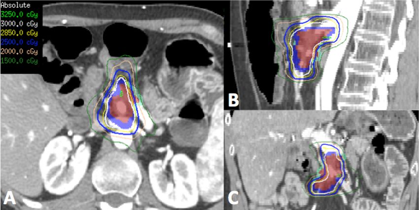
Cancers 2018, 10, 75                                                                                2 of 12

highlight the emerging role of stereotactic body radiotherapy (SBRT) and particle therapy in the
treatment of pancreatic cancer.

2. Stereotactic Body Radiotherapy (SBRT) for Locally Advanced Pancreatic Cancer
      SBRT is an advanced technique of radiation planning and delivery where it delivers a highly
conformal dose to the target with very steep dose gradient falloff at the edge of the target.
This technique enables highly precise delivery of a high dose of radiation to the target in 1–5 fractions.
As SBRT employs a hypofractionated regimen, particular care is required during planning and delivery
because any geographic miss of the target could be a calamity, not only because the tumor was
not adequately treated but also because the adjacent surrounding normal structures may receive
higher than intended doses, thereby increasing the risk of toxicity. In a patient with pancreatic cancer,
the adjacent normal structures include the duodenum, stomach and/or small bowel. Exceeding dose
tolerance to these structures could cause gastrointestinal perforation and/or haemorrhage which can be
fatal. Therefore, when considering SBRT, patient selection and adequate image guidance is paramount.
      In the past decade, there is increasing interest in utilizing SBRT when treating patients with
pancreatic cancer. SBRT allows for the delivery of higher doses to the tumor and as most regimens
are 5 fractions or less, patients only have to be off systemic treatment for a short period of time, as
compared to conventional dose/fractionation regimens, where patients may be without adequate
systemic therapy for up to 6 weeks.
      Table 1 summarizes the current literature. As depicted in Table 1, most studies demonstrated that
local control with SBRT was approximately 80% at 1 year after treatment. However, survival rates
remained poor as these patients were predominantly dying of distant disease. Hence, this provides
further emphasis that these patients should have only limited time without systemic treatment.

2.1. Radiobiological Reasoning for SBRT
     Various institutional and phase II study dose/fractionation has been reported. The current
dose required to ablate the tumor has remained unknown. As the surrounding bowel structures are
relatively radiosensitive, the total dose that can be delivered to the tumor is currently limited by the
tolerance dose of adjacent normal structures.
     The effect of escalated dose to the tumor on patient outcomes have been demonstrated by the
MD Anderson group. Krishnan et al. [4] have reported, in a group of 200 patients with LAPC
treated, using fractionated intensity modulated radiotherapy (IMRT), those who received the biological
equivalent dose (BED) of >70 Gy had superior overall survival and locoregional relapse free survival,
as compared to those who had BED ≤70 Gy, regardless of tumor size or frequency of surgical resection.
Only 1 patient developed Grade 3 acute toxicity (diarrhea) in this analysis.
     As SBRT is a highly conformal treatment with rapid dose falloff beyond the target,
hypofractionation has been employed to deliver high dose per fraction (>2 Gy) to the tumor with
limited dose to the normal structures, thereby maximizing the therapeutic ratio. Furthermore,
delivering an intended dose of treatment in a shorter period of time was thought to have greater
biological kill in a tumor that is theorized to have a low alpha/beta ratio of 3 [5]. Fractionated SBRT
is preferred due to better tolerability of treatment, reduced late effect toxicity, and allowing time
for reoxygenation of hypoxic tumor cells and redistribution of resistant tumor cells into a more
radiosensitive cell cycle phase [5].

2.2. Dose/Fractionation and Toxicities
     The optimal dose/fractionation for SBRT has yet to be established. Initial studies have shown that
fractionated (3–5 fractions) regimens are, in general, better tolerated than single fraction treatments.
Although the safety of single fraction SBRT was initially established by the Stanford group in
6 patients with locally advanced pancreatic cancer (LAPC), subsequent larger trials showed excellent
efficacy but with high rates of late toxicity (20% rate of Grade ≥2 late toxicity) [6,7]. Subsequently,
Cancers 2018, 10, 75 3 of 12
a multi-institutional phase II study reported by Herman et al. [8] demonstrated equivalent efficacy
with 1-year freedom from local progression of 78% with acceptable toxicity profile (11% rate of Grade
≥2 late toxicity). Other studies using fractionated regimens, as described in Table 1, have similar late
toxicity profile.
Given the location of the pancreas to the small bowel, stomach and biliary structures, the most
common subacute and late toxicities include gastrointestinal ulcer, which may result in perforation
and/or bleeding, and fibrosis of the region causing biliary or duodenal stricture leading to obstruction.
Although the rates of Grade ≥3 toxicities are low, these radiation-induced toxicities can potentially
cause significant morbidity and/or fatal. Therefore, the dose tolerance of normal structures must be
respected during treatment planning and delivery, with adequate daily on-board imaging to identify
where these structures are located daily in proximity to the tumor.Cancers 2018, 10, 75 4 of 12
Table 1. Summary of studies reporting on outcomes of patients treated with Stereotactic Body Radiotherapy (SBRT).
Study Nature Number of Patients Dose/Fractionation Outcomes Toxicity (≥Grade 3)
1-year FFLP: 84%, Acute: Gastric ulcer (1),
77, unresectable (included
Chang et al., 2009 [9] Retrospective 25 Gy/1 fraction 1-year PFS: 9%. Late: Gastric ulcer (3), duodenal stricture (1),
metastatic patients)
1 year OS: 21% biliary stricture (2)
1-year PFS: 42.8% (borderline),
41% (LAPC),
Chuong et al., 2013 [10] Retrospective 73, LAPC and borderline 25 Gy/5 fractions Late: GI bleeding (3), anorexia (1)
1-year OS: 72.2% (borderline),
68.1% (LAPC)
2-year FFLP: 90%,
Comito et al., 2017 [11] Phase II, single institution 45, LAPC 45 Gy/6 fractions None
Median OS: 23.5 months
6-month FFLP: 82%, Late: Gastric outlet obstruction (1),
Gurka et al., 2017 [12] Retrospective 38, LAPC and borderline 25–30 Gy/5 fractions Median PFS: 6.8 months, biliary obstruction (1),
Median OS: 12.3 months GI bleeding (1—grade 5)
1-year FFLP: 78%,
Acute: Duodenal ulcer (1), elevated liver
1-year PFS: 32%,
function tests (5),
Herman et al., 2015 [8] Phase II, multi-institutional 49, LAPC 33 Gy/5 fractions 2-year PFS: 10%,
Late: fistula (1),
1-year OS: 59%,
ulcer (3)
2-year OS: 18%
Local control 85% (median follow
up: 21 months), Late: GI bleeding (2),
Mahadevan et al., 2011 [13] Retrospective 39, LAPC 24–36 Gy/3 fractions
Median DFS: 15 months, gastric outlet obstruction (1)
Median OS: 20 months
Median OS: 19.2 months
Mellon et al., 2015 [14] Retrospective 159 (110 borderline + 49 LAPC) 28–30 Gy/5 fractions Late: GI bleeding (6)
(borderline), 15 months (LAPC)
Acute: GI bleed (1), gastric ulcer (1),
25 Gy/1 fraction (76), Late: Duodenal perforation (3), biliary
Pollom et al., 2014 [15] Retrospective 167, unresectable 1-year OS: 33.1%
25–45 Gy/5 fraction (91) stricture (1), gastric ulcer (4), GI bleed (1),
duodenal ulcer (2), duodenal stricture (2)
Acute: gastric outlet obstruction (1),
1-year OS: 50%
Schellenberg et al., 2008 [6] Phase II, single institution 16, LAPC 25 Gy/1 fraction Late: duodenal perforation (1), duodenal
Median OS: 11.4 months
stenosis (1)
1-year FFLP: 77%,
30, (21 LAPC + 9 recurrence post 2-year FFLP: 77%,
Tozzi et al., 2013 [16] Prospective, single institution 45 Gy/6 fractions None
surgery) Median PFS: 8 months,
1-year OS: 47%
1-year FFLP: 48.5%,
(73% in those who received ≥24
71 (40 LAPC, 11 recurrence post Acute: nausea (1), enteritis (1), gastroparesis
Gy vs 45% in those who had
Rwigema et al., 2011 [17] Retrospective, single institution surgery, 8 with metastatic disease, 18–25 Gy/1–4 fractions (1),
lower doses),
12 adjuvant treatment) Late: None
1-year OS: 41%,
Median OS: 10.3 months
LAPC: Locally advanced pancreatic cancer; FFLP: Freedom from local progression; PFS: Progression free survival; OS: Overall survival; DFS: Disease free survival.Cancers
Cancers 2018, 2018,
10, 7510, x FOR PEER REVIEW 5 of 12
5 of 12
2.3. Reirradiation
2.3. Reirradiation
Although
Althoughdistant relapse
distant remained
relapse the primary
remained cause ofcause
the primary pancreatic cancer-related
of pancreatic death, thedeath,
cancer‐related local the
progression of disease contributed to approximately 30% of pancreatic cancer-related
local progression of disease contributed to approximately 30% of pancreatic cancer‐related deaths deaths [3,18].
Standard
[3,18].ofStandard
care is toofevaluate
care is tothese patients
evaluate for patients
these further surgery.
for further However,
surgery. this subgroup
However, of subgroup
this patients of
is frequently
patients non-surgical
is frequentlycandidates
non‐surgical duecandidates
to patient-related
due to factors (age, comorbidities,
patient‐related factors (age, performance
comorbidities,
status) and/or treatment-related
performance factors (technicallyfactors
status) and/or treatment‐related unresectable). In patients
(technically with limited
unresectable). distantwith
In patients
disease burden,
limited radiotherapy
distant can beradiotherapy
disease burden, re-considered canto be
delay disease progression
re‐considered that canprogression
to delay disease potentially that
cause significant morbidity such as pain and/or obstruction [18,19].
can potentially cause significant morbidity such as pain and/or obstruction [18,19].
In patients whowho
In patients havehavehad had
previous radiotherapy,
previous radiotherapy, it is imperative
it is imperativeto obtain previous
to obtain treatment
previous treatment
plans to assess
plans to assessdose in in
dose relation
relationtotosurrounding
surroundingnormalnormal structures,
structures, in inparticular
particularthetheduodenum,
duodenum, small
smallbowel
boweland andstomach
stomach(Figure
(Figure 1). Reirradiation in this setting could potentially cause significant
Reirradiation in this setting could potentially cause significant late late
effects, depending
effects, dependingon the
on cumulative
the cumulative doses to the
doses to normal
the normal structures. SBRT
structures. is becoming
SBRT is becomingincreasingly
increasingly
accepted when
accepted whenpatients areare
patients considered
considered for
forreirradiation
reirradiationin in the attempt to
the attempt tomaximize
maximizedose doseto to
thethe
tumor
tumor whilst limiting the dose to surrounding normal organs thereby
whilst limiting the dose to surrounding normal organs thereby reducing the risk of reducing the risk of significant late
late effects.
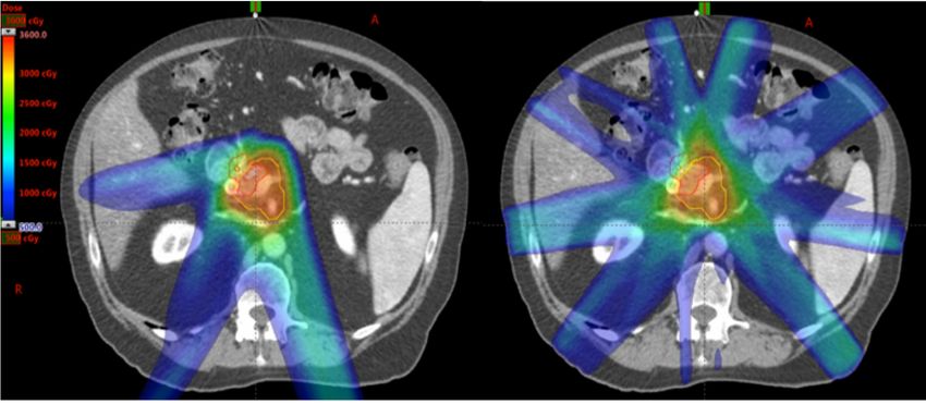
Figure 1. Representative transverse (A), sagittal (B) and coronal (C) images of the reirradiation
Figure 1. Representative transverse (A), sagittal (B) and coronal (C) images of the reirradiation
treatment plan using stereotactic body radiotherapy (SBRT) for a patient who had previous 3D
treatment plan using stereotactic body radiotherapy (SBRT) for a patient who had previous 3D
conformal radiotherapy to the region 5 years ago to 50.4 Gy in 28 fractions. (Red—gross tumor
conformal radiotherapy to the region 5 years ago to 50.4 Gy in 28 fractions. (Red—gross tumor
volume, Dark blue—target for 30 Gy, Light blue—target for 25 Gy due to proximity to surrounding
volume, Dark blue—target for 30 Gy, Light blue—target for 25 Gy due to proximity to surrounding
bowel and stomach). Patient tolerated treatment well with Grade 1 nausea and was alive 12 months
bowel and stomach). Patient tolerated treatment well with Grade 1 nausea and was alive 12 months
post-treatment with with
post‐treatment no significant
no significant late toxicity.
late toxicity.
SBRT is potentially a feasible and safe option in patients with local recurrence. Table 2
SBRT is potentially
summarizes a feasible
the studies where and safe was
SBRT option in patients
utilized in thewith localofrecurrence.
setting Table 2with
local recurrence summarizes
the majority
the studies where SBRT was utilized in the setting of local recurrence with the majority
of publications on patients who received previous radiotherapy. As this group of patients is small, of publications
on patients who received
the evidence is limitedprevious radiotherapy.
to retrospective studiesAs this group
alone. of patients
Nevertheless, fromis Table
small,2,the evidence
SBRT is
can achieve
limited to retrospective studies alone. Nevertheless, from Table 2, SBRT can achieve
good local control (1‐year freedom from local progression of 62–91%). Although the incidence of good local control
(1-year freedom
grade from local
3 or higher progression
late toxicity of great
is rare, 62–91%).care Although
should bethe incidence
taken of grade
at treatment 3 or higher
planning late
and delivery
toxicity is rare, great care should be taken at treatment planning and delivery
with regards to the stomach and bowel dose as gastrointestinal toxicity may cause significant with regards to the
stomach and bowel dose as gastrointestinal toxicity may
morbidity to this group of patients with limited life expectancy. cause significant morbidity to this group of
patients with limited life expectancy.Cancers 2018, 10, 75 6 of 12
Table 2. Summary of studies utilizing SBRT for treatment of local recurrence.
Study Nature Number of Patients Dose/Fractionation Outcomes Toxicity (≥Grade 3)
1-year FFLP: 91%,
2-year FFLP: 82%,
Comito et al., 2017 [20] Retrospective 31, after R0 surgery 45 Gy/6 fractions None
Median PFS: 9 months,
Median OS: 18 months
1-year FFLP: 78%, Acute: GI bleeding (1),
Median dose: 25 Gy (24–36),
Dagoglu et al., 2016 [21] Retrospective 30, reirradiation 2-year FFLP: 78%, Vomiting (1), pain (1),
Median no of fractions: 5 (3–5)
Median OS: 14 months Late: Bowel obstruction (2)
9 patients had 25 Gy/1 fraction; 1-year FFLP: 81%, Acute: Gastric fistula (1),
Koong et al., 2017 [22] Retrospective 23, reirradiation
14 patients had 20–33 Gy/1–5 fractions Median OS: 8.5 months gastric ulcer (1)
Median: 22.5 Gy (20–30), 1-year FFLP: 70%, Late: Bowel obstruction (1),
Lominska et al., 2012 [23] Retrospective 28, reirradiation
Median no of fractions: 3 (3–5) Median OS: 5.9 months gastric perforation (1)
For reirradiated group,
25 out of 51 patients received Acute: Bowel obstruction (1),
Median: 25 Gy 1-year FFLP: 37%,
Ryan et al., 2018 [24] Retrospective reirradiation, Late: Bowel obstruction (2),
No of fractions: 5 fractions Median PFS: 7 months,
all patients had previous surgery GI bleeding (1)
Median OS: 11 months
1-year FFLP: 62%,
Wild et al., 2013 [19] Retrospective 18, reirradiation Median dose: 25 Gy (20–27)/5 fractions Late: Small bowel obstruction (1)
Median OS: 8.8 monthsCancers 2018, 10,
Cancers 2018, 10, 75
x FOR PEER REVIEW 7 of 12
3. Particle
3. Particle Therapy
Therapy for
for Pancreatic
Pancreatic Cancer
Cancer
3.1. Proton Therapy
3.1. Proton Therapy
The proton beam, a particle therapy, has the benefit of delivering dose to the target with no
The proton beam, a particle therapy, has the benefit of delivering dose to the target with no exit
exit dose in the beam path (Figure 2); thereby potentially reducing dose to the normal tissues within
dose in the beam path (Figure 2); thereby potentially reducing dose to the normal tissues within the
the exit beam path [25]. This, theoretically, may reduce both acute and late toxicities of treatment.
exit beam path [25]. This, theoretically, may reduce both acute and late toxicities of treatment. In
In addition, with minimal dose in the exit beam path, proton therapy opens the door to possible
addition, with minimal dose in the exit beam path, proton therapy opens the door to possible dose
dose escalation studies. As proton therapy is a relatively new player in the clinical radiation oncology
escalation studies. As proton therapy is a relatively new player in the clinical radiation oncology
domain, the evidence for the use in pancreatic cancer remains limited. Table 3 summarizes the current
domain, the evidence for the use in pancreatic cancer remains limited. Table 3 summarizes the current
literature for particle therapy in pancreatic cancer.
literature for particle therapy in pancreatic cancer.
A dosimetric study comparing IMRT and proton plans in 13 patients with unresectable pancreatic
A dosimetric study comparing IMRT and proton plans in 13 patients with unresectable
cancer, planned to 55 Gy in 25 fractions, by Thompson et al. [26] demonstrated that proton plans,
pancreatic cancer, planned to 55 Gy in 25 fractions, by Thompson et al. [26] demonstrated that proton
utilizing either passive scattering or pencil beam scanning, yielded significantly lower doses to the
plans, utilizing either passive scattering or pencil beam scanning, yielded significantly lower doses
stomach, duodenum and small bowel in the intermediate to low dose regions (defined as volume
to the stomach, duodenum and small bowel in the intermediate to low dose regions (defined as
receiving at least 20 Gy) compared to IMRT. However, in the intermediate to high dose regions (defined
volume receiving at least 20 Gy) compared to IMRT. However, in the intermediate to high dose
as volume receiving at least 45 Gy), proton plans had significantly higher dose to those structures
regions (defined as volume receiving at least 45 Gy), proton plans had significantly higher dose to
compared to IMRT. However, proton plans did yield significant reduction in mean liver (50% reduction)
those structures compared to IMRT. However, proton plans did yield significant reduction in mean
and kidney (18% reduction) doses. The biological significance of reduced dose to the low dose regions
liver (50% reduction) and kidney (18% reduction) doses. The biological significance of reduced dose
remained to be investigated.
to the low dose regions remained to be investigated.
Figure 2. An
Figure 2. An example
example of
of proton (Left) and
proton (Left) and photon
photon (Right)
(Right) SBRT
SBRT plans
plans on
on the
the same
samepatient.
patient. Note
Note that
that
there is less low dose scatter in the proton plan due to the inherent physical properties of proton
there is less low dose scatter in the proton plan due to the inherent physical properties of proton
particles compared
particles compared to
to photon.
photon.
As proton is a charged particle, it has a higher linear energy transfer [27]. Therefore, proton
As proton is a charged particle, it has a higher linear energy transfer [27]. Therefore, proton
therapy can potentially deliver higher relative biological effectiveness than photon therapy [27]. In
therapy can potentially deliver higher relative biological effectiveness than photon therapy [27].
theory, proton therapy could result in greater cell killing than photon therapy given the same dose/
In theory, proton therapy could result in greater cell killing than photon therapy given the same
fractionation. Although this is a desirable effect on cancer cells, this may also indicate that greater
dose/fractionation. Although this is a desirable effect on cancer cells, this may also indicate that
care is required when delivering proton therapy as a small increase in dose to normal structures could
greater care is required when delivering proton therapy as a small increase in dose to normal structures
potentially translate to greater risk of late toxicity compared to photon therapy. The exact
could potentially translate to greater risk of late toxicity compared to photon therapy. The exact
‘conversion’ of proton therapy dose effect to photon therapy equivalence remained under
‘conversion’ of proton therapy dose effect to photon therapy equivalence remained under investigation.
investigation. As image guidance in proton therapy is being developed and implemented, careful
As image guidance in proton therapy is being developed and implemented, careful planning and
planning and motion management should be taken when delivering proton therapy to a moving
motion management should be taken when delivering proton therapy to a moving target such as
target such as the pancreas.
the pancreas.Cancers 2018, 10, 75 8 of 12
3.2. Carbon Ion Therapy
It has been theorized that heavy ion particle therapy, such as carbon ion therapy, provides greater
biological effect due to even higher linear energy transfer of the particle which translates to greater
relatively biological effectiveness. Currently, there are no carbon ion facilities in North America.
Our Japanese colleagues, Shinoto et al. [28] first reported on the use of carbon ion therapy in patients
with pancreatic cancer. In this phase I dose-escalation trial, a cohort of 26 patients were treated with
pre-operative carbon ion therapy in 8 fractions over 2 weeks (total dose: 30 Gray equivalents [GyE] to
36.8 GyE). Twenty-one patients had surgery, with 19 patients achieving an R0 resection. There was no
local recurrence reported in this cohort and only 2 patients developed regional recurrence. The 5-year
overall survival for this cohort was 42%. Only one patient developed late Grade 4 toxicity with portal
vein stenosis and deranged liver function.
In a cohort of patients with locally advanced pancreatic cancer treated with a dose-escalation
protocol up to 55.2 GyE, Shinoto et al. [29] reported the treatment was well-tolerated and that those
who received ≥45.6 GyE had overall better clinical outcomes in terms of local disease control and
overall survival than those who received lower doses.Cancers 2018, 10, 75 9 of 12
Table 3. Summary of particle therapy studies.
Dose/Fractionation, Concurrent
Study Nature Number of Patients Outcomes Toxicity (≥Grade 3)
Chemotherapy
11 patients did not have surgery,
Out of 48 patients:
PFS: 10.4 months,
OS: 17.3 months, Acute: colitis (1),
Hong et al., 2014 [30] Prospective; Neoadjuvant (Proton) 50 25 Gy/5 fractions, Capecitabine
2-year OS: 42%, chest wall pain (1)
Out of 37 patients who had surgery:
PFS: 14.5 months,
OS: 27 months
2-year PFS: 14%,
OS: 18.4 months,
Sachsman et al., 2014 [31] Prospective; Definitive (Proton) 11 59.4 Gy/33 fractions, Capecitabine None
2-year OS: 31%,
2-year FFLP: 69%
Overall:
1-year PFS: 64.3%,
P1 and P2:,
P1: 50 Gy/25 fractions (5), 1-year OS: 76.8%,
Acute GI bleeding (1),
P2: 70.2 Gy/26 fractions (5), 1-year FFLP: 81.7%,
Terashima et al., 2012 [32] Prospective; Definitive (Proton) 50 P3: GI ulcer treated with
P3: 67.5 Gy/25 fractions (40), P3 patients:
medications (3); death from GI
All with concurrent gemcitabine 1-year PFS: 60.8%,
bleed (1)
1-year OS: 78.8%,
1-year FFLP: 79.9%
No local recurrence,
1-year PFS: 40%, Acute: Liver abscess (1),
Prospective, Phase I, 30–36.8 GyE/5 fractions,
Shinoto et al., 2013 [28] 21 1-year OS: 69%, Late: Deranged liver function
neoadjuvant (Carbon) No concurrent chemotherapy
5-year OS: 42%, due to portal vein stenosis (1)
Median OS: 18.6 months
1-year OS: 73%,
2-year OS: 35%,
43.2–55.2 GyE/12 Acute (non-hematologic):
Shinoto et al., 2016 [29] Prospective, Phase I, LAPC (Carbon) 71 Median OS: 19.6 months,
fractions, Gemcitabine Anorexia (6), GI bleed (1)
Better outcomes in those who had
≥45.6 GyECancers 2018, 10, 75 10 of 12
4. Conclusions
Overall, SBRT and proton therapy are emerging novel radiotherapy techniques/modalities that
could potentially revolutionize treatment for patients with pancreatic cancer. These techniques allow
the gain of local control benefit with minimal toxicity in patients with LAPC whilst minimizing their
time away from optimal effective systemic treatment. With advancements in imaging and radiation
treatment planning, current efforts to improve clinical outcomes in those with LAPC in the realm
of radiation therapy includes dose-escalation trials to maximize local and/or regional control and
MR-guided radiation therapy.
Conflicts of Interest: The authors declare no conflict of interest.
References
1. American Cancer Society. Facts & Figures 2018. 2018. Available online: https://cancerstatisticscenter.cancer.
org/module/yg6E0ZLc (accessed on 21 February 2018).
2. Hammel, P.; Huguet, F.; van Laethem, J.L.; Goldstein, D.; Glimelius, B.; Artru, P.; Borbath, I.; Bouché, O.;
Shannon, J.; André, T.; et al. Effect of Chemoradiotherapy vs. Chemotherapy on Survival in Patients with
Locally Advanced Pancreatic Cancer Controlled after 4 Months of Gemcitabine with or without Erlotinib:
The LAP07 Randomized Clinical Trial. JAMA 2016, 315, 1844–1853. [CrossRef] [PubMed]
3. Iacobuzio-Donahue, C.A.; Fu, B.; Yachida, S.; Luo, M.; Abe, H.; Henderson, C.M.; Vilardell, F.; Wang, Z.;
Keller, J.W.; Banerjee, P.; et al. DPC4 gene status of the primary carcinoma correlates with patterns of failure
in patients with pancreatic cancer. J. Clin. Oncol. 2009, 27, 1806–1813. [CrossRef] [PubMed]
4. Krishnan, S.; Chadha, A.S.; Suh, Y.; Chen, H.C.; Rao, A.; Das, P.; Minsky, B.D.; Mahmood, U.;
Delclos, M.E.; Sawakuchi, G.O.; et al. Focal Radiation Therapy Dose Escalation Improves Overall Survival
in Locally Advanced Pancreatic Cancer Patients Receiving Induction Chemotherapy and Consolidative
Chemoradiation. Int. J. Radiat. Oncol. Biol. Phys. 2016, 94, 755–765. [CrossRef] [PubMed]
5. Stauder, M.C.; Miller, R.C. Stereotactic Body Radiation Therapy (SBRT) for Unresectable Pancreatic
Carcinoma. Cancers 2010, 2, 1565–1575. [CrossRef] [PubMed]
6. Schellenberg, D.; Goodman, K.A.; Lee, F.; Chang, S.; Kuo, T.; Ford, J.M.; Fisher, G.A.; Quon, A.; Desser, T.S.;
Norton, J.; et al. Gemcitabine chemotherapy and single-fraction stereotactic body radiotherapy for locally
advanced pancreatic cancer. Int. J. Radiat. Oncol. Biol. Phys. 2008, 72, 678–686. [CrossRef] [PubMed]
7. Koong, A.C.; Christofferson, E.; Le, Q.T.; Goodman, K.A.; Ho, A.; Kuo, T.; Ford, J.M.; Fisher, G.A.; Greco, R.;
Norton, J.; et al. Phase II study to assess the efficacy of conventionally fractionated radiotherapy followed by
a stereotactic radiosurgery boost in patients with locally advanced pancreatic cancer. Int. J. Radiat. Oncol.
Biol. Phys. 2005, 63, 320–323. [CrossRef] [PubMed]
8. Herman, J.M.; Chang, D.T.; Goodman, K.A.; Dholakia, A.S.; Raman, S.P.; Hacker-Prietz, A.;
Iacobuzio-Donahue, C.A.; Griffith, M.E.; Pawlik, T.M.; Pai, J.S.; et al. Phase 2 multi-institutional trial
evaluating gemcitabine and stereotactic body radiotherapy for patients with locally advanced unresectable
pancreatic adenocarcinoma. Cancer 2015, 121, 1128–1137. [CrossRef] [PubMed]
9. Chang, D.T.; Schellenberg, D.; Shen, J.; Kim, J.; Goodman, K.A.; Fisher, G.A.; Ford, J.M.; Desser, T.; Quon, A.;
Koong, A.C. Stereotactic radiotherapy for unresectable adenocarcinoma of the pancreas. Cancer 2009, 115,
665–672. [CrossRef] [PubMed]
10. Chuong, M.D.; Springett, G.M.; Freilich, J.M.; Park, C.K.; Weber, J.M.; Mellon, E.A.; Hodul, P.J.; Malafa, M.P.;
Meredith, K.L.; Hoffe, S.E.; et al. Stereotactic body radiation therapy for locally advanced and borderline
resectable pancreatic cancer is effective and well tolerated. Int. J. Radiat. Oncol. Biol. Phys. 2013, 86, 516–522.
[CrossRef] [PubMed]
11. Comito, T.; Cozzi, L.; Clerici, E.; Franzese, C.; Tozzi, A.; Iftode, C.; Navarria, P.; D’Agostino, G.; Rimassa, L.;
Carnaghi, C.; et al. Can Stereotactic Body Radiation Therapy Be a Viable and Efficient Therapeutic Option
for Unresectable Locally Advanced Pancreatic Adenocarcinoma? Results of a Phase 2 Study. Technol. Cancer
Res. Treat. 2017, 16, 295–301. [CrossRef] [PubMed]
12. Gurka, M.K.; Kim, C.; He, A.R.; Charabaty, A.; Haddad, N.; Turocy, J.; Johnson, L.; Jackson, P.; Weiner, L.M.;
Marshall, J.L.; et al. Stereotactic Body Radiation Therapy (SBRT) Combined with Chemotherapy for
Unresected Pancreatic Adenocarcinoma. Am. J. Clin. Oncol. 2017, 40, 152–157. [CrossRef] [PubMed]Cancers 2018, 10, 75 11 of 12
13. Mahadevan, A.; Miksad, R.; Goldstein, M.; Sullivan, R.; Bullock, A.; Buchbinder, E.; Pleskow, D.; Sawhney, M.;
Kent, T.; Vollmer, C.; et al. Induction gemcitabine and stereotactic body radiotherapy for locally advanced
nonmetastatic pancreas cancer. Int. J. Radiat. Oncol. Biol. Phys. 2011, 81, e615–e622. [CrossRef] [PubMed]
14. Mellon, E.A.; Hoffe, S.E.; Springett, G.M.; Frakes, J.M.; Strom, T.J.; Hodul, P.J.; Malafa, M.F.; Chuong, M.D.;
Shridhar, R. Long-term outcomes of induction chemotherapy and neoadjuvant stereotactic body radiotherapy
for borderline resectable and locally advanced pancreatic adenocarcinoma. Acta Oncol. 2015, 54, 979–985.
[CrossRef] [PubMed]
15. Pollom, E.L.; Alagappan, M.; von Eyben, R.; Kunz, P.L.; Fisher, G.A.; Ford, J.A.; Poultsides, G.A.; Visser, B.C.;
Norton, J.A.; Kamaya, A.; et al. Single-versus multifraction stereotactic body radiation therapy for pancreatic
adenocarcinoma: Outcomes and toxicity. Int. J. Radiat. Oncol. Biol. Phys. 2014, 90, 918–925. [CrossRef]
[PubMed]
16. Tozzi, A.; Comito, T.; Alongi, F.; Navarria, P.; Iftode, C.; Mancosu, P.; Reggiori, G.; Clerici, E.; Rimassa, L.;
Zerbi, A.; et al. SBRT in unresectable advanced pancreatic cancer: Preliminary results of a mono-institutional
experience. Radiat. Oncol. 2013, 8, 148. [CrossRef] [PubMed]
17. Rwigema, J.C.; Parikh, S.D.; Heron, D.E.; Howell, M.; Zeh, H.; Moser, A.J.; Bahary, N.; Quinn, A.; Burton, S.A.
Stereotactic body radiotherapy in the treatment of advanced adenocarcinoma of the pancreas. Am. J.
Clin. Oncol. 2011, 34, 63–69. [CrossRef] [PubMed]
18. Myrehaug, S.; Sahgal, A.; Russo, S.M.; Lo, S.S.; Rosati, L.M.; Mayr, N.A.; Lock, M.; Small, W., Jr.; Dorth, J.A.;
Ellis, R.J.; et al. Stereotactic body radiotherapy for pancreatic cancer: Recent progress and future directions.
Expert Rev. Anticancer Ther. 2016, 16, 523–530. [CrossRef] [PubMed]
19. Wild, A.T.; Hiniker, S.M.; Chang, D.T.; Tran, P.T.; Khashab, M.A.; Limaye, M.R.; Laheru, D.A.; Le, D.T.;
Kumar, R.; Pai, J.S.; et al. Re-irradiation with stereotactic body radiation therapy as a novel treatment
option for isolated local recurrence of pancreatic cancer after multimodality therapy: Experience from two
institutions. J. Gastrointest. Oncol. 2013, 4, 343–351. [CrossRef] [PubMed]
20. Comito, T.; Cozzi, L.; Zerbi, A.; Franzese, C.; Clerici, E.; Tozzi, A.; Iftode, C.; Navarria, P.; D’Agostino, G.;
Fogliata, A.; et al. Clinical results of stereotactic body radiotherapy (SBRT) in the treatment of isolated local
recurrence of pancreatic cancer after R0 surgery: A retrospective study. Eur. J. Surg. Oncol. 2017, 43, 735–742.
[CrossRef] [PubMed]
21. Dagoglu, N.; Callery, M.; Moser, J.; Tseng, J.; Kent, T.; Bullock, A.; Miksad, R.; Mancias, J.D.; Mahadevan, A.
Stereotactic Body Radiotherapy (SBRT) Reirradiation for Recurrent Pancreas Cancer. J. Cancer 2016, 7,
283–288. [CrossRef] [PubMed]
22. Koong, A.J.; Toesca, D.A.S.; von Eyben, R.; Pollom, E.L.; Chang, D.T. Reirradiation with stereotactic
body radiation therapy after prior conventional fractionation radiation for locally recurrent pancreatic
adenocarcinoma. Adv. Radiat. Oncol. 2017, 2, 27–36. [CrossRef] [PubMed]
23. Lominska, C.E.; Unger, K.; Nasr, N.M.; Haddad, N.; Gagnon, G. Stereotactic body radiation therapy for
reirradiation of localized adenocarcinoma of the pancreas. Radiat. Oncol. 2012, 7, 74. [CrossRef] [PubMed]
24. Ryan, J.F.; Groot, V.P.; Rosati, L.M.; Hacker-Prietz, A.; Narang, A.K.; McNutt, T.R.; Jackson, J.F.; Le, D.T.;
Jaffee, E.M.; Zheng, L.; et al. Stereotactic Body Radiation Therapy for Isolated Local Recurrence After Surgical
Resection of Pancreatic Ductal Adenocarcinoma Appears to be Safe and Effective. Ann. Surg. Oncol. 2018, 25,
280–289. [CrossRef] [PubMed]
25. Mendenhall, N.P.; Malyapa, R.S.; Su, Z.; Yeung, D.; Mendenhall, W.M.; Li, Z. Proton therapy for head
and neck cancer: Rationale, potential indications, practical considerations, and current clinical evidence.
Acta Oncol. 2011, 50, 763–771. [CrossRef] [PubMed]
26. Thompson, R.F.; Mayekar, S.U.; Zhai, H.; Both, S.; Apisarnthanarax, S.; Metz, J.M.; Plastaras, J.P.; Ben-Josef, E.
A dosimetric comparison of proton and photon therapy in unresectable cancers of the head of pancreas.
Med. Phys. 2014, 41, 081711. [CrossRef] [PubMed]
27. Paganetti, H. Relative biological effectiveness (RBE) values for proton beam therapy. Variations as a function
of biological endpoint, dose, and linear energy transfer. Phys. Med. Biol. 2014, 59, R419–R472. [CrossRef]
[PubMed]
28. Shinoto, M.; Yamada, S.; Yasuda, S.; Imada, H.; Shioyama, Y.; Honda, H.; Kamada, T.; Tsujii, H.; Saisho, H.;
Working Group for Pancreas Cancer. Phase 1 trial of preoperative, short-course carbon-ion radiotherapy for
patients with resectable pancreatic cancer. Cancer 2013, 119, 45–51. [CrossRef] [PubMed]Cancers 2018, 10, 75 12 of 12
29. Shinoto, M.; Yamada, S.; Terashima, K.; Yasuda, S.; Shioyama, Y.; Honda, H.; Kamada, T.; Tsujii, H.; Saisho, H.;
Working Group for Pancreas Cancer. Carbon Ion Radiation Therapy with Concurrent Gemcitabine for
Patients with Locally Advanced Pancreatic Cancer. Int. J. Radiat. Oncol. Biol. Phys. 2016, 95, 498–504.
[CrossRef] [PubMed]
30. Hong, T.S.; Ryan, D.P.; Borger, D.R.; Blaszkowsky, L.S.; Yeap, B.Y.; Ancukiewicz, M.; Deshpande, V.;
Shinagare, S.; Wo, J.Y.; Boucher, Y.; et al. A phase 1/2 and biomarker study of preoperative short course
chemoradiation with proton beam therapy and capecitabine followed by early surgery for resectable
pancreatic ductal adenocarcinoma. Int. J. Radiat. Oncol. Biol. Phys. 2014, 89, 830–838. [CrossRef] [PubMed]
31. Sachsman, S.; Nichols, R.C.; Morris, C.G.; Zaiden, R.; Johnson, E.A.; Awad, Z.; Bose, D.; Ho, M.W.; Huh, S.N.;
Li, Z.; et al. Proton Therapy and Concomitant Capecitabine for Non-Metastatic Unresectable Pancreatic
Adenocarcinoma. Int. J. Part. Ther. 2014, 1, 692–701. [CrossRef]
32. Terashima, K.; Demizu, Y.; Hashimoto, N.; Jin, D.; Mima, M.; Fujii, O.; Niwa, Y.; Takatori, K.; Kitajima, N.;
Sirakawa, S.; et al. A phase I/II study of gemcitabine-concurrent proton radiotherapy for locally advanced
pancreatic cancer without distant metastasis. Radiother. Oncol. 2012, 103, 25–31. [CrossRef] [PubMed]
© 2018 by the authors. Licensee MDPI, Basel, Switzerland. This article is an open access
article distributed under the terms and conditions of the Creative Commons Attribution
(CC BY) license (http://creativecommons.org/licenses/by/4.0/).You can also read