Abdominal Compartment Syndrome in a Patient Suspected of Coronavirus 2019 (COVID-19)
←
→
Page content transcription
If your browser does not render page correctly, please read the page content below
ISSN: 2474-3658
                                                                                               Shah et al. J Infect Dis Epidemiol 2020, 6:139
                                                                                                         DOI: 10.23937/2474-3658/1510139
                                                                                                                           Volume 6 | Issue 4
                  Journal of                                                                                                    Open Access

                  Infectious Diseases and Epidemiology
Case Report

Abdominal Compartment Syndrome in a Patient Suspected of
Coronavirus 2019 (COVID-19)
Rony Shah, MD*, Linda Klumpp, MD, Naeem Syed, DO, Parth Patel, MD and Jeffrey                                                    Check for
                                                                                                                                 updates
Jordan, MD
Healthcare/USF Morsani College of Medicine, GME Programs at Citrus Memorial Hospital, Inverness, Florida, USA

*Corresponding author: Rony Shah, MD, Healthcare/USF Morsani College of Medicine, GME Programs at Citrus Memorial
Hospital, Inverness, Florida, USA

 Abstract                                                              a worldwide pandemic with more than 2.5 million cas-
                                                                       es reported and more than 175,000 confirmed deaths
 Severe acute respiratory syndrome coronavirus (SARS-
 CoV-2) is a novel coronavirus which has become a world-
                                                                       [1]. The first case was reported in early December 2019
 wide pandemic, the other common coronaviruses to cause                in Wuhan, China and the first case in the United States
 deadly respiratory disease are SARS-CoV and MERS-CoV.                 was reported on January, 22, 2019 in the state of Wash-
 SARS-CoV-2, SARS-CoV, and MERS-CoV are commonly                       ington [2-4]. The coronaviruses are enveloped RNA vi-
 transmitted through respiratory droplets and direct contact.
 Patients with SARS-CoV-2 can present with varying sever-
                                                                       ruses that are found in humans, mammals, and other
 ity from asymptomatic to multi-organ failure. We present a            birds and they can cause respiratory, hepatic, enteric,
 rare case of abdominal compartment syndrome in a patient              and neurologic diseases [3]. The other two strains of the
 suspected of coronavirus 2019 (COVID-19).                             coronavirus, severe acute respiratory syndrome corona-
 A 59-year-old male presented to the emergency room (ER)               virus (SARS-CoV) and Middle East respiratory syndrome
 complaining of abdominal pain, urinary retention, and ab-             coronavirus (MERS-CoV) have been known to cause fa-
 dominal distension. He gave a past medical history of hy-
 pertension, nephrolithiasis, and recent outpatient treatment
                                                                       tal disease as well [3]. SARS-CoV was responsible for the
 for acute bronchitis. The following day patient developed             outbreaks in China in 2002 and 2003 [3]. MERS-CoV was
 fever, altered mental status, and shortness of breath. Pa-            the pathogen that caused severe respiratory disease in
 tient underwent surgery and was found to have abdominal               the Middle East in 2012 [3]. SARS-CoV-2, SARS-CoV, and
 compartment syndrome. Daily chest X-ray demonstrated
 worsening bilateral pulmonary infiltrates concerning for an
                                                                       MERS-CoV are commonly transmitted through respira-
 underlying COVID-19 infection. Patient’s wife was positive            tory droplets and direct contact [5]. We present a rare
 for COVID-19.                                                         case of abdominal compartment syndrome in a patient
 Coronaviruses are known to cause respiratory or intestinal            suspected of coronavirus 2019 (COVID-19).
 infections in humans and animals. Patient’s onset of symp-
 toms is about four to five days after being infected. Our pa-         Case Description
 tient may have mistakenly been diagnosed with bronchitis                  A 59-year-old male presented to the emergency
 instead of COVID-19 prior to his hospitalization. The com-
 bination of a COVID-19 infection with abdominal compart-              room (ER) complaining of abdominal pain, urinary reten-
 ment syndrome gave our patient a poor prognosis. We are               tion, and abdominal distension. He gave a past medical
 hoping our case report will be able to bring more awareness           history of hypertension and nephrolithiasis. Patient re-
 and highlight a rare presentation of COVID-19.                        ported worsening abdominal distension with decreased
                                                                       urination and bowel movements over the past five days.
Introduction                                                           He took over-the-counter laxatives to help with his con-
   Severe acute respiratory syndrome coronavirus                       stipation but had minor improvement in bowel move-
(SARS-CoV-2) is a novel coronavirus which has become                   ments, his last bowel movement was four days ago. Pa-

                                       Citation: Shah R, Klumpp L, Syed N, Patel P, Jordan J (2020) Abdominal Compartment Syndrome in a Pa-
                                       tient Suspected of Coronavirus 2019 (COVID-19). J Infect Dis Epidemiol 6:139. doi.org/10.23937/2474-
                                       3658/1510139
                                       Received: May 08, 2020: Accepted: July 01, 2020: Published: July 03, 2020
                                       Copyright: © 2020 Shah R, et al. This is an open-access article distributed under the terms of the
                                       Creative Commons Attribution License, which permits unrestricted use, distribution, and reproduction
                                       in any medium, provided the original author and source are credited.

Shah et al. J Infect Dis Epidemiol 2020, 6:139                                                                             • Page 1 of 4 •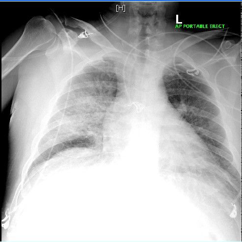
DOI: 10.23937/2474-3658/1510139 ISSN: 2474-3658
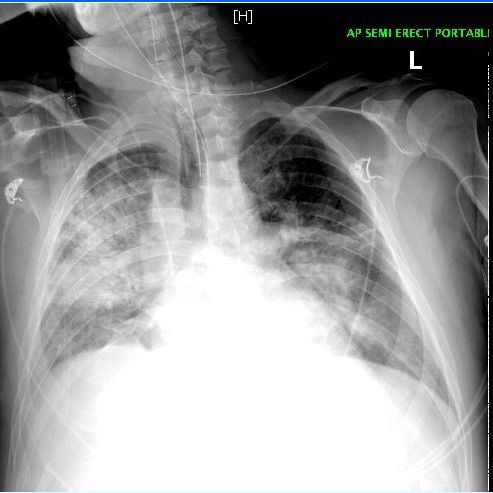
Figure 1: Chest X-ray revealed poor inspiratory effort with Figure 2: Chest X-ray demonstrated increased bilateral
no evidence of acute pathology. interstitial and alveolar opacities with findings suspicious
for pneumonia or pulmonary edema.
tient went to his primary care physician two days prior
to coming to the ER and was prescribed doxycycline for nary edema or infiltrates, hepatic steatosis, mild sple-
bronchitis. Vitals were temperature: 98.1 F, blood pres- nomegaly, bilateral nonobstructing nephrolithiasis, and
sure: 151/88 mmHg, heart rate: 119 beats per minute, colonic diverticulosis. Chest X-ray demonstrated in-
respiratory rate: 16 breaths per minute, oxygen satura- creased bilateral interstitial and alveolar opacities with
tion: 97%. Physical examination revealed a distended findings suspicious for pneumonia or pulmonary edema
abdomen with diffuse tenderness. Laboratory results (Figure 2). Patient underwent a diagnostic laparoscopy
were significant for white blood count (WBC): 39.6, he- for suspected ischemic bowel which revealed a signif-
moglobin (Hg): 13.6, platelet: 16, blood urea nitrogen icant amount of pressure in the abdomen which limit-
(BUN)/creatinine: 36 mg/dL/2.1 mg/dL, aspartate ami- ed visualization. There were no signs of necrotic bowel,
notransferase (AST): 137 U/L, alanine aminotransferase perforation, abdominal fluid, or any other pathology.
(ALT): 281 U/L, alkaline phosphatase: 121 U/L, amylase: The peak airway pressure remained greater than 40
38 U/L, lipase: 25 U/L, lactic acid: 1.91 mmol/L. Urinaly- mmHg and the small bowel appeared pale on diagnostic
sis was negative. Chest X-ray revealed poor inspiratory laparoscopy so the surgery was converted to an explor-
effort with no evidence of acute pathology (Figure 1). atory laparotomy. The small bowel initially appeared to
Right upper quadrant ultrasound demonstrated hep- be pale but became pink with adequate perfusion and
atomegaly with fatty infiltration. Computed tomogra- an intact mesenteric pulse following the decompressive
phy (CT) of abdomen and pelvis revealed possible acute laparotomy. The combination of elevated peak airway
pancreatitis, a 5 mm calculous at the right ureteropel- pressure and improvement of the bowel appearance
vic junction without any evidence of hydronephrosis. following the decompressive laparotomy, abdominal
Patient was started on ceftriaxone and metronidazole compartment syndrome was suspected. Due to the el-
for empiric coverage. The next day he developed fever, evated intra-abdominal pressure it was left open with
altered mental status, shortness of breath, worsening an open abdomen negative pressure therapy wound
tachycardia, and severe abdominal distention with hy- vacuum-assisted closure (VAC) device. The peak airway
peractive bowel sounds. Patient was transferred to the pressure improved to 25 mmHg at the end of the pro-
medical intensive care unit (ICU) for closer monitoring. cedure but did not return to normal. Repeat chest X-ray
Patient was being treated for severe sepsis with aggres- following the procedure demonstrated worsening bilat-
sive fluid resuscitation and empiric antibiotic coverage. eral consolidative airspace opacity since the previous
The source of infection was unknown but there was a chest X-ray (Figure 3). Antibiotic coverage was changed
high suspicion for intra-abdominal pathology. His anti- to vancomycin and meropenem following the proce-
biotic coverage was changed to meropenem and met- dure. Patient was hemodynamically unstable following
ronidazole for broader coverage. Patient was evaluated the procedure requiring mechanical ventilation, vaso-
for his thrombocytopenia present since admission and pressor support, and abdominal wound VAC. Patient
it was believed to be secondary to early disseminated went into cardiac arrest with the initial rhythm being
intravascular coagulation (DIC). Repeat CT abdomen/ pulse electrical activity (PEA) and immediate advanced
pelvis demonstrated development of bilateral pulmo- cardiovascular life support treatment was started. The
Shah et al. J Infect Dis Epidemiol 2020, 6:139 • Page 2 of 4 •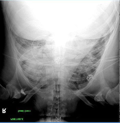
DOI: 10.23937/2474-3658/1510139                                                                         ISSN: 2474-3658

                                                             intestinal infections in humans and animals [6]. SARS-
                                                             CoV-2 virus is related to the bat coronaviruses and to
                                                             SARS-CoV-1, it enters human cells through the angio-
                                                             tensin-converting-enzyme 2 (ACE2) receptors [7]. SARS-
                                                             CoV-2 virus is primarily spread from person to person
                                                             through respiratory droplets, which are typically re-
                                                             leased by an infected person coughing or sneezing [7].
                                                             Transmission typically does not occur through inhala-
                                                             tion of aerosols, which are virions suspended in the air,
                                                             but the virus may linger in aerosols for more than three
                                                             hours which increases the risk of exposure [7]. Our pa-
                                                             tient’s wife was at bedside throughout his hospitaliza-
                                                             tion which increased her risk of exposure. SARS-CoV-2
                                                             RNA has been detected in blood and stool, but oral fe-
                                                             cal route has not been documented [7]. Reports suggest
                                                             that patients may be infectious one to three days before
                                                             the onset of symptoms and up to 40 to 50% of cases are
                                                             due to transmission from asymptomatic or presymp-
  Figure 3: Repeat chest X-ray following the procedure       tomatic patients [7]. The time period from exposure to
  demonstrated worsening bilateral consolidative airspace    symptom onset is approximately four to five days and
  opacity since the previous chest X-ray.                    97.5% of symptomatic patients will have symptoms
                                                             within 11.5 days after infection [7]. Common symptoms
                                                             are fever, cough, sore throat, malaise, myalgias, loss
                                                             of smell, loss of taste, and gastrointestinal symptoms
                                                             such as anorexia, nausea, and diarrhea [7]. Shortness
                                                             of breath may develop five to eight days after onset of
                                                             the initial symptoms [7]. Our patient may have been
                                                             mistakenly diagnosed with bronchitis prior to his hos-
                                                             pitalization. His symptoms of bronchitis and abdominal
                                                             pain in addition to developing altered mental status, fe-
                                                             ver, and shortness of breath during his hospitalization,
                                                             within five to eight days of his initial symptoms makes a
                                                             SARS-CoV-2 infection very likely. Risk factors associated
                                                             with COVID-19 complications are age (> 65), cardiovas-
                                                             cular disease, chronic lung disease, hypertension, dia-
                                                             betes, and obesity [7]. Other conditions with presumed
                                                             increased risk of complications are kidney disease, im-
                                                             munosuppression, malignancy, and uncontrolled hu-
                                                             man immunodeficiency virus (HIV) [7]. In our case the
                                                             patient’s only risk factor was hypertension. The diagno-
  Figure 4: Repeat chest X-ray prior to the cardiac arrest   sis of COVID-19 is made by detection of SARS-CoV-2 by
  demonstrated worsening bilateral airspace opacities sug-   polymerase-chain-reaction (PCR) assay [7]. Evaluation
  gestive of pulmonary edema or pulmonary infiltrates.
                                                             and management of COVID-19 is guided by the severity
                                                             of the disease. Severe disease was defined as marked
rhythm converted to Torsades and eventually asysto-          tachypnea (respiratory rate > 30 breaths per minute),
le. Patient was unable to be resuscitated and bedside        hypoxemia (Oxygen saturation < 93%), ratio of partial
ultrasound confirmed cardiac standstill. Repeat chest        pressure of arterial oxygen to fraction of inspired oxy-
X-ray prior to the cardiac arrest demonstrated worsen-       gen (< 300), and lung infiltrates (> 50% of the lung field
ing bilateral airspace opacities suggestive of pulmonary     within 24 to 48 hours) [7]. Our patient had severe dis-
edema or pulmonary infiltrates (Figure 4). On further        ease exacerbated by his underlying abdominal compart-
review of the chest X-ray the findings may be suggestive     ment syndrome. A case series by Bhatraju, et al. found
of SARS-CoV-2 disease. There was increased suspicion         that 96% of ICU patient’s chest radiographs showing
of this because the patient’s wife presented with SARS-      bilateral pulmonary opacities [2]. In our case the pa-
CoV-2 symptoms two days after his death requiring hos-       tient’s chest X-ray showed worsening bilateral pulmo-
pitalization. She tested positive for SARS-CoV-2.            nary opacities daily. Hypotension requiring vasopressor
Discussion                                                   support in patients without a secondary infection was
                                                             found in 71% of the patients reported in a case series
    Coronaviruses are known to cause respiratory or          by Bhatraju, et al. [2]. In our case vasopressor support

Shah et al. J Infect Dis Epidemiol 2020, 6:139                                                          • Page 3 of 4 •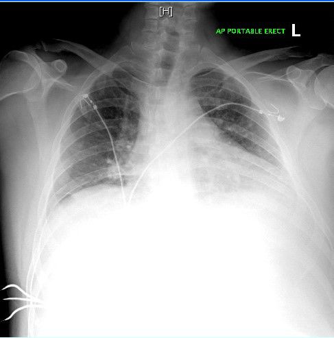
DOI: 10.23937/2474-3658/1510139                                                                                 ISSN: 2474-3658

was needed to maintain a mean arterial pressure > 65          severity, and complications associated with the disease.
mmHg. A recent study showed that critical patients with       Focusing on treating other medical conditions such as
COVID-19 showed signs of organ dysfunction, including         cardiovascular disease, renal disease, gastrointestinal
ARDS in 67%, acute kidney injury in 29%, cardiac inju-        disease, and coagulopathy can lead to a missed diag-
ry in 23%, and liver dysfunction in 29% [6]. Our patient      nosis of COVID-19 as seen in our patient. Recent data
exhibited multi-organ dysfunction such as acute kidney        is suggesting that increased mortality rate of all medi-
injury, liver dysfunction, hypoxemic respiratory failure,     cal conditions may have been driven by an underlying
and eventual cardiac injury. Viral infections may trigger     COVID-19 infection. We hope our case report will bring
arrhythmias due to a combination of significant system-       more awareness and highlight a rare presentation of
ic inflammatory response and vascular inflammation            COVID-19.
at the arterial plaque level [6]. Our patient went into
sudden cardiac arrest for multiple reasons but one of
                                                              Consent
the main reasons could be his underlying COVID-19                The patient provided informed consent.
infection. As reported by Shi, et al. cardiac injury is a
common condition among hospitalized patients with
                                                              Disclaimer
COVID-19 and is associated with a higher risk of in hos-          This research was supported in part by HCA Health-
pital mortality [8]. Another study reported that 44% of       care and/or an HCA Healthcare affiliated entity. The
patients with COVID-19 were transferred to the ICU due        views expressed in this publication represent those of
to arrhythmias [6].                                           the author(s) and do not necessarily represent the offi-
                                                              cial views of HCA Healthcare or any of its affiliated en-
    Our patient had abdominal compartment syndrome
                                                              tities.
due to an unknown etiology except for possible severe
septic shock. His abdominal compartment syndrome              References
may have contributed to his ventilation defects, de-          1. Center for Disease Control and Prevention (2020) Corona-
creased cardiac output, oliguria, and altered mental             virus (COVID-19).
status [9,10]. The pathogenesis for abdominal com-
                                                              2. Bhatraju P, Ghassemieh B, Nichols M, Kim R, Jerome KR,
partment syndrome due to shock occurs from vasocon-              et al. (2020) COVID-19 in critically ill patients in the Seattle
striction mediated by the sympathetic nervous system             region- case series. N Engl J Med 382: 2012-2022.
leading to decreased blood flow to the skin, muscles,         3. Zhu N, Zhang D, Wang W, Li X, Yang B, et al. (2020) A
kidneys, and gastrointestinal tract to preserve blood            novel coronavirus from patients with pneumonia in China,
flow to the brain and heart [10]. Decreased splanchnic           2019. N Engl J Med 382: 727-733.
circulation and intestinal tissue hypoxia is associated       4. Holshue M, DeBolt C, Lindquist S, Lofy K, Wiesman J, et
with three critical factors (cytokine release, free radical      al. (2020) First case of 2019 novel coronavirus in the United
formation, and decreased ATP production) which lead              States. N Engl J Med 382: 929-936.
to intra-abdominal hypertension and abdominal com-            5. Guan W, Zheng-yi N, Hu Y, Liang W, Ou C, et al. (2020)
partment syndrome [10]. It is possible that our patient’s        Clinical characteristics of coronavirus disease 2019 in Chi-
symptoms may have been exacerbated by his underly-               na. N Engl J Med 382: 1708-1720.
ing abdominal compartment syndrome but the over-              6. Madjid M, Safavi-Naeini P, Solomon S, Vardeny O (2020)
whelming evidence makes a COVID-19 infection very                Potential effects of coronavirus on the cardiovascular sys-
                                                                 tem. JAMA Cardiol, E1-E10.
likely. To the best of our knowledge no other cases of
coronavirus with abdominal compartment syndrome               7. Gandhi R, Lynch J, Rio C (2020) Mild or moderate
have been previously reported.                                   COVID-19. N Engl J Med.
                                                              8. Shi S, Qin M, Shen B, Cai Y, Liu T, et al. (2020) Association
Conclusion                                                       of cardiac injury with mortality in hospitalized patients with
                                                                 COVID-19 in Wuhan, China. JAMA Cardiol, E1-E8.
   The SARS-CoV-2 virus has become one of the most
deadly worldwide pandemics in recent history. Ev-             9. Rogers W, Garcia L (2018) Intrabdominal hypertension, ab-
                                                                 dominal compartment syndrome, and the open abdomen.
ery day we are gaining a deeper understanding of the
                                                                 CHEST 153: 238-250.
symptoms and diseases associated with COVID-19 pa-
tients. Physicians need to be able to recognize unusual       10. Pereira B (2019) Abdominal compartment syndrome and
                                                                  intra-abdominal hypertension. Curr Opin Crit Care 25: 688-
presentations of COVID-19 because at this time we do              696.
not have enough information to predict clinical course,

Shah et al. J Infect Dis Epidemiol 2020, 6:139                                                                  • Page 4 of 4 •
You can also read