Non-union of Intertrochanteric Fracture Right Femur with Limb Shortening
←
→
Page content transcription
If your browser does not render page correctly, please read the page content below
International Journal of Medical and Pharmaceutical
                            Case Reports

                            13(1): 7-15, 2020; Article no.IJMPCR.55942
                            ISSN: 2394-109X, NLM ID: 101648033

Non-union of Intertrochanteric Fracture Right Femur
                               with Limb Shortening
     Htay Lwin1*, Htoo Htoo Kyaw Soe2, Adinegara Lutfi Abas2, Mila Nu Nu Htay2,
                                             Kaung Khant Zaw3 and Soe Moe2
         1
          Department of Community Medicine, Family Medicine Unit, Melaka-Manipal Medical College,
                                         Manipal Academy of Higher Education, Melaka, Malaysia.
                            2
                             Department of Community Medicine, Melaka-Manipal Medical College,
                                         Manipal Academy of Higher Education, Melaka, Malaysia.
                                                       3
                                                        Orthopedic Hospital, Mandalay, Myanmar.

                                                                                              Authors’ contributions

  This work was carried out in collaboration among all authors. Author HL did substantial contributions
 to conception and design, acquisition of data, drafting the article, performed the analysis of the study,
   wrote the protocol, wrote the first draft of the manuscript, revised it critically for important intellectual
         content, final approval of the version to be published. Authors HHKS, ALA and MNNH equally
  managed the analyses of the study and equally contributed the literature searches. Authors KKZ and
     SM equally managed the literature searches. All authors read and approved the final manuscript.

                                                                                                   Article Information

                                                                                   DOI: 10.9734/IJMPCR/2020/v13i130111
                                                                                                                   Editor(s):
                                                            (1) Dr. Hayrettin Ozturk, Abant Izzet Baysal University, Turkey.
                                                                                                                Reviewers:
                                                                      (1) Mohit Kumar Patralekh, GGS IP University, India.
                                                                        (2) Preksha Barot, GMERS Medical College, India.
                                                             (3) Kastanis Grigorios, General Hospital of Heraklion, Greece.
                                             Complete Peer review History: http://www.sdiarticle4.com/review-history/55942

                                                                                        Received 08 February 2020
                                                                                           Accepted 13 April 2020
   Case Report
                                                                                           Published 21 April 2020

ABSTRACT

 Nonunion of intertrochanteric fracture is uncommon because there are excellent blood supply and
 good cancellous bone in the intertrochanteric region of the femur. A diagnosis of primary
 intertrochanteric nonunion is made when at least 15 weeks after the fracture there is radiological
 evidence of a fracture line, with either no callus (atrophic) or with callus that does not bridge the
 fracture site (hypertrophic). The patient was subsequently treated successfully with an open
 reduction procedure. Open reduction and internal fixation with right DHS (Dynamic Hip Screw) with
 8 screw holes plate inserted first and then 6 screws inserted under the guidance of Image Intensifier
 (C Arm). We would like to report a case of right intertrochanteric fracture with non-union. A 60 years
_____________________________________________________________________________________________________

*Corresponding author: E-mail: drlwin2003@gmail.com;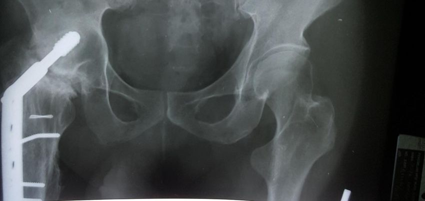
Lwin et al.; IJMPCR, 13(1): 7-15, 2020; Article no.IJMPCR.55942

old patient presented with the history of pain in the right hip, off and on for many years after the
operation. The pain was associated with stiffness of the right hip joint. This case is selected for
reporting because it’s a relatively rare incidence.

Keywords: Hip disorder; intertrochanteric fracture; hip pain; old age.

1. INTRODUCTION                                            He lived with his daughter. Physical examination
                                                           was done in the A&E department immediately.
The incidence of hip fracture has been increasing          GCS was full, 15/15, and no cervical spine
with the aging population and about 50% of hip             tenderness. Pelvic spring test was positive, chest
fracture are intertrochanteric fracture [1].               and clavicle spring test was negative. The patient
                                                           was afebrile, BP 100/60, P/R 78/min, SpO2 95%
Nonunion of intertrochanteric fracture is                  on air. In local examination; tenderness was
uncommon because there are excellent blood                 present at the right hip, no other bony
supply and good cancellous bone in the                     tenderness, peripheral pulses were palpable,
intertrochanteric region of the femur [2]. A               sensation intact, capillary refill time (CRT) > 2
diagnosis of primary intertrochanteric nonunion is         sec, no foot drop was found. The respiratory
made when at least 15 weeks after the fracture             system was normal and equal air entry, vesicular
there is radiological evidence of a fracture line,         breath sound was heard. The cardiovascular
with either no callus (atrophic) or with callus that       system was dual rhythm, no murmur. In
does not bridge the fracture site (hypertrophic)           abdominal examination, liver and spleen were
[3]. Most intertrochanteric fractures treated by           not palpable and there was no tenderness. The
conservative methods or internal fixation heal [4,         right lower limb was examined and saw
5]. Occasionally, nonunion or early failure of             shortening gait, limb length shortening of 6 cm
fracture fixation occurs, the reasons being                with restricted ROM (Range of movement) right
delayed treatment, unfavorable fracture patterns,          hip joint. Neurovascular was intact.
poor bone quality, or suboptimal internal fixation
[6-10]. Nonunion results in pain and functional            The      diagnosis     was      sustained   closed
disability [8].                                            intertrochanteric fracture with extending to
                                                           subtrochanteric region of the right femur with a
In the elderly, hip arthroplasty (Total Hip                history of TB right hip joint (Completed treatment
Replacement surgery) is the preferred treatment            at 2008).
for intertrochanteric nonunion with the damaged
articular surface or inadequate bone stock, but in         Renal profile and liver function test were normal.
the physiologically young with good bone quality           Hb was12.0, Platelet 320, total WBC 11.2 was
preservation of the femoral head is preferred [2].         seen.

2. CASE REPORT                                             Swab Culture & Sensitivity test result was shown
                                                           that Pseudomonas aeruginosa cod negative
A 60 years old male came to the emergency                  Staphylococcus aureus sensitive to Fusidic acid
department with complained of alleged Motor                and Rocephine.
Vehicle Accident (skidded motorbike as he tried
                                                           Pelvic and Right hip X-ray showed that fracture
to avoid being hit by a car) on 5th February 2010.
                                                           intertrochanteric extending to subtrochanteric
He fell on the ground, and then the right hip was
                                                           right femur. Chest X-ray was normal.
hit on the road divider. No loss of consciousness,
no retrograde amnesia, no ear, nose, throat                Management was started with DHS (Dynamic
bleeding. The right hip was painful, swollen and           Hip Screw) right femur, Tablet Fusidic acid
unable to ambulate or stand. Past medical                  (Fucidin tablet) 500 mg tds for one week,
history had a history of Pulmonary Tuberculosis            morphine, and Rocephin given for one week after
in 1975 and completed the treatment. Moreover,             operation.
he was treated for the right hip Tuberculosis and
completed in 1994. Since then he had shorting of           The suture was taken out on day 14.
the right lower limb due to the destruction of the
right hip joint. He was initially smoker and               Preoperative      diagnosis    was     closed
stopped already for 10 years. He was self-                 intertrochanteric fracture with extension to
employed as a car wash.                                    subtrochanteric right femur. The name of the

                                                       8
Lwin et al.; IJMPCR, 13(1): 7-15, 2020; Article no.IJMPCR.55942
Fig. 1. Pre-operative radiograph of the right femur, anteroposterior view, (05/02/2010)
th
operation was Dynamic Hip Screw (DHS) Right On 17 March 2010, Knee flexion 0 to 90
femur. The patient was in a supine position under degrees and hip flexion was 0 to 45 degrees, not
spinal analgesia. A vertical incision was made at tender, no swelling and no inflammation. But the
the lateral aspect of the right thigh. The patient patient still complained of unable to bear weight
was subsequently treated successfully with open without crutches. Unable to flex the knee and hip
reduction and internal fixation procedure. Open fully. Wound was clean, the suture was intact, no
reduction and internal fixation with right DHS gapping and well healed. X-ray right hip showed
(Dynamic Hip Screw) with 8 screw holes plate that good alignment and no obvious callus seen.
was inserted first and then 6 screws were
inserted to keep the bone in their places, under The patient suffered off and on slight hip pain.
the guidance of Image Intensifier (C Arm). However, it was not too severe. He could tolerate
it. The patient was ambulating independently and
The patient was discharged after 7 days when wearing a shoe raise. The right lower limb and
wound healing was good. the right hip was not tender, no swelling, reduced
range of movement in flexion. The patient’s hip
Rehabilitation was initiated immediately after mobility was not too much improved. The knee
surgery, and weight-bearing was deferred for 3 joint had a full range of movement. The patient
months. No weight-bearing was advised was on regular follow up until 18 months after the
postoperatively for six weeks. Partial weight- operation.
bearing was allowed after the seventh week and
started full weight-bearing walk after three At January 2014, the patient came to regular
months of surgery. follow up in our ortho clinic. The case was noted
9Lwin et al.; IJMPCR, 13(1): 7-15, 2020; Article no.IJMPCR.55942
Fig. 2. Pre-operative radiograph of the right femur, anteroposterior view, (05/02/2010)
with non-union intertrochanteric fracture of the fracture line, with either no callus (atrophic) or
right femur with limb shortening and fusion at the with callus that does not bridge the fracture site
right hip joint. We had a plan to do an operation (hypertrophic) and mobility of the fragments on
again for this patient. Plan of operation was the examination under an image intensifier [10].
Total Hip Replacement (THR) (Hip Arthroplasty).
But the patient was not keen on surgical The factors resulting in primary nonunion have
intervention. not been dealt with in any study due to the rarity
of nonunion and because of ethical issues. Only
In 2016, the patient can walk without aid, no one series exclusively describes seven primary
numbness, no muscle weakness in both lower nonunion of intertrochanteric fractures [10], five
limbs. But the right hip flex deformity and of the patients in that series had Tronzo type 4
shortening of the right lower limb were still fractures, with a large posteromedial fragment
present. [10].
Most of the reported nonunion have followed
3. DISCUSSION unsuccessful attempts at operative stabilization
of fractures [8,9,11,15,16].
Nonunion of intertrochanteric fractures is
uncommon as these fractures tend to occur The critical point in the surgery is the insertion of
through well-vascularized cancellous bone the lag screw. It should be preferably in the
[2,9,11-14]. poster inferior sector of the femoral head, where
we think the best bone stock is available.
A diagnosis of primary intertrochanteric nonunion Release of the medial soft tissues (mainly the
is made when at least 15 weeks after the articular capsule and iliopsoas tendon) at the
fracture. There is radiological evidence of a level of lesser trochanter considerably facilitates
10Lwin et al.; IJMPCR, 13(1): 7-15, 2020; Article no.IJMPCR.55942
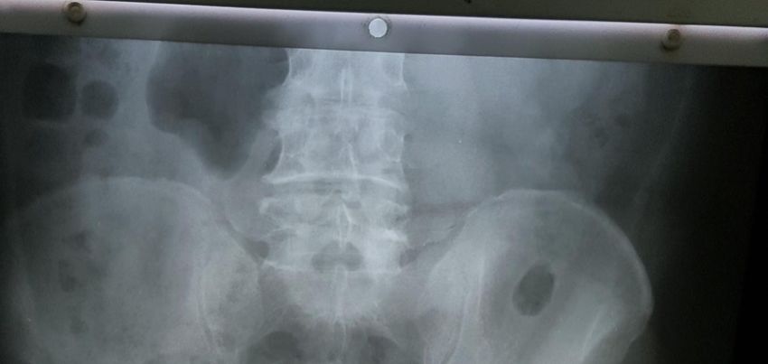
Fig. 3. Post-operative radiograph of the right femur, anteroposterior view, (15/02/2010)
the reduction of the fragments, including lateral In this case, the intramedullary nail option was
displacement. If this medial soft tissue release is not considered due to the poor muscle status
done carefully, close to the insertion, it does not around the hip and undisplaced nature of the
impair vascularization of the femoral head. The intertrochanteric fracture.
medial opening after valgisation is filled with
corticocancellous bone graft always. Although the weight-bearing walk was started
later than the intramedullary case, the healing of
Subsequent operative treatment for the non- the fracture and other rehabilitation of this patient
union consists of end prostheses, total hip went uneventful. We considered that a dynamic
arthroplasties, and repeated attempts at ORIF hip screw was also an option for the
(Open Reduction and Internal Fixation). The intertrochanteric fracture with the ankylosed hip.
patient can be achieved union following removal Plain x ray (Fig. 5) showing “stage of arthritis”,
of internal fixation and bone grafting (five pathology involving articular surface. Irregular
months) and sometimes require a total hip and hazy joint margins with diminished joint
arthroplasty. space on the right side. So, this is one of the
causes may effect on the non-union fracture after
Total hip arthroplasty (THA) commonly post operation. Patient might have TB hip joint
used for fused hip joints but it is technically with hip arthrodesis because patient already
demanding because of the lack of surgical complaint of shortening of right hip joint before
landmarks [17]. the accident.
There are reported cases of application of intra- The TB of hip is still a common condition in
medullary nail [18,19] and cannulated screws developing countries. Early presentations are
only with a good outcome [20]. pain around hip and limp. Later the patient
11Lwin et al.; IJMPCR, 13(1): 7-15, 2020; Article no.IJMPCR.55942
Fig. 4. Post-operative radiograph of the right femur, oblique view, (15/02/2010)
presents with deformities, shortening of limb and viscera. Through the hematogenous route, the
restriction of movements. Histological proof may bacteria reach either to synovium or bone. When
not be necessary in all the cases in the endemic it lodges first in synovium, the synovial
zones for TB. The management depends upon membrane becomes swollen and congested. The
the stage of clinical presentation and the severity granulation tissue from the synovium extends
of destruction as visible radiologically. From over the bone resulting in necrosis of sub
conservative therapy in the form of ATT and chondral bone, sequestra and may be kissing
traction to debridement and joint replacement, a lesion on either side of joint [22].
variety of surgical procedures have been
described. On an average 2-5% of patient's LIPUS (low-intensity pulsed ultrasound)
report back with reactivation of the disease within treatment was associated with a high rate of
about 20 years after the apparent clinical healing healing (86.2%) in a registry cohort of 767 non-
of the first lesion [21]. union fractures that had failed to heal for at least
one year prior to treatment [23]. The LIPUS heal
Osteoarticular TB is secondary to primary rate is comfortably within the range of heal rates
pathology in lungs, lymph nodes or any of the reported after surgical revision, suggesting that
12Lwin et al.; IJMPCR, 13(1): 7-15, 2020; Article no.IJMPCR.55942
Fig. 5. Post-operative radiograph of pelvis and both hip joints, anteroposterior view
(15/02/2010)
Plain x ray showing “stage of arthritis”, pathology involving articular surface. Irregular and hazy joint margins with
diminished joint space on the right side
LIPUS treatment may provide comparable heparin infusion through an implanted arterial
benefit to surgery. So that LIPUS therapy may catheter [24].
represent an effective, low-risk alternative to
surgical revision in the setting of impaired 4. CONCLUSION
fracture healing. So, we can consider for the
The treatment of intertrochanteric nonunion is
LIPUS treatment if patient has non-union
guided by the age of the patient. In older patients
fracture.
with low-demand activities and poor bone quality
or a damaged hip articular surface, arthroplasty
Surgical modalities for posttraumatic septic non- allows earlier patient mobilization and greater
union would be treated with a free vascularized certainty of outcome.
fibular graft (FVFG) including three strategies:
This patient also has a known case of TB hip
i) a two-staged operation; ii) a flow-through joint with limb shortening. This might be one of
anastomosis to conserve blood flow in the major the causes of the nonunion of intertrochanteric
vessels in the lower leg; and iii) continuous fractures.
13Lwin et al.; IJMPCR, 13(1): 7-15, 2020; Article no.IJMPCR.55942
The right hip joint was also seen with destruction 9. Mariani EM, Rand JA. Nonunion of
and fusion present before the operation. intertrochanteric fractures of the femur
following open reduction and internal
That’s why THR is the best for this patient. But fixation: Results of second attempts to gain
the patient refused for operation. union. Clin Orthop Relat Res. 1987;218:
81–9.
Cases with good bone stock, union in primary 10. Sarathy MP, Madhavan P, Ravichandran
nonunion of intertrochanteric fractures can be KM. Nonunion of intertrochanteric fractures
achieved with internal fixation, valgization, and of the femur. J Bone Joint Surg Br. 1994;
grafting procedures. 77:90–2.
11. Haidukewych GJ, Berry DJ. Salvage of
CONSENT AND ETHICAL APPROVAL failed internal fixation of intertrochanteric
hip fractures. Clin Orthop Relat Res. 2003;
As per university standard guideline, participant 412:184–8.
consent and ethical approval have been 12. Fracture and dislocation compendium.
collected and preserved by the authors. Orthopedic Trauma Association
Committee for Coding and Classification. J
COMPETING INTERESTS Orthop Trauma. 1996;10(Suppl 1):1–
154.
Authors have declared that no competing 13. Harris WH. Traumatic arthritis of the hip
interests exist. after dislocation and acetabular fractures:
Treatment by mold arthroplasty: An end-
REFERENCES result study using a new method of result
evaluation. J Bone Joint Surg Am. 1969;
1. Bhandari M, Schemitsch E, Onsson AJ, 51:737–55.
Zlowodzki M,Haidukewych GJ. Gamma 14. Altner PC. Reasons for failure in treatment
nails revisited: Gamma nails versus of intertrochanteric fractures. Orthop Rev.
compression hip screws in the 1982;11:117.
management of intertrochanteric fractures 15. Wu CC, Shih CH, Chen WJ, Tai CL.
of the hip: A meta-analysis. J. Orthop Treatment of cutout of a lag screw of a
Trauma. 2009;23(6):460–4.2 dynamic hip screw in an intertrochanteric
2. Angelini M, McKee MD, Waddell JP, fracture. Arch Orthop Trauma Surg. 1998;
Haidukewych G, Schemitsch EH. Salvage 117:193–6.
of failed hip fracture fixation. J Orthop 16. Bartonícek J, Skála-Rosenbaum J, Dousa
Trauma. 2009;23:471–8. P. Valgus intertrochanteric osteotomy for
3. Dhammi IK, Jain AK, Singh AP. Primary malunion and nonunion of trochanteric
nonunion of intertrochanteric fractures of fractures. J Orthop Trauma. 2003;17:606–
femur: An analysis of results of valgization 12.
and bone grafting. Indian Journal of 17. Kilgus DJ, Amstutz HC, Wolgin MA, Dorey
Orthopaedics. 2011;45(6):514. FJ. Joint replacement for ankylosed hips. J
4. Baumgartner MR, Solberg BD. Awareness Bone Joint Surg Am. 1990;72(1):45–
of tipapex distance reduces failure of 54.
fixation of trochanteric fractures of the hip. 18. Ishimaru D, Nozawa S, Maeda M, Shimizu
J Bone Joint Surg Br. 1997;79:969–71. M. Intertrochanteric fracture of the
5. Kyle RF, Gustilo RB, Premer RF. Analysis ankylosed hip joint treated by a gamma
of 622 intertrochanteric hip fractures. J nail: A case report. Case Rep Orthop.
Bone Joint Surg Am. 1979;61:216–21. 2012;278156.3.
6. Baker HR. Ununited intertrochanteric 19. Manzotti A, Confalonieri N, Pullen C.
fractures of the femur. Clin Orthop Relat Intertrochanteric fracture of an arthrodesis
Res. 1960;18:209–20. hip. J Bone Joint Surg Br. 2007;89(3):
7. Boyd HB, Lipinski SW. Nonunion of 390–2.
trochanteric and subtrochanteric fractures. 20. Font-Vizcarra L, Carre˜no AM, Prat S,
Surg Gynecol Obstet. 1957;104:463–70. Mu˜noz-Mahamud E, Camacho P,
8. Haidukewych GJ, Israel TA, Berry DJ. Casanova L. Less invasive fixation of an
Reverse obliquity of fractures of the intertrochanteric fracture in an ankylosed
intertrochanteric region of the femur. J hip with cannulateds crews: A case report.
Bone Joint Surg Am. 2001;83:643–50. Hip Int. 2010;20(4):565–7.
14Lwin et al.; IJMPCR, 13(1): 7-15, 2020; Article no.IJMPCR.55942
21. Saraf SK, Tuli SM. Tuberculosis of hip: A 24. Kawakami R, Ejiri S, Hakozaki M,
current concept review. Indian J Orthop. et al. Surgical treatment options for
2015;49(1):1–9. septic non-union of the tibia:
DOI: 10.4103/0019-5413.143903 Two staged operation, Flow-through
22. Tuli SM. General principles of anastomosis of FVFG and continuous
osteoarticular tuberculosis. Clin Orthop local intraarterial infusion of heparin.
Relat Res. 2002;398:11–9. Fukushima J Med Sci. 2016;62(2): 83–
23. Zura R, et al. Injury, Int. J. Care Injured. 89.
2015;46:2036–2041 DOI: 10.5387/fms.2016-5
_________________________________________________________________________________
© 2020 Lwin et al.; This is an Open Access article distributed under the terms of the Creative Commons Attribution License
(http://creativecommons.org/licenses/by/4.0), which permits unrestricted use, distribution, and reproduction in any medium,
provided the original work is properly cited.
Peer-review history:
The peer review history for this paper can be accessed here:
http://www.sdiarticle4.com/review-history/55942
15You can also read