How to interpret your sleep study - Anita Bhola, MD, FCCP Clinical Director ABIM Board Certified Sleep Specialist
←
→
Page content transcription
If your browser does not render page correctly, please read the page content below
How to interpret your sleep study

           Anita Bhola, MD, FCCP
               Clinical Director
      ABIM Board Certified Sleep Specialist

            Lexington Medical Services, PLLC
                 Sleep Disorders Center
                  200A East 62nd Street
                 New York, NY 10065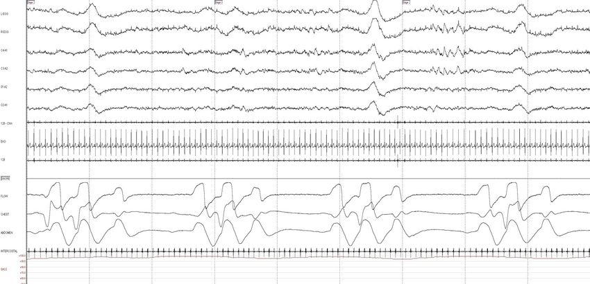
• You go to your doctor/sleep specialist for consultation (or
  your bed partner tells you to), because of symptoms of
     –   Snoring, gasping/choking, stop breathing at night
     –   Daytime sleepiness or tiredness
     –   Drowsy driving
     –   Morning headaches, dry mouth, sore throat
     –   Decreased sex drive, waking up often to urinate
     –   Mood, memory, attention problems, drowsy driving
     –   Twitching and jerking in limbs at night
     –   Difficulty falling or staying sleep
•   Your doctor/sleep specialist recommends an overnight sleep study
    (polysomnogram)
    •    Intimidating test: fill out questionnaires, sleep in a strange environment (sleep
         lab/center), no family members allowed, hooked up to electrodes and wires, feel
         you are being watched, woken up early, made to fill out more questionnaires and
         kicked out
•   A few days later, your doctor/sleep specialist call you to give you the
    results and what to do next
•   You may or may not get a copy of your sleep study report, which is
    as intimidating as the test itself
•   The goal of my talk is to help you understand what is going on and
    how to make sense your report………………..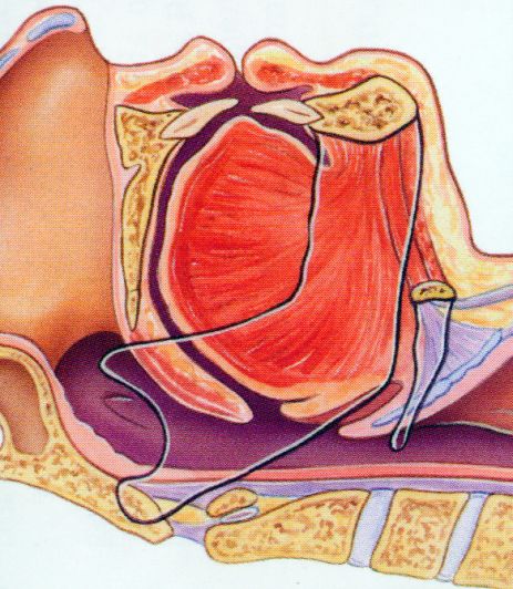
Indications for sleep studies
                  (polysomnogram or PSG)
•   Commonest indication:
    • Sleep disordered breathing (obstructive sleep apnea, upper airway
       resistance syndrome, primary snoring)
•   Less common indications:
    •   Severe parasomnias
    •   REM behavior disorder
    •   Nocturnal seizures
    •   Narcolepsy
    •   Periodic limb movement disorder
    •   Bruxism
    •   Occasionally insomnia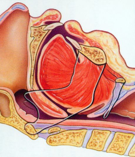
Sleep Study (Polysomnogram)
• Noninvasive, pain-free procedure that usually requires spending a night or
two in a sleep facility. The testing bedrooms are designed to resemble a
typical bedroom, with décor and televisions to help make you feel as
relaxed as possible.
• Occasionally daytime studies are performed (night shift workers, MSLT and
MWT)
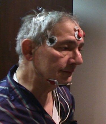
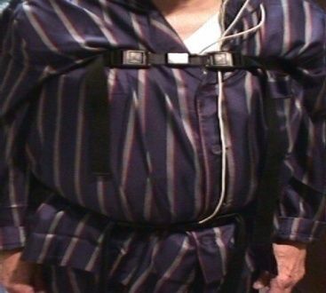
• During a polysomnogram, a sleep technologist, will hook you up to
electrodes and wires so that they can simultaneously record multiple
biological functions during your sleep, on a digital recording
• Depending on the physician’s orders, patients may be given therapy during
the course of the study, which may include device called continuous positive
airway pressure therapy (CPAP), oxygen, oral appliance or allowed to take
their own medication.
• After a full night’s sleep is recorded, the data will be tabulated by a
technologist, scored by a registered polysomnographic technologist and
presented to for interpretation.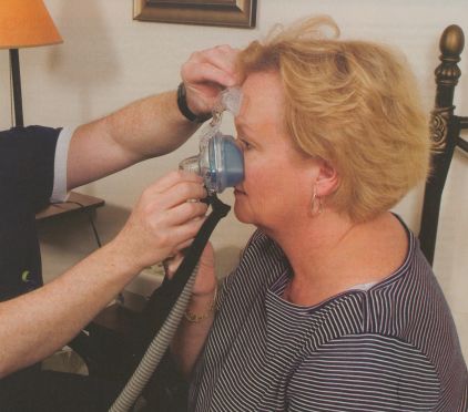
Interpretation of sleep studies

•   The interpreting physician is a board certified sleep physician who
    must
     – Review the entire history, techs and scorers notes, patient questionnaires
     – Review entire scored digital recording (raw data).
     – Simultaneously review tabulated data

•   Form a summary, impression and recommendations

•   The clinical judgment of the sleep specialist is a key element in the
    interpretation.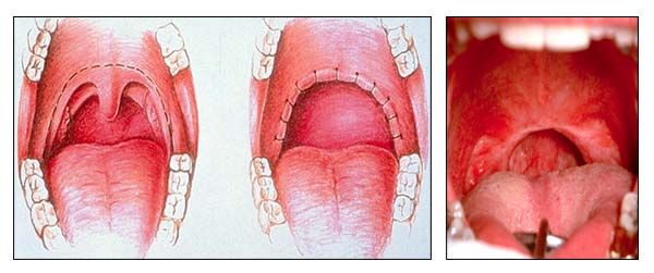
Channels commonly recorded during a PSG

–   Brain wave activity (EEG),
–   Eye movement (EOG),
–   Muscle tone (chin EMG),
–   Airflow via thin catheters placed in front of nostrils and mouth
–   Breathing effort via belts placed over chest and abdomen
–   Snoring (microphone placed over the neck)
–   heart rhythm (EKG)
–   Oxygen level (SpO2)
–   Leg muscle activity (PLM)
–   Body position
–   Video recording

Intercom to communicate with technician
Snoring sound transducer

    Nasal and Oral Airflow

Respiratory Effort

 Leg muscle activity
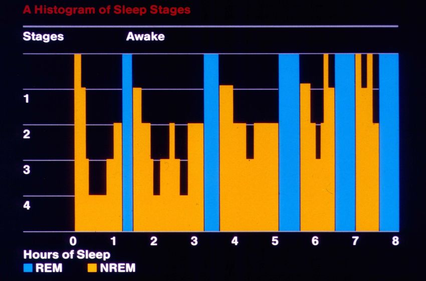
Stages of Sleep
• Stage W (Wakefullness)
• Stage NREM
  – Stage N1
  – Stage N2
  – Stage N3
• Stage R (REM)
Sleep Cycles
• Normal sleep consists of 4 - 6 cycles of NREM
  sleep alternating with REM sleep every 90-120
  minutes

• First two cycles are predominantly NREM

• Later stages (early am) are predominantly REM
Hypnogram of Normal Sleep Stages
Polysomnogram recording at normal speed
At fast speed
Snoring
Apneas

– Obstructive
– Central
– Mixed
Obstructive Apnea
Complete airway collapse causing cessation of airflow at nose and
  mouth for at least 10 secs, with drop in oxygen level and arousal
  from sleep
Central Apnea
•   Complete absence of airflow for >10 secs accompanied by a complete
    absence of respiratory effort
•   Signal from the brain to breathe is delayed
•   Types: idiopathic, high altitude, narcotic induced, CSR

     ECG

     Airflow

     Thor.
     Effort

     Abd.
     Effort

     SAO2
Cheyne-Stokes respiration • A crescendo-decrescendo respiratory pattern • Usually associated with CHF, neurologic disease • Atrial fibrillation may be present
Mixed apnea
•   A complete absence of airflow for >10 secs accompanied by
    complete absence of respiratory effort at the beginning of the event,
    followed by a gradual increase in effort which eventually breaks the
    apnea
Hypopnea:
• Partial airway collapse causing shallow breathing (airflow reduced by at least
30-50%) during sleep, lasting 10 seconds or longer, usually associated with a fall in
blood oxygen saturation.
• Physiologically same as an apneaRespiratory Effort Related Arousal (RERA)
• A sequence of breaths characterized by marked decreased in airflow for at
least 10 secs, with increased respiratory effort, no desaturation and which
leads to an arousal from sleepBruxism (teeth grinding) • In sleep, jaw contraction frequently occurs. • Detected on chin EMG electrodes • Additional electrodes may be placed
Leg movements (Periodic Leg Movements)
Also look for

– EEG abnormalities
   • alpha intrusion (seen in pain syndromes)

   • seizure waveforms

– Behavioral abnormalities (sleep talking etc)

– Parasomnias: defined as undesirable motor, or verbal
  phenomena that arise from sleep or sleep-wake
  transition (confusional arousals, night terrors, REM
  behavior disorder)
Seizures
Rem Behavior Disorder
characterized by recurrent violent nighttime awakenings/dream enacting
Review of Sleep Study Times, formulas and
                      calculations:

• Sleep statistics
   –   Lights Out
   –   Light On
   –   Total Recording Time
   –   Total Sleep Time
   –   Sleep Latency
   –   Sleep Efficiency
   –   Rem Latency
   –   WASO (wake after sleep onset)
   –   Time and percentage in each sleep stage
Sleep architecture Distribution (percentage) of Sleep Stages during Normal Sleep NREM stage Stage N1 Stage N2 Stage N3 (slow wave sleep) Stage REM Absent or decreased N3 and/or REM often seen in obstructive sleep apnea Rebound often seen with CPAP titration
Distribution of Sleep Stages
         during Normal Sleep
• 75-80% of total sleep is typically NREM

NREM stage            % Total sleep time
Stage I                    3-8
Stage 2                    45-55
Stage 3/4                  15-20

• 20-25% of total sleep is in stage REM
Arousals
• “Micro-awakenings”
• Abrupt change of EEG from a deeper stage of NREM sleep to a lighter
stage, or from REM sleep toward wakefulness, with the possibility of
awakening as the final outcome.
• An arousal may be accompanied by increased chin (EMG) activity and
heart rate, as well as by an increased number of body movements
• Minimum duration is 3 secs
• Types: respiratory, PLMs, spontaneous
• Increased arousals are associated with increased daytime sleepiness and
decreased performance, similar to that seen in sleep deprivation
• Arousal index=the number of arousals per hour=measure of sleep
fragmentation
• Age dependent
• 10-12/hr are normal at age 20
• 20-22/hr are normal at age 50-60
• Increase in Insomnia and severe sleep apneaRespiratory Events – Number of obstructive apneas – Number of mixed apneas – Number of central apneas – Number of hypopneas – Respiratory effort related arousals (RERAs)
Apnea Hypopnea index (AHI)

AHI= All apneas+hypopneas/Total sleep
 time (hrs)
 supine AHI
 REM and NREM AHI

RDI=All apneas+hypopneas+RERAs/Total
 sleep time (hrs)
EKG abnormalities during sleep

•   Heart rate too fast (tachycardia) or too slow (bradycardia)
•   Heart rhythm irregular
•   Pauses
Oxygen saturation
– Baseline oxygen saturation (at the start of the study)
– Lowest oxygen saturation during sleep
   • Above 90% normal
   • 85-90% mild
   • 75-85% moderate
   •
Sleep Disordered
              Breathing

              UARS               OSA
Snoring
           (+/-snoring)      (+/-snoring)
Primary Snoring • Incidence: at least 24% adult women, 40-50% of adult men and 10% children • Bed partner reporting • Not associated with airflow limitation, arousals from sleep, or oxygen desaturation • Risk of developing OSA with age or weight gain • Higher prevalence of CV disease (hypertension, stroke, ischemic heart disease) in snorers, on epidemiological studies
Upper Airway Resistance Syndrome

• Now included as part of the OSA
  diagnosis
• UARS: > 5 RERA’s per hour of sleep
Obstructive Sleep Apnea
• The disorder is characterized by
   – recurrent episodes of upper airway obstruction
   – episodic oxyhemoglobin desaturation during sleep
   – recurrent arousals

   Typically worse in supine, REM
Diagnosis of OSA:

AHI > 5/hr

Severity of Obstructive Sleep Apnea
AHI/RDI 5-15 mild
AHI/RDI 15-30 moderate
AHI/RDI > 30 severe
Central Sleep Apnea Syndrome

• Central apneas ≥ 5/hr and symptoms of either excessive
  sleepiness or disrupted sleep
• Central apneas frequently co-exist with obstructive or
  mixed apneas and hypopneas
• If > 50% of events are purely central = CSAS
When do we treat OSA
CMMS’s* Definition of Obstructive Sleep Apnea (OSA)
CPAP will be covered for adults with sleep disordered breathing if:
AHI/RDI > 15 OR
AHI/RDI 5-15 with
Daytime sleepiness
Hypertension
Stroke
Ischemic heart disease
Insomnia
Mood disorders
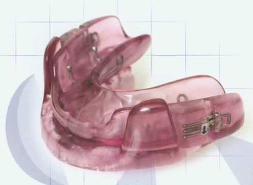
*Center for Medicare and Medicaid ServicesOSA Rx • CPAP • Dental Appliance • ENT Surgery • Positional Rx • Wt loss • Avoid sedatives/ETOH • Maximize nasal patency
CPAP
• Pneumatic splint-keeps airway open
• Non invasive
• Continuous pressure
Types of Sleep Reports

•   Diagnostic PSG Reports
•   CPAP Titration Reports
•   Split Night Reports
•   MSLT/MWT Reports
Lexington Medical Services, PLLC
                                                                         Sleep Disorders Center
                                                                             New York, NY
                                                                 DIAGNOSTIC SLEEP REPORT
     MEDICAL                            Name:                          John Doe                 Birthdate:            10/1/1956                                    SYMPTOMS
    CONDITIONS                          Test Date:                     11/28/2008               Age                   52 yrs                                      CONSISTENT
    ASSOCIATED                          Interpreting Physician:        Anita Bhola, MD          Gender:               Male                                          WITH OSA
     WITH OSA                           Scoring Technologist:          T. Oram RPSGT            Test Description:     NPSG
                                        Brief Summary of Statistics: Lights Out: 10:12:25 PM ~ Lights On: 5:12:25 AM
      Mr. Doe is a 52 year-old male referred by Dr. Sleepy with symptoms of snoring witnessed apneas and daytime sleepiness. His physical examination is
      significant for a Mallampati class 3 oropharynx. His Epworth Sleepiness Score is 8. The patient is 73 inches in height and 225 pounds in weight, with a Body
      Mass Index (BMI) of 30. Medical history provided by the patient and/or referral sheet from the referring physician, is significant for
      hypertension, coronary artey disease and he is s/p CABG in 2003. His current medications are metroprolol, fosinopril, Lipitor, amlodipine, isorbide and
      hydrochlorothiazide. The patient’s normal bedtime is 10:00 pm. Nocturnal Polysomnography (NPSG) was ordered to rule out the diagnosis of obstructive
      sleep apnea (OSA).

       During this the following were monitored: frontal, central and occipital EEG, electrooculogram (EOG), submentalis EMG, nasal and oral airflow, thoracic wall motion, anterior
       tibialis EMG, and electrocardiogram. Arterial oxygen saturation was monitored with a pulse oximeter. Patient was monitored by video. The tracing was scored using 30 second
       epochs. Sleep latency was defined as lights out to the first epoch of any sleep (AASM). Hypopneas were scored per AASM definition
                              DECREASED                                                                            Arousal
                                SLEEP                                                                              Statistics
Sleep Architecture            EFFICIENCY
                                                                                                                                                     Limb
                                                                                      Wake After                                 Total Respiratory          Snore Spontaneous
                                                                                                                                                   Movement
                                                                                      Sleep Onset          58.5                    #
Total Recording Time (min)          420       N1:       39.9% (139.0 min.)            (WASO):              min.    REM            62       60          0      0        2
                                                                                      Sleep Latency                NREM           173        158            0           2              13
Sleep Period Time (min)            406.5      N2:       36.4% (126.5 min.)            (min)                13.5
                                                                                      REM Latency                  WAKE            0          0             0           0              0
Total Sleep Time (min)              348       N3:       0.0% (0.0 min.)               (min)                 80     TOTAL          235        218            0           2              15
                                              Stage
                                                                                                                   INDEX         40.5       37.6            0         0.3              2.6
Sleep Efficiency (%)               82.9       R:        23.7% (82.5 min.)
                                                                                                   SpO2 Statistics
     INCREASED                Respiratory Events                                                   Baseline Saturation:                        97%                  DECREASED
         AHI                  # Obstr. Apnea:               1      RDI (/hr):        38.6                                                                             OXYGEN
                                                                                                   Lowest Sat During Sleep:                    78%
                                                                                                                                                                    SATURATION
                              # of Mixed Apnea:             0      AHI (/hr):        38.3          PLM Events
   AHI
                                                                   AHI NREM
ELEVATED                      # of Hypopneas:             221      (/hr):            36.4          PLM:                                         12
 IN REM                                                            AHI REM (/                      PLM Index (/hr):                          2.2 (/hr)
                                                                                                                                                                       INSIGNIFICANT #
                              # of Central Apnea:           0      hr):              44.4          PLM Arousals:                                6                          OF LEG
   AHI ELEVATED                                                    AHI supine                      PLM Arousal Index (/hr):                  0.8 (/hr)
   IN THE SUPINE                                                                                                                                                         MOVEMENTS
                              # RERA:                       2      ( /hr):           59.2          :
      POSITION
Obstructive sleep apnea syndrome,
severe, with desaturation in REM sleep
The patient is advised to return for a
CPAP titration study.
RESPIRATORY EVENTS
AND OXYGEN
DESATURATIONS ARE
INCREASED IN REMCPAP Titration Study
• Overnight study with same hook up as NPSG, with the addition of CPAP
• Occasionally a non-benzodiazepine sleep aid may be prescribed if poor
sleep efficiency is detected on the PSG
• Prior to starting study, mask fitting and desensitization with lowest level of
CPAP and humidification
• CPAP increased, once asleep, to eliminate airway collapse, snoring and
correct drop in oxygenation (goal is AHILexington Medical Services, PLLC
Sleep Disorders Center
New York, NY
CPAP TITRATION REPORT
Name: John Doe Birthdate: 3/12/1959
Test Date: 1/9/2009 Age 49 yrs
Interpreting Physician: Anita Bhola, MD Gender: Male
Scoring Technologist: T. Oram RPSGT Test Description: CPAP Titration HISTORY OF
SEVERE OSA
Brief Summary of Statistics: Lights Out: 10:28:46 PM ~ Lights On: 5:20:46 AM
Mr. Doe is a 49 year-old male, initially referred by Dr. Sleepy with symptoms of snoring witnessed apnea and daytime sleepiness. His physical
examination is significant for a Mallampati class 3 oropharynx. His Epworth Sleepiness Score is 8. The patient is 73 inches in height and 225 pounds
in weight, with a Body Mass Index (BMI) of 30. Medical history provided by the patient and/or referral sheet from the referring physician, is significant
for hypertension, coronary artey disease and he is s/p CABG in 2003. His current medications are metroprolol, fosinopril, Lipitor, amlodipine, isorbide
and hydrochlorothiazide.
The patient’s normal bedtime is 10:00 pm. Mr. Doe had a previous NPSG study done on 11/28/2008 at Lexington Medical Service’s Sleep Disorders
Center, which revealed an AHI of 38.3 and lowest saturation of 78% in REM sleep, consistent with a diagnosis of severe obstructive sleep apnea with
desaturation. CPAP Titration was ordered to determine if CPAP is an effective treatment for the patient’s condition and to determine the optimal
CPAP pressure.
During this the following were monitored: frontal, central and occipital EEG, electrooculogram (EOG), submentalis EMG, nasal and oral airflow, thoracic wall motion,
anterior tibialis EMG, and electrocardiogram. Arterial oxygen saturation was monitored with a pulse oximeter. Patient was monitored by video. The tracing was scored
using 30 second epochs. Sleep latency was defined as lights out to the first epoch of any sleep (AASM). Hypopneas were scored per AASM definition
Sleep
Architecture
Arousals
Total Recording Time Wake After Sleep Onset 71.0
Limb
(min) 412 N1: 29.5% (100.5 min.) (WASO): min. Total Respiratory Snore Spontaneous
Movement
Sleep Period Time #
(min) 406.5 N2: 43.8% (149.5 min.) Sleep Latency (min) 0 REM 19 8 0 3 8
Total Sleep Time NREM 42 15 3 2 22
(min) 341 N3: 0.0% (0.0 min.) REM Latency (min) 119
WAKE 0 0 0 0 0
Stage
Sleep Efficiency (%) 82.8 R: 26.7% (91.0 min.) TOTAL 61 23 3 5 30
INDEX 10.7 4 0.5 0.9 5.3
AHI Respiratory
Events
SpO2
Sta7s7cs
WITHIN # Obstr. Apnea: 2 RDI (/hr): 8.4 SPO2
NORMAL Baseline Saturation: 98% WITHIN
# of Mixed Apnea: 0 AHI (/hr): 2.6
RANGE Lowest Sat During Sleep: 90% NORMAL
# of Hypopneas: 12 AHI NREM (/hr): 2.6 RANGES
# of Central Apnea: 1 AHI REM (/hr): 2.6 PLM Events
# RERA: 33 AHI supine ( /hr): 2.5 PLM: 12
INSIGNIFICANT
AHI at Final Pressure: PLM Index (/hr): 2.1 LEG
PLM Arousals: 2 MOVEMENTS
PLM Arousal Index (/hr): 0.4Summary: (CPAP TITRATION)
1.Prior to hook-up, the patient was fitted with a large Res Med
Micro Mirage mask and desensitized to CPAP. The patient
tolerated the fitting and desensitizing well.
2.During the present study, sleep latency was zero as sleep
occurred immediately upon Lights Out. This is likely
secondary to severe sleepiness. Sleep efficiency was mildly
secondary to frequent arousals and awakenings.
3.Following application and titration of CPAP from 4 to 8.0
cmH2O, using warm humidification, sleep architecture mildly
Compared to the NPSG, slow wave sleep remained absent.
REM rebound was noted. Respiratory disturbances were .
The optimal CPAP pressure was 8.0 cmH2O, with in overall
AHI to 1.1 events/hour of sleep in the supine position.
Sustained REM sleep achieved in the supine position.
Snoring eliminated and were noted at this pressure.
4.EKG monitoring showed with occasional PVCs.
5.There were 12 periodic limb movements (PLM’s) during the
study, resulting in a PLM index of 2.1 per hour of sleep. This is
ELIMINATION OF consistent with PLM activity.
RESPIRATORY 6.The patient tolerated the procedure
Impression:
EVENTS DURING 1. 327.23 Obstructive sleep apnea syndrome, severe
SUPINE REM Recommendations:
SLEEP •Treatment of severe obstructive sleep apnea syndrome is
recommended with the nightly use of CPAP.
•The patient is advised to use CPAP nightly at a pressure of
8.0 cmH2O pressure with warm humidification and a large
Res Med Micro Mirage mask.
•CPAP follow up is recommended in 2-4 weeks.
•Weight loss is recommended.
•General recommendations include maximization of nasal
patency, avoidance of the supine position, and avoiding
alcohol, nicotine, benzodiazepine sedative-hypnotics and
sleep deprivation as these can make sleep apnea worse.
•The patient is advised not to drive or operate any large
machinery while sleepy, until the daytime sleepiness is
adequately treated.
•Follow up with referring ENT physician is recommended.
% AHI WITHIN
Duration % RE NORMAL
CPAP (min) Sleep M % SWS #CA #OA #MA #Hyp #RERA AHI RDI RANGE
4 34.2 59.1 0.0 0.0 0 0 0 0 3 0.0 8.9 WHILE
5 42.1 48.9 0.0 0.0 0 0 0 1 7 2.9 23.3 ON 7-8
6 69.2 88.4 23.3 0.0 0 0 0 10 1 9.8 10.8 CMH2O
7 15.4 87.0 83.8 0.0 0 0 0 0 1 0.0 4.5
8 244.3 91.8 25.1 0.0 1 2 0 1 21 1.1 6.7Split Night Study
• Initial diagnostic PSG followed by CPAP titration during the same
night
• Typically need 2 hours of sleep prior to applying CPAP followed by 3
hours of CPAP titration
• Compares the 2 sections of the test side by side
• Is an alternative to one full night of diagnostic PSG followed by a
second full night of titration if
– high likelihood for severe OSA
– patient reluctant or unable to undergo 2 full night studies
• Most labs have protocols for performing emergency Split night
studies based on AHI and lowest oxygen level observedLexington Medical Services, PLLC
200 A East 62nd Street
New York, NY 10065
SPLIT NIGHT REPORT
Brief Summary of Sleep Diagnostic Therapy Mr. Doe is a 65 year-old male referred by Dr. Sleepy with symptoms of snoring,
Statistics Total Study Portion Portion witnessed apneas and daytime sleepiness/fatigue. His physical examination is
significant for a Mallampati class 3 oropharynx. His Epworth Sleepiness Score
is 3. The patient is 67 inches in height and 205 pounds in weight, with a Body
Total Recording Time (min) 443 149 294 Mass Index (BMI) of 32. Medical history provided by the patient and/or referral
Sleep Period Time (min) 439 135 280.5 sheet from the referring physician, is significant for allergies/nasal obstruction,
hypertension, mild obesity, enlarged prostrate and hypercholestremia. His
Total Sleep Time (min) 400 123.5 276.5 current medications are Flomax, Avodart, simvastatin and Ecotrin. Split Night
Sleep Efficiency (%) 90.3 82.9 94 Nocturnal Polysomnography was ordered to rule out the diagnosis of
obstructive sleep apnea and to perform CPAP titration, if indicated, to
Sleep Latency (min) 4 4 13.5 determine if CPAP is an effective treatment for the patient’s condition.
REM Latency (min) 175 N/A N/A CPAP FOR > 3 hrs.
Awake (WASO) (min): 39 N/A N/A
During this the following were monitored: frontal, central and occipital EEG, electrooculogram
N1: 8.50% 2.70% (EOG), submentalis EMG, nasal and oral airflow, thoracic wall motion, anterior tibialis EMG, and
N2: 91.50% 40.30% electrocardiogram. Arterial oxygen saturation was monitored with a pulse oximeter. Patient
was monitored by video. The tracing was scored using 30 second epochs. Sleep latency was
N3: 0.00% 25.10% defined as lights out to the first epoch of any sleep (AASM). Hypopneas were scored per AASM
Stage R: 0.00% 31.80% definition
Diagnostic Therapy
Respiratory Statistics Total Study Portion Portion SLOW WAVE N3 AND REM SLEEP REBOUND
# Obstr. Apnea: 0 0
# of Mixed Apnea: 0 0
# of Hypopneas: 123 22
# of Central Apnea: 0 0
# RERA: 0 2
AHI (/hr): 59.8 4.8
Baseline Saturation: 96% 96%
Lowest Sat During Sleep: 88% 92%
Diagnostic Therapy
Leg Movement Statistics Total Study Portion Portion
PLM: N/A N/A
PLM Index (/hr): N/A N/A
PLM Arousals: N/A N/A
SpO2 IMPROVED AHI DECREASED TOSummary (Split Night): 1.During the present study, sleep latency was reduced. Sleep efficiency was within the normative range. 2.During Diagnostic Portion of the study, sleep architecture was severely abnormal with a high percentage of N1 at 8.5%, a high percentage of N2 at 91.5%, and absent slow wave N3 and REM sleep. Snoring was mild and present for 0.8 minutes or 0.6% of total sleep time. There were a severe number of respiratory disturbances seen during the study. The overall apnea/hypopneas index (AHI) was 59.8 events/hour, predominantly in the supine position. Oximetry showed a baseline SaO2 of 96%. The mean saturation during respiratory events was 92%, and the lowest observed desaturation was 88%. This is consistent with mild desaturation during the study. *Desaturations based on 3% or greater drop from baseline. 3.The Therapy Portion of the study, consitsted of the application and titration of CPAP from 4 to 9.0 cmH2O, using warm humidification and a Medium Respironics Comfort Gel nasal mask. 4. Sleep architecture and slow wave N3 and REM sleep rebound was noted. Respiratory disturbances were . The optimal CPAP pressure was 8.0 cmH2O, with in overall AHI to 0.5 events/hour of sleep in the supine position. Sustained REM sleep achieved in the supine position. Snoring eliminated and were noted at this pressure. 5.EKG monitoring showed . 6.There were no significant PLMs during the study. 7.The patient tolerated the procedure very Impression: 1.327.23 Obstructive Sleep Apnea syndrome, severe Recommendations: •Treatment of obstructive sleep apnea is recommended with the nightly use of CPAP. •The patient is advised to use CPAP nightly at a pressure of 8.0 cm H2O pressure with warm humidification and a Medium Respironics Comfort Gel nasal mask. •CPAP follow-up is recommended in 2-4 weeks. •Weight loss is recommended. •General recommendations include maximization of nasal patency, avoidance of the supine position, and avoiding alcohol, nicotine, benzodiazepine sedative-hypnotics and sleep deprivation as these can make sleep apnea worse. •The patient is advised not to drive or operate any large machinery while sleepy, until the daytime sleepiness is adequately treated. •Follow up with referring ENT physician is recommended.
CPAP compliance is extremely important

• Only ~ 35% of patients use CPAP three years after
  prescription-Adherance and compliance are a problem
• Heated humidifiers and patient education have helped
• After 12 weeks meet face to face with doctor
• Usage of >4 hours/night, 70% of nights, over 30
  consecutive days of therapy
Multiple Sleep Latency Test (MSLT)
• Indications: R/O narcolepsy or other hypersomnias
   –   Severe excessive daytime sleepiness
   –   Fragmented nocturnal sleep
   –   Cataplexy
   –   Hypnagogic hallucinations
   –   Sleep paralysis

• Multiple sleep latency test:
   – gold standard test for objective assessment of EDS
   – assess ability to fall asleep in a quiet environment
   – 4-5 daytime naps (20 mins) after an overnight PSG

• Criteria for narcolepsy:
   – mean sleep latency of 8 minutes or less and
   – two or more sleep-onset REM periods (SOREMPS)
Maintenance of wakefulness test (MWT)
• Validated measure of an individual's degree of alertness
  during waking hours
• Most commonly used when an individual's ability to
  remain awake becomes a personal or professional/public
  safety issue.
• The Federal Aviation Administration requires pilots with
  treated obstructive sleep apnea to undergo an MWT to
  assess alertness prior to return to work
• Typically preceded by overnight sleep study during which
  the patient wears their CPAP, followed by four 40-minute
  daytime sessions conducted at 2-hour intervals to
  assess response to therapy.
• Mean sleep latencies less than 8 minutes are considered
  abnormal
…………Thank You
You can also read