Usefulness of Home Overnight Pulse Oximetry in Patients with Suspected Sleep-Disordered Breathing
←
→
Page content transcription
If your browser does not render page correctly, please read the page content below
Hindawi
Canadian Respiratory Journal
Volume 2020, Article ID 1891285, 6 pages
https://doi.org/10.1155/2020/1891285

Research Article
Usefulness of Home Overnight Pulse Oximetry in Patients with
Suspected Sleep-Disordered Breathing

          Cristina Esteban-Amarilla ,1 Silvia Martin-Bote ,2 Antonio Jurado-Garcia ,3
          Ana Palomares-Muriana ,4 Nuria Feu-Collado ,5,6 and Bernabe Jurado-Gamez                                              5,6,7

          1
            Department of Respiratory Medicine, Pitie Salpetriere University Hospital, Paris, France
          2
            Department of Respiratory Medicine, Infanta Leonor University Hospital, Madrid, Spain
          3
            Physiotherapy Unit, San Juan de Dios Hospital, Cordoba, Spain
          4
            Department of Respiratory Medicine, Hospital de Alta Resolución, Puente Genil, Cordoba, Spain
          5
            Department of Respiratory Medicine, Reina Sofı́a University Hospital, Córdoba, Spain
          6
            Maimónides Biomedical Research Institute of Cordoba (IMIBIC), Córdoba, Spain
          7
            School of Medicine, University of Córdoba, Córdoba, Spain

          Correspondence should be addressed to Silvia Martin-Bote; silviamartinbote@gmail.com

          Received 16 June 2020; Revised 28 September 2020; Accepted 28 October 2020; Published 12 November 2020

          Academic Editor: Andrea S. Melani

          Copyright © 2020 Cristina Esteban-Amarilla et al. This is an open access article distributed under the Creative Commons
          Attribution License, which permits unrestricted use, distribution, and reproduction in any medium, provided the original work is
          properly cited.
          Background and Objective. To determine the diagnostic yield of nocturnal oximetry versus polygraphy for the diagnosis and
          classification of sleep apnea hypopnea syndrome (SAHS). Methods. Prospective study conducted in a university hospital. Subjects
          with a clinical suspicion of SAHS were included. All of them underwent home polygraphy and oximetry on the same night. A
          correlation was made between the apnea-hypopnea index (AHI) and the oximetry variables. The variable with the highest
          diagnostic value was calculated using the area under the curve (AUC), and the best cut-off point for discriminating between
          patients with SAHS and severe SAHS was identified. Results. One hundred and four subjects were included; 73 were men (70%);
          mean age was 52 ± 10.1 years; body mass index was 30 ± 4.1, and AHI � 29 ± 23.2/h. A correlation was observed between the AHI
          and oximetry variables, particularly ODI3 (r � 0.850; P < 0.001) and ODI4 (r � 0.912; P < 0.001). For an AHI ≥ 10/h, the ODI3 had
          an AUC � 0.941 (95% confidence interval (CI) � 0.899–0.982) and the ODI4, an AUC � 0.984 (95% CI � 0.964–1), with the ODI4
          having the best cut-off point (5.4/h). Similarly, for an AHI ≥ 30/h, the ODI4 had an AUC � 0.922 (95% CI � 0.859–0.986), with the
          best cut-off point being 10.5/h. Conclusion. Nocturnal oximetry is useful for diagnosing and evaluating the severity of SAHS. The
          ODI4 variable was most closely correlated with AHI for both diagnosis.

1. Introduction                                                         considered the gold standard for sleep studies [6], although it
                                                                        is limited by a lack of availability in all hospitals, long waiting
Sleep apnea-hypopnea syndrome (SAHS) is a major public                  lists, and high cost. Cardiorespiratory polygraphy, a tech-
health problem due to its high prevalence, which can reach              nique validated for the diagnosis of SAHS, is therefore in-
up to 25% among middle-aged adults [1]. It is also associated           creasingly used [7–9]. Nevertheless, SAHS is still highly
with increased vascular morbidity and mortality [2, 3], and             underdiagnosed, and this has sparked interest in other more
fragmented sleep results in nonrestorative sleep and ex-                simplified alternative techniques. Nocturnal oximetry re-
cessive daytime sleepiness. It is important to make an early            quires no previous training, correctly detects peripheral
diagnosis, as continuous positive airway pressure (CPAP)                oxygen saturation (SpO2), and shows a typical pattern of
treatment is effective for controlling SAHS and vascular risk            intermittent hypoxemia in patients with SAHS. For these
factors [4, 5]. Full-night polysomnography is currently                 reasons, it has been used in both sleep laboratories [10] and
2 Canadian Respiratory Journal
home [11–14]. Moreover, it could be a tool available to Table 1: Demographic and clinical information for the 104 subjects
primary care medicine. Results obtained with this technique, included in the study.
however, are highly variable [13], and it is not recommended Values
as a general practice in clinical practice guidelines [15]. Some Age (years) 52 ± 10.1
authors have suggested that nocturnal oximetry could be a BMI (kg/m2) 30 ± 4.1
diagnostic alternative in subjects with symptoms of SAHS Male (number, %) 73 (70%)
and could also aid in therapeutic decision-making [10, 11]. Epworth sleepiness scale 9 ± 4.1
However, these aspects have not been well studied in clinical Diabetes (number, %) 23 (22%)
practice conditions. In this study, the main aim was to assess COPD (number, %) 22 (21%)
the diagnostic yield of home nocturnal oximetry versus Dyslipidemia (number, %) 34 (33)
polygraphy in patients with symptoms suggestive of SAHS. Vascular disease (number, %) 22 (21%)
Hypertension (number, %) 49 (47%)
Cardiac arrhythmia (number, %) 14 (13%)
2. Materials and Methods AHI (events/h) 28 ± 23.2
Baseline SpO2 (%) 95 ± 1.6
2.1. Design. This is an investigator-blinded prospective study
Mean SpO2 (%) 94 ± 2.2
comparing diagnostic tests and evaluation. ODI3 (events/h) 40 ± 26
The protocol for this study was approved by the Cordoba ODI4, (events/h) 25 ± 20.4
Research Ethics Committee (ref: 2965). T90, (%) 5 ± 9.7
Results are presented as mean ± standard deviation for continuous variables
and number (%) for categorical variables. BMI: body mass index; COPD:
2.2. Study Population. Individuals with clinical suspicion of
chronic obstructive pulmonary disease; AHI: apnea-hypopnea index; SpO2:
SAHS were assessed in the sleep unit of a university hospital. oxygen saturation measured by nocturnal oximetry; ODIs: mean number of
Individuals who consecutively referred to the sleep unit for oxygen desaturations ≥ 3% and 4% (ODI3 and ODI4) per hour of analyzed
polygraphy and who met the following criteria were in- recording; T90: time spent with SpO2 < 90%.
cluded: (i) aged between 18 and 75 years; (ii) willing and able
to perform the diagnostic tests in the home; (iii) signed ODI4), defined as the number of times SpO2 fell by ≥3% or
informed consent. The exclusion criteria were (i) thoraco- 4%, respectively, and the T90 or percentage time recorded
genic (severe kyphoscoliosis) or neuromuscular disease, or with SpO2 < 90%. The test was inconclusive if there was a
chronic respiratory failure resulting from any cause >25% loss in basic signals (respiratory flow and SpO2).
(breathing room air, an SpO2 < or � 92%), (ii) drug user, Clinically relevant SAHS was diagnosed if the test showed an
including alcoholism, and (iii) specific sleep disorder other AHI ≥ 10/h and severe SAHS if the AHI was ≥30/h [6, 15].
than respiratory disorders, especially insomnia. Nocturnal oximetry was performed with a pulse oxi-
meter (Konica-Minolta Pulsox-300i; Software: Data Analysis
2.3. Methods. All patients who met the inclusion criteria DS-5). According to the manufacturer’s recommendations,
underwent home nocturnal oximetry and polygraphy on the the sampling frequency of the Pulsox-300i is set at 1 Hz with
same night, following clinical practice guidelines [6]. The an averaging duration of 3 sec and a resolution of 0.1% SpO2.
following variables were recorded of patient history: sex, Absence of variability in the SpO2 measurement in the index
body mass index (BMI), alcohol and medication use, finger of each hand was verified in the sleep unit, and the
smoking habits, general, and sleep-related symptoms patient was taught how to place the pulse oximeter. Oxi-
(snoring, gasping, witnessed apnea, nocturia, nonrestorative metry data were automatically scored and then visually
sleep, morning headaches, and excessive daytime sleepi- checked by the physician, and obvious artifacts in the
ness), comorbidities (diabetes, dyslipemia, vascular disease, oximetry signal were excluded.
hypertension, and cardiac arrhythmia), and respiratory Manual interpretation of the polygraph and oximetry
diseases other than SAHS (bronchial asthma and chronic was always performed by the same physicians, and the
obstructive pulmonary disease (COPD). patient’s information was anonymized by means of an al-
Polygraphy was carried out in the patient’s home phanumeric code, thus preventing the investigator from
influencing the results.
®
(Somnea , Compumedics Ltd., Abbottsford, Australia)
following the established recommendations [6, 16]. The
methodology has been previously described and validated in
our sleep unit [11]. Briefly, all the studies included recording 2.4. Statistical Analysis. Categorical variables are expressed
of the oronasal flow and pressure, respiratory movements, as absolute numbers and percentages, and statistical sig-
and SpO2. Apnea was defined as a significant decrease nificance was tested using the Chi2 test or Fisher’s exact test.
(>90%) in oronasal flow of >10 s and hypopnea as an evident To compare the severity groups, the Kruskal–Wallis test was
decrease in airflow >30%, butCanadian Respiratory Journal 3
Table 2: Number and percentage of the subjects reporting each of the symptoms stratified by severity.
Symptom No SAHS (n � 30) Mild-to-moderate SAHS (n � 34) Severe SAHS (n � 40) P value
Snoring (number, %) 27 (90%) 33 (97%) 38 (95 %) 0.468
Gasping (number, %) 4 (13, 3%) 4 (11, 8%) 12 (30%) 0.130
Witnessed apneas (number, %) 1 (3, 3%) 21 (61, 8%) 35 (87, 5%) 0 < 001≠
Nocturia (number, %) 15 (50%) 16 (47, 1) 26 (65%) 0.252
Unrefreshing sleep (number, %) 19 (63, 3%) 20 (58, 8%) 22 (55%) 0.784
Morning headaches (number, %) 6 (20%) 12 (35, 3%) 7 (17, 5%) 0.171
Excessive daytime sleepiness ‡ (number, %) 1 (3, 3%) 1 (2, 9%) 3 (7, 5%) 0.266
∗
P value of the Kruskal–Wallis test comparing subgroup classified for severity. ≠P value of no SAHS vs. severe SAHS groups. ‡ � Epworth sleepiness scale ≥15.
60 r = 0.525, p < 0.01 100 r = 0.912, p < 0.001
50
80
40
60
ODI4
30
T90
40
20
20
10
0 0
0 20 40 60 80 100 0 20 40 60 80 100
AHI AHI
(a) (b)
r = –0.415, p < 0.01 120
97,5 r = 0.850, p < 0.001
96,0 100
94,5
80
SpO2 mean
93,0
ODI3
60
91,5
90,0 40
88,5
20
87,0
0
85,5
0 20 40 60 80 100 0 20 40 60 80 100
AHI AHI
(c) (d)
Figure 1: Correlations of apnea-hypopnea index with oxygen desaturation index and percentage of recording with SpO2 < 90% measured by
nocturnal oximetry. The correlation in ODI3 (good correlation, r > 0.8) and ODI4 (excellent correlation, r > 0.9) is shown. AHI: apnea-
hypopnea index; ODIs: mean number of oxygen desaturations ≥3% and 4% (ODI3 and ODI4) per hour of recording; T90: time spent with
SpO2 < 90%.
polygraph and those observed in the oximetry was analyzed, calculated for the overnight SpO2 variables to determine
after which a receiver operator characteristic (ROC) curve which were more accurate in diagnosing AHI ≥ 10/h and
was constructed and the area under the curve (AUC) was AHI ≥ 30/h. All statistical tests were two-tailed, accepting a P4 Canadian Respiratory Journal
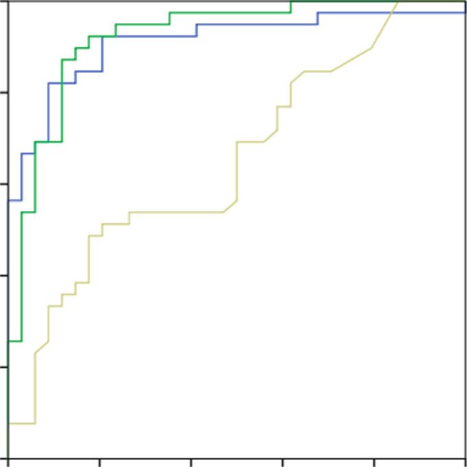
Table 3: Receiver operating characteristic calculations using the polygraphy apnea-hypopnea index as the reference standard.
ROC for AHI ≥ 10 AUC 95% confidence limits ROC for AHI ≥ 30 AUC 95% confidence limits
AHI vs. ODI3 0.941 0.899–0.982 0.911 0.841–0.981
AHI vs. ODI4 0.984 0.964–1 0.922 0.859–0.986
AHI vs. T90 0.759 0.662–0.856 0.653 0.527–0.779
The best discrimination for both mild-to-moderate SAHS and severe SAHS was the ODI3 and ODI4. AHI: apnea-hypopnea index from the polygraphy; AUC:
area under the curve; ODIs: mean number of oxygen desaturations ≥3% and 4% (ODI3 and ODI4) per hour of recording; T90: time spent with SpO2 < 90%;
ROC: receiver-operator characteristic.
1,0 1,0
0,8 0,8
0,6 0,6
Sensitivity
Sensitivity
0,4 0,4
0,2 0,2
0,0 0,0
0,0 0,2 0,4 0,6 0,8 1.0 0,0 0,2 0,4 0,6 0,8 1.0
1 – specificity 1 – specificity
ODI3 ODI3
ODI4 ODI4
T90 T90
(a) (b)
Figure 2: Receiver operating characteristic curves for oxygen desaturation index and percentage of recording with SpO2 < 90% with
thresholds. (a) AHI ≥ 10/h and (b) AHI ≥ 30/h, respectively. The discrimination degree between AHI ≥ 10/h and ODI3 (very good,
AUC � 0.941) and ODI4 (excellent, AUC � 0.984) (a) and between AHI ≥ 30/h and ODI3 (very good, AUC � 0.911) and ODI4 (very good,
AUC � 0.922) is shown. AHI: apnea-hypopnea index; ODIs: mean number of oxygen desaturations ≥3% and 4% (ODI3 and ODI4) per hour
of recording; AUC: area under the curve.
value 85%; the cut-off point was 5.4/h. For an AHI ≥ 30/h, in
final sample was therefore composed of 104 subjects: 74 Figure 2(b), the ODI4 also showed good diagnostic validity;
(71%) were classified as SAHS, while 39 (37%) presented the best cut-off point was 10.5/h for a sensitivity � 1 and a
with severe SAHS (AHI ≥ 30/h). Following the Global Ini- specificity > 85%.
tiative for Chronic Obstructive Lung Disease 2017, COPD
patients showed mild (n � 16), moderate (n � 5), severe 4. Discussion
(n � 1), and very severe (n � 0) functional grade, with the
latter patient having FEV1 � 44% and a baseline SpO2 of We evaluated the role of home nocturnal oximetry versus
95%. The baseline characteristics of the study subjects and polygraphy in patients referred for suspected SAHS. The
symptoms are shown in Tables 1 and 2. main study findings were that the ODI4 and ODI3 measured
The Pearson statistic was applied in order to determine by nocturnal oximetry showed a correlation with AHI.
the correlation between the AHI values and the pulse oxi- Therefore, results suggested a diagnostic approximation
metry variables. The results are shown in Figure 1 and, as can defined by the gold standard for SAHS [6, 16] and alsoCanadian Respiratory Journal 5
provide relevant information for therapeutic management of the best yield for diagnosing the severity of SAHS (AHI ≥ 30/
severe SAHS and CPAP indication [6]. These results are h), however, with little advantage over the ODI3. When
clinically applicable, given the availability and ease of use of included in a clinical protocol, this finding would be an
home overnight pulse oximetry. In clinical practice, pol- indication for CPAP treatment to be initiated, followed by
ygraphy is a diagnostic tool used extensively in the patient’s split-night polysomnography. It should be remembered that
home [7–9] if there is clinical suspicion of sleep-disordered diagnosis and treatment is very important in vascular disease
breathing and is ideal as a reference for comparison with [2–5], occupational health [26], and elderly men [25]. In
home nocturnal oximetry. The variability in the nocturnal these circumstances, nocturnal oximetry may be useful for
oximetry methodologies used in different studies must be prioritizing polygraphy or polysomnography studies or for
considered when comparing our results with previous identifying patients with SAHS and indicating the best
findings [10–14, 17]. In some studies, nocturnal oximetry therapeutic strategy on an individual basis. We have shown
was not done on the same night as the sleep test (poly- that home nocturnal oximetry can be useful in clinical
somnography or polygraphy) [12, 13, 17], while in others, practice for the diagnosis and treatment of SAHS, always
the tests were carried out in a sleep laboratory [11, 18, 19]. taking into account the patient’s medical history and signs
These differences, therefore, make it impossible to generalize and symptoms that could indicate a higher risk of SAHS
the conclusions of these authors to the out-of-hospital en- [27]. Furthermore, the low cost, ease of use, and availability
vironment. In order to avoid variability in the results be- of pulse oximetry make it a good alternative in the primary
tween different nights, in our study, both polygraphy and care setting [28, 29]. The study had some limitations. It was
nocturnal oximetry were performed on the same night. It is conducted in a tertiary reference hospital, and subjects were
also relevant to highlight the importance of the significant previously assessed in a specific sleep clinic, so the results
desaturation criterion [19]; Ho et al. [20] showed that the should be limited to this clinical context.
percentage decrease in SpO2 used to define hypopnea sig-
nificantly influences the diagnosis and severity classification 5. Conclusions
of SAHS. This could explain the variations in sensitivity and
specificity of nocturnal oximetry compared to polygraphy. In conclusion, in patients with suspected SAHS, home
In our study, we followed current recommendations for the nocturnal oximetry shows acceptable correlation with AHI
interpretation of polygraphy results [6, 16] and defined determined by polygraphy. The variable with best capacity
hypopnea as a ≥3% drop in SpO2 [6, 21, 22]. Taking into for discriminating between subjects with or without SAHS
account the AHI observed in the polygraph, the ODI4 gave and establishing severity was the ODI4. These results
the most accurate diagnosis and severity classification of therefore support the home nocturnal oximetry, especially in
SAHS in the nocturnal analysis of SpO2. However, the ODI3 primary care medicine, which could be a screening tool for
also shows a high diagnostic performance. Both parameters the diagnosis of SAHS, helping therapeutic management.
have more than a 90% probability that the diagnosis made in
patients with SAHS is correct. Therefore, it would allow Data Availability
prioritizing the diagnosis in patients with SAHS and in those
with severe SAHS, starting treatment with CPAP [6]. This is All data that were used to support the findings of this study
a key finding and could help define overall strategies for the are included within the article.
management SAHS. The specificity increases in patients with
moderate-to-severe disease [11–13], although it also cor- Conflicts of Interest
relates well in subjects with mild-to-moderate SAHS [23]. It
The authors declare that there are no conflicts of interest
has been observed that an OID4 > 15 has a positive pre-
regarding the publication of this paper.
dictive value of 100%, which could avoid having to perform
sleep tests [10–13]. In these studies, the criterion for clas-
sifying hypopnea was a drop in SpO2 > 4%. However, our References
work shows that the ODI3 is also equally useful to correctly [1] J. Durán, S. Esnaola, R. Rubio, and Á. Iztueta, “Obstructive
classify patients with SAHS and with severe SAHS. In ad- sleep apnea-hypopnea and related clinical features in a
dition, the ODI and the T90 report on the hypoxemic population-based sample of subjects aged 30 to 70 yr,”
burden in the patient with SAHS and correctly determine American Journal of Respiratory and Critical Care Medicine,
intermittent hypoxemia [10–13, 17]. Desaturation severity vol. 163, no. 3, pp. 685–689, 2001.
predicts all-cause and cardiovascular mortality [24, 25]. [2] K. M. Hla, T. Young, E. W. Hagen et al., “Coronary heart
Therefore, the additional information on SpO2 is clinically disease incidence in sleep disordered breathing: the Wis-
important and may modify the therapeutic strategy in pa- consin sleep cohort study,” Sleep, vol. 38, no. 5, pp. 677–684,
tients with severe OSA and vascular risk factors or advanced 2015.
[3] J. M. Marin, S. J. Carrizo, E. Vicente, and A. G. Agusti, “Long-
age. These results are consistent with our findings and
term cardiovascular outcomes in men with obstructive sleep
support the use of overnight pulse oximetry when SAHS is apnoea-hypopnoea with or without treatment with contin-
suspected. In a recent study, the authors concluded that, for uous positive airway pressure: an observational study,” The
a disordered breathing rate of 15/h, an OID4 ≥ 7/h obtained Lancet, vol. 365, no. 9464, pp. 1046–1053, 2005.
in pulse oximetry had a positive predictive value of 97% [13]. [4] F. Campos-Rodriguez, M. A. Martinez-Garcia, N. Reyes-
As previously mentioned, in our study, the ODI4 provided Nuñez, I. Caballero-Martinez, P. Catalan-Serra, and6 Canadian Respiratory Journal
C. V. Almeida-Gonzalez, “Role of sleep apnea and continuous [18] L. W. Hang, H. L. Wang, J. H. Chen et al., “Validation of
positive airway pressure therapy in the incidence of stroke or overnight oximetry to diagnose patients with moderate to
coronary heart disease in women,” American Journal of Re- severe obstructive sleep apnea,” BioMed Cental Pulmonary
spiratory and Critical Care Medicine, vol. 189, no. 12, Medicine, vol. 15, p. 24, 2015.
pp. 1544–1550, 2014. [19] U. J. Magalang, J. Dmochowski, S. Veeramachaneni et al.,
[5] M.-A. Martı́nez-Garcı́a, F. Capote, F. Campos-Rodrı́guez “Prediction of the apnea-hypopnea index from overnight
et al., “Effect of CPAP on blood pressure in patients with pulse oximetry,” Chest, vol. 124, no. 5, pp. 1694–1701, 2003.
obstructive sleep apnea and resistant hypertension,” Journal of [20] V. Ho, C. M. Crainiceanu, N. M. Punjabi, S. Redline, and
the American Medical Association, vol. 310, no. 22, D. J. Gottlieb, “Calibration model for apnea-hypopnea in-
pp. 2407–2415, 2013. dices: impact of alternative criteria for hypopneas,” Sleep,
[6] P. Lloberes, J. Durán-Cantolla, M. Á. Martı́nez-Garcı́a et al., vol. 38, no. 12, pp. 1887–1892, 2015.
“Diagnosis and treatment of sleep apnea-hypopnea syn- [21] R. B. Berry, R. Budhiraja, D. J. Gottlieb et al., “Rules for
drome,” Archivos de Bronconeumologı́a, vol. 47, no. 3, scoring respiratory events in sleep: update of the 2007 AASM
pp. 143–156, 2011. Manual for the scoring of sleep and associated event. De-
[7] J. F. Masa, J. Corral, R. Pereira et al., “Effectiveness of home liberations of the sleep apnea definitions task force of the
respiratory polygraphy for the diagnosis of sleep apnoea and American Academy of Sleep Medicine,” Journal of Clinical
hypopnoea syndrome,” Thorax, vol. 66, no. 7, pp. 567–573, Sleep Medicine, vol. 8, no. 5, pp. 595–619, 2012.
2011. [22] S. Vat, J. Haba-Rubio, M. Tafti, N. Tobback, D. Andries, and
[8] J. Corral, M.-Á. Sánchez-Quiroga, C. Carmona-Bernal et al., R. Heinzer, “Scoring criteria for portable monitor recordings:
“Conventional polysomnography is not necessary for the a comparison of four hypopnoea definitions in a population-
management of most patients with suspected obstructive sleep based cohort,” Thorax, vol. 70, no. 11, pp. 1047–1053, 2015.
apnea. Noninferiority, randomized controlled trial,” Ameri- [23] A. Dawson, R. T. Loving, R. M. Gordon et al., “Type III home
can Journal of Respiratory and Critical Care Medicine, vol. 196, sleep testing versus pulse oximetry: is the respiratory dis-
no. 9, pp. 1181–1190, 2017. turbance index better than the oxygen desaturation index to
[9] B. Jurado Gámez, J. Redel Montero, L. Muñoz Cabrera et al., predict the apnoea-hypopnoea index measured during lab-
“Coste-eficiencia y grado de satisfacción de la poligrafı́a oratory polysomnography?” Bio Med Journal Open, vol. 5,
domiciliaria en pacientes con sı́ntomas de apnea del sueño,” no. 6, Article ID e007956, 2015.
Archivos de Bronconeumologı́a, vol. 43, no. 11, pp. 605–610, [24] A. Muraja-Murro, A. Kulkas, M. Hiltunen et al., “Adjustment
2007. of apnea-hypopnea index with severity of obstruction events
[10] N. Netzer, A. H. Eliasson, C. Netzer, and D. A. Kristo, enhances detection of sleep apnea patients with the highest
“Overnight pulse oximetry for sleep-disordered breathing in risk of severe health consequences,” Sleep and Breathing,
adults,” Chest, vol. 120, no. 2, pp. 625–633, 2001. vol. 18, no. 3, pp. 641–647, 2014.
[11] E. Chiner, J. Signes-Costa, J. M. Arriero, J. Marco, I. Fuentes, [25] A. Azarbarzin, S. A. Sands, K. L. Stone et al., “The hypoxic
and A. Sergado, “Nocturnal oximetry for the diagnosis of the burden of sleep apnoea predicts cardiovascular disease-related
sleep apnoea hypopnoea syndrome: a method to reduce the mortality: the osteoporotic fractures in men study and the
number of polysomnographies?” Thorax, vol. 54, no. 11, sleep heart health study,” European Heart Journal, vol. 40,
pp. 968–971, 1999. no. 14, pp. 1149–1157, 2018.
[12] S. Gyulay, L. G. Olson, M. J. Hensley, M. T. King, K. M. Allen, [26] B. Jurádo-Gámez, O. Guglielmi, F. Gude-Sampedro, and
and N. A. Saunders, “A comparison of clinical assessment and G. Buela-Casal, “Effect of CPAP therapy on job productivity
home oximetry in the diagnosis of obstructive sleep apnea,” and psychosocial occupational health in patients with mod-
American Review of Respiratory Disease, vol. 147, no. 1, erate to severe sleep apnea,” Sleep and Breathing, vol. 19, no. 4,
pp. 50–53, 1993. pp. 1293–1299, 2015.
[13] K. M. Kunisaki, O. A. Bohn, E. E. Wetherbee, and T. S. Rector, [27] I. M. Rosen, D. B. Kirsch, R. D. Chervin et al., “Clinical use of a
“High-resolution wrist-worn overnight oximetry has high home sleep apnea test: an American academy of sleep
positive predictive value for obstructive sleep apnea in a sleep medicine position statement,” Journal of Clinical Sleep
study referral population,” Sleep and Breathing, vol. 20, no. 2, Medicine, vol. 13, no. 10, pp. 1205–1207, 2017.
pp. 583–587, 2016. [28] C. L. Chai-Coetzer, N. A. Antic, L. S. Rowland et al., “A
[14] S. F. Hussain and J. A. Fleetham, “Overnight home oximetry: simplified model of screening questionnaire and home
can it identify patients with obstructive sleep apnea-hypopnea monitoring for obstructive sleep apnoea in primary care,”
who have minimal daytime sleepiness?” Respiratory Medicine, Thorax, vol. 66, no. 3, pp. 213–219, 2011.
vol. 97, no. 5, pp. 537–540, 2003. [29] M. J. Epton, P. T. Kelly, B. I. Shand et al., “Development and
[15] V. K. Kapur, D. H. Auckley, S. Chowdhuri et al., “Clinical outcomes of a primary care-based sleep assessment service in
practice guideline for diagnostic testing for adult obstructive Canterbury, New Zealand,” NPJ Primary Care Respiratory
sleep apnea: an American academy of sleep medicine clinical Medicine, vol. 27, p. 26, 2017.
practice guideline,” Journal of Clinical Sleep Medicine, vol. 13,
no. 3, pp. 479–504, 2017.
[16] N. A. Collop, W. M. Anderson, B. Boehlecke et al., “Clinical
guidelines for the use of unattended portable monitors in the
diagnosis of obstructive sleep apnea in adult patients,” Journal
of Clinical Sleep Medicine, vol. 3, pp. 737–747, 2007.
[17] T. Gumb, A. Twumasi, S. Alimokhtari et al., “Comparison of
two home sleep testing devices with different strategies for
diagnosis of OSA,” Sleep and Breathing, vol. 22, no. 5,
pp. 139–147, 2017.You can also read