Tandem Specialty Management of Complex Lower Extremity Wounds: A Report of Three Cases - Cureus
←
→
Page content transcription
If your browser does not render page correctly, please read the page content below
Open Access Case
Report DOI: 10.7759/cureus.6193
Tandem Specialty Management of Complex
Lower Extremity Wounds: A Report of
Three Cases
Adam Carr 1 , Joseph Long 2 , Ralph J. Napolitano Jr. 3 , Nicholas A. Cheney 4 , Jeffrey Gittins 4
1. Orthopedic Surgery, McLaren Greater Lansing, Lansing, USA 2. Medicine, Ohio State University,
Columbus, USA 3. Podiatry and Wound Care, OrthoNeuro, Columbus, USA 4. Foot and Ankle Surgery,
OrthoNeuro, Columbus, USA
Corresponding author: Adam Carr, arcarr14@gmail.com
Abstract
Complex lower extremity wounds present a unique problem to foot and ankle clinicians, with
many obstacles to achieving a successful outcome. The decreased vasculature of the lower
extremities creates environments where wounds lack the resources to properly heal on their
own. Conditions such as diabetes mellitus and smoking can exacerbate these issues by further
decreasing vascular flow providing resources to the wound. For physicians trained in
orthopedic foot and ankle surgery, they often do not receive training in advanced wound care,
whereas podiatric surgeons can obtain fellowship training in wound care management. This
dynamic presents a unique opportunity for tandem management of complex lower extremity
wounds, which can decrease patient morbidity and the costs associated with care. We present
three cases of complex wounds managed in a tandem fashion that achieved optimal outcomes
after both orthopedic surgery and podiatric surgery were involved. These cases illustrate the
potential benefits associated with tandem wound management in foot and ankle surgery,
Categories: Endocrinology/Diabetes/Metabolism, Quality Improvement, Orthopedics
Keywords: complex wounds, lower extremity wounds, tandem specialty, diabetic ulcers, podiatry,
orthopedic surgery, team approach, gastrocnemius recession
Introduction
In patients with a decrease in blood circulation, wounds to the lower extremities can become
difficult to properly heal [1]. These wounds often arise due to complications in diseases such as
Charcot-Marie-Tooth, diabetes mellitus, and rheumatoid arthritis or after an invasive surgical
procedure [2-3]. Wound healing presents a serious issue due to the increased risk of infection
when the skin barrier is disrupted for a prolonged period of time. An infection could produce
Received 05/08/2019 many adverse effects in the area, including deep bone infections, which may require amputation
Review began 05/27/2019
[4].
Review ended 11/17/2019
Published 11/19/2019
Patients exhibiting diseases with peripheral neuropathy are at risk for ulcers forming on the
© Copyright 2019
Carr et al. This is an open access
bottom of the foot [3,5]. These ulcers can go unnoticed for extended periods of time, allowing
article distributed under the terms of them to progress or become infected. One cause of these ulcers is intense pressure on the skin
the Creative Commons Attribution [3]. In cases where complex ulcers form under the forefoot, a gastrocnemius recession can
License CC-BY 3.0., which permits provide an offloading of weight, to allow the wound to properly heal [5].
unrestricted use, distribution, and
reproduction in any medium, provided
the original author and source are After extensive midfoot or forefoot reconstruction, patients with poor circulation, secondary to
credited. disease, can have difficulty with wound healing. These patients may need extraordinary
How to cite this article
Carr A, Long J, Napolitano R J, et al. (November 19, 2019) Tandem Specialty Management of Complex
Lower Extremity Wounds: A Report of Three Cases. Cureus 11(11): e6193. DOI 10.7759/cureus.6193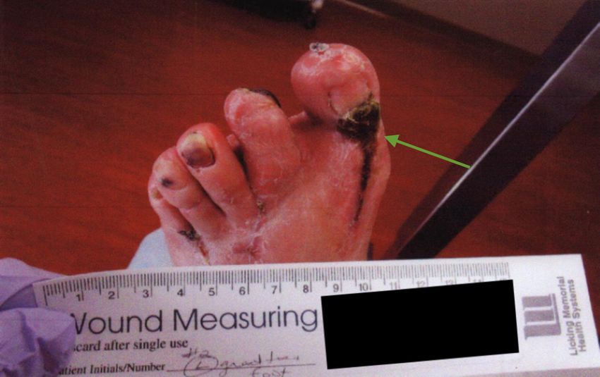
measures to begin the healing process or an extended period of time for the wound to properly
                                   heal.

                                   Orthopedic surgeons trained in foot and ankle surgery and podiatric surgeons trained in wound
                                   care can work together to provide resources and appropriate care for complicated wounds.
                                   Therefore, a tandem management approach using both specialties provides patients with the
                                   appropriate treatment plan to prevent further complications during their recovery.

                                   Case Presentation
                                   Case 1
                                   A 75-year-old female presented to her orthopedic surgeon in May 2015 with a complaint of
                                   bilateral foot pain at the great toe joint. The patient had undergone previous surgeries to
                                   correct the pain but all were unsuccessful. After the consultation, a left gastrocnemius recession
                                   and a left midfoot reconstruction were performed to offload excess weight through the ball of
                                   the foot.

                                   At the postoperative appointment one week after surgery, the skin around the incision
                                   appeared to be ecchymotic without signs of breakdown or infection. Two weeks
                                   postoperatively, there were large blisters with wound dehiscence. The skin appeared broken
                                   down, although the incision was well-approximated. At this time, the patient was referred to a
                                   podiatric surgeon with advanced wound training by her orthopedic surgeon (Figures 1-3).

                                      FIGURE 1: Gross image of initial presentation showing
                                      swelling blisters and wound dehiscence

2019 Carr et al. Cureus 11(11): e6193. DOI 10.7759/cureus.6193                                                                 2 of 10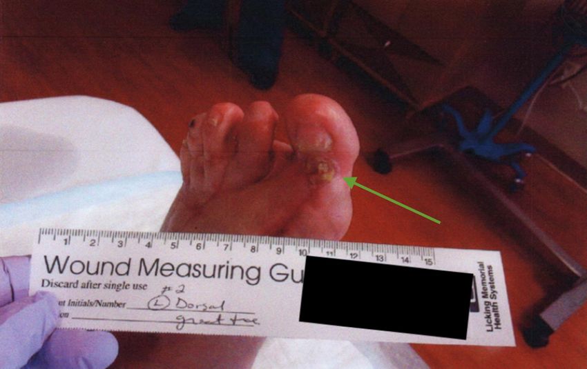
FIGURE 2: Gross image of progress at three weeks showing
                                      healthy granular tissue after necrotic tissue removal

                                      FIGURE 3: Gross image of progress at six months showing
                                      continued healing of the dorsal wound

                                   The wound care protocol for this patient used Adaptic dressing (Johnson and Johnson, NJ, US)
                                   along the staple lines together with Aquacel silver (ConvaTec Group PLC, Deeside, United

2019 Carr et al. Cureus 11(11): e6193. DOI 10.7759/cureus.6193                                                              3 of 10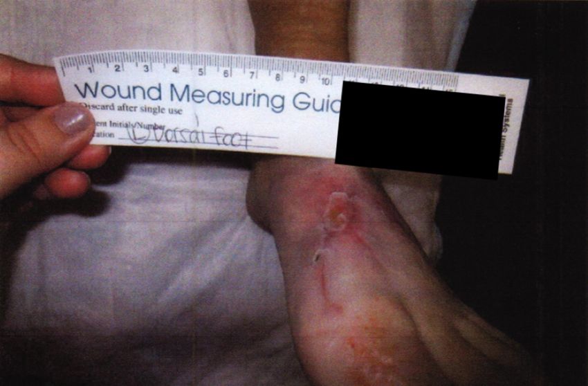
Kingdom) and a non-bordered film dressing to be changed every other day or as needed with
                                   strict non-weight bearing. Five weeks postoperatively, necrotic tissue was removed and healthy
                                   granular tissue was appreciated underneath as well as subtle fibrinogen. In the next visit,
                                   during postoperative week six, negative pressure therapy began and was continued for one
                                   month. Ten weeks postoperatively, silver nitrate was placed on some of the hypertrophic areas
                                   that remained. Eleven weeks postoperatively, a skin graft was placed over the remaining wound
                                   and curettage was performed to debulk some of the remaining slightly thick hypertrophic
                                   tissue. Over the course of the next three months, the wound was cared for by the certified
                                   wound specialist. Debridement and cleaning the wound happened on a weekly basis until the
                                   wound healed and the patient was discharged from care.

                                   Case 2
                                   A 64-year-old man with a past medical history of rheumatoid arthritis underwent an extensive
                                   forefoot reconstruction of his left lower extremity in January 2015 at the hands of his
                                   orthopedic surgeon. The immediate postoperative course was uneventful, but he gradually
                                   developed wound-healing issues. The patient was referred to a podiatric surgeon with advanced
                                   wound training in February 2015 with eschar on the dorsal left foot incision and a second toe
                                   distal eschar with some fibrinogen and suggestion of granular tissue. The patient was previously
                                   shown to have adequate perfusion in the extremity via noninvasive arterial studies (Figures
                                   4-6).

                                      FIGURE 4: Gross image of the initial presentation showing
                                      eschar on the dorsal left foot incision

2019 Carr et al. Cureus 11(11): e6193. DOI 10.7759/cureus.6193                                                               4 of 10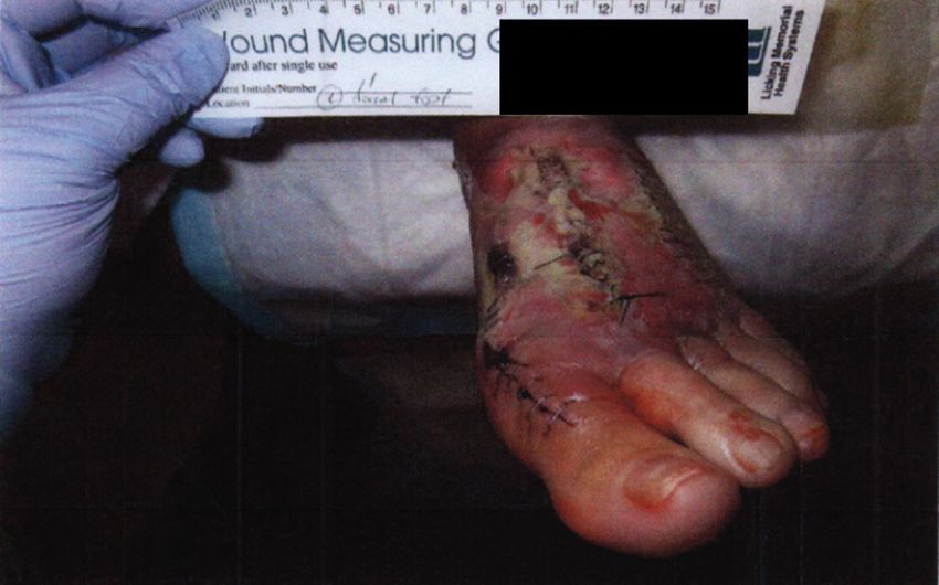
FIGURE 5: Gross image of progress at two months showing
                                      diminished eschar

                                      FIGURE 6: Gross image of progress at three months showing
                                      complete epithelialization

                                   Initially, the patient was treated with clean dressings utilizing AMD gauze (Cardinal Health,
                                   Ohio, US), changed as needed. He was also instructed to continue elevating the leg above heart
                                   level as much as possible. Multiple surgical debridements were performed between late

2019 Carr et al. Cureus 11(11): e6193. DOI 10.7759/cureus.6193                                                               5 of 10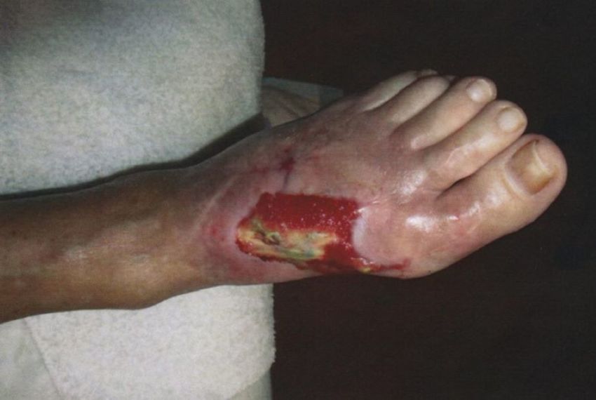
February and early March 2015. Treatment following debridement consisted of antibiotic-
loaded bone cement pellets, daily dressing changes using gentamicin cream, and gauze for the
second toe; Aquacel silver with saline covered by gauze for the great toe was changed every two
or three days in addition to oral antibiotics. Follow-up appointments took place weekly
through March. All wounds were completely epithelialized by May 2015, with some tissue loss.
The patient was discharged and advised to keep the area clean and dry, check it daily, and
follow up with his orthopedic surgeon.
Case 3
A 58-year-old man with a history of diabetic foot disease and Charcot-Marie-Tooth disease
presented to the wound clinic in August 2014 for the further management and surveillance of a
left foot plantar ulceration. At this time, the wound was approximately 6 cm in diameter and
the large, granular deep defect was without significant periwound hyperkeratosis. He
experienced episodes of improvement and worsening throughout the course of this wound.
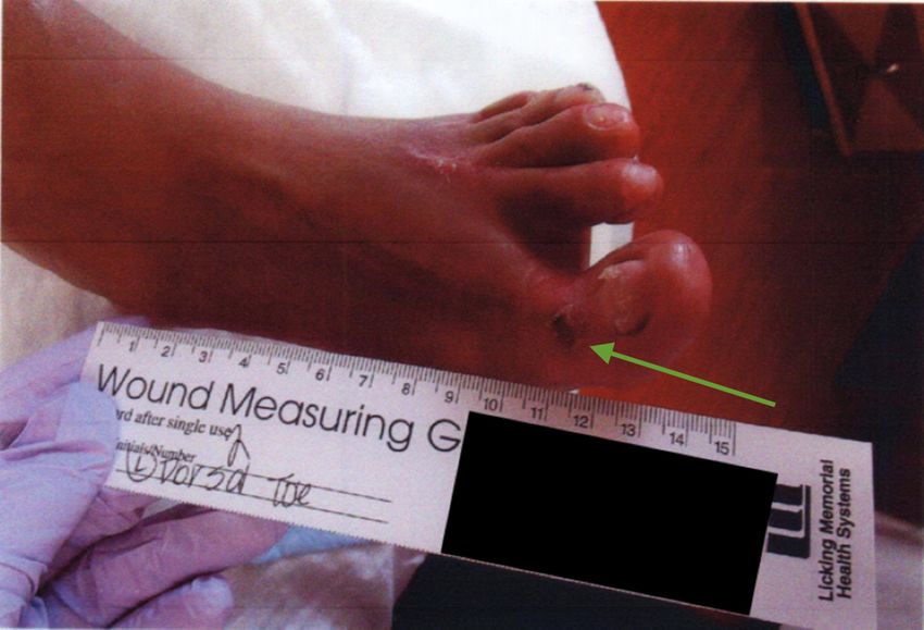
Perfusion was noted and obvious neuropathy existed (Figures 7-9).
FIGURE 7: Gross image showing 6 cm left foot plantar
ulceration
2019 Carr et al. Cureus 11(11): e6193. DOI 10.7759/cureus.6193 6 of 10FIGURE 8: Gross image of progress at three months showing
minimal decrease in ulcer size before gastrocnemius recession
FIGURE 9: Gross image of progress two months after the
tandem operation showing significant improvement in healing
The wound was treated with dressing changes every other day using AMD gauze covered with
Kerlix roll (Cardinal Health). He was also instructed to elevate his legs and to wear an offloading
2019 Carr et al. Cureus 11(11): e6193. DOI 10.7759/cureus.6193 7 of 10shoe with SpandaGrip (Meditech International, FL, US). A Crowe boot (Crowe Boot & Shoe, IL,
US) was also prescribed. Debridement took place in September 2014, in which periwound
hyperkeratosis was taken down. In November 2014, augmentin was prescribed to prevent
infection and CelluTome skin grafting (Acelity, Texas, US) was offered. The patient wished to
proceed with the skin grafting and the appropriate procedure was performed. After the
procedure, wound dressing was applied solely at the wound clinic, and the patient was
instructed to leave dressings on between visits, with follow-up appointments every three to
four days through mid-December. In mid-December, the ulcer was continuing to improve; it
was now approximately 3 cm in diameter. Wound care was allowed to be transferred back to the
patient’s responsibility, with instructions to pack with Aquacel silver and wrap with
gauze. Negative pressure therapy was suggested in February 2015 due to delayed wound
healing and was begun with a wound vacuum at that time.
By mid-March 2015, a plateau phase had been reached in the patient’s wound healing. It was at
this point that a prophylactic surgery was discussed. The patient was a prime candidate for this
procedure because his wound was no longer making significant gains, but he had shown the
capability to heal. The patient had also developed several ulcerations on the second and third
toes of his right foot due to hammertoe contractures. It was suggested to the patient that
hammertoe correction with distal phalangectomy will help offload the distal aspect of the right
toes. At the same time, it was recommended that a gastrocnemius recession be performed on
the left leg to help further offload the patient’s forefoot. The patient agreed to both surgical
interventions.
The patient requested the surgery be delayed until June 2015. Wound management continued
with Aquacel silver wrapped with gauze and the wound vacuum, up to the day of the procedure.
The surgery was performed in a tandem fashion with both an orthopedic surgeon and a wound
specialist performing specific interventions. The patient’s equinus contracture and hammertoe
deformities were successfully corrected surgically, with no intraoperative complications. The
patient developed some stasis ulcerations and toe infections postoperatively, which were
managed with compression therapy, Coban 2 (3M Health Care, Minnesota, US), and Keflex.
Surgical intervention was successful in continuing the progression of the overall healing
process of his left foot plantar ulceration. By mid-August 2015, the plantar ulceration,
hammertoe, and gastrocnemius recession incisions were completely healed and the stasis
ulceration was improving. The patient was placed into a walking boot to prevent further
breakdown until his diabetic shoes were available.
Discussion
The complex nature of chronic wound management provides an opportunity for implementing
a tandem specialty approach. Two specialties that have significant potential for improving
outcomes in patients battling complex lower extremity wounds are podiatric surgeons with
advanced wound training and orthopedic foot and ankle surgeons.
Lower extremity wounds are exceedingly common in patients with comorbidities such as
diabetes mellitus and Charcot-Marie-Tooth disease, with some prevalence estimates
approaching nearly 25% [6]. The effective management of these wounds can ultimately be the
difference between saving or losing a limb [7]. The literature contains multiple examples of the
interdisciplinary management of foot and ankle wounds [7-11], however, we were unable to find
any examples in PubMed of wound cases being managed exclusively between wound specialists,
such as a podiatric surgeon with advanced wound training, and orthopedic surgeons. The three
cases presented in the current case report are a few examples of what can be expected when
patients are efficiently managed within separate, but related, specialties to achieve the most
desirable outcome.
2019 Carr et al. Cureus 11(11): e6193. DOI 10.7759/cureus.6193 8 of 10The most promising feature of the system implemented for these patients is its bidirectional
nature. One direction is best illustrated by patients who developed surgical dehiscence or
significantly delayed postoperative healing (Cases 1-2). Orthopedic surgeons operating on the
foot and ankle are capable of handling many wound complications. However, understanding
that referring the patients who develop major complications to wound specialists who are
experts in the management of complex wounds was the catalyst these patients needed in order
to overcome their wounds. The second direction is demonstrated by the wound specialist
requesting assistance from the orthopedic surgeon to perform surgical interventions as part of
the wound treatment protocol. Case 3 illustrates a patient obviously suffering from forefoot
overloading. There was little likelihood of this patient healing the wound permanently without
finding a way to offload the forefoot region. Performing a gastrocnemius recession was a vital
step in healing the wound.
Conclusions
With complex lower extremity wounds, patient morbidity is greatly affected, and the high risk
of further complications makes management an urgent concern. In this study, tandem
management between orthopedic and podiatric surgeons handling complex lower extremity
wounds resulted in favorable outcomes. This case series serves as an example of how the
interprofessional management of complex medical problems can lead to better outcomes for
patients in foot and ankle surgery.
Additional Information
Disclosures
Human subjects: Consent was obtained by all participants in this study. Conflicts of interest:
In compliance with the ICMJE uniform disclosure form, all authors declare the following:
Payment/services info: All authors have declared that no financial support was received from
any organization for the submitted work. Financial relationships: Dr. Napolitano, Dr. Cheney
declare(s) personal fees from BESPA. Dr. Cheney declare(s) personal fees from Flower
Orthopedics. Dr. Gittins declare(s) personal fees from Stryker Orthopedics. Dr. Napolitano
declare(s) personal fees from Acelity/KCI. Dr. Napolitano declare(s) personal fees from
Organogenesis. Other relationships: All authors have declared that there are no other
relationships or activities that could appear to have influenced the submitted work.
Acknowledgements
The authors thank Nancy Summers, Medical Records Secretary at Licking Memorial Hospital,
for her work in collecting patient records. We also thank Orthopedic & Neurological
Consultants, Inc. and New Albany Podiatry for their support of this project.
References
1. Folk JW, Starr AJ, Early JS: Early wound complications of operative treatment of calcaneus
fractures: analysis of 190 fractures. J Orthop Trauma. 1999, 13:369-372.
2. Forsythe RO, Hinchliffe RJ: Assessment of foot perfusion in patients with a diabetic foot
ulcer. Diabetes Metab Res Rev. 2015, 32:232-238. 10.1002/dmrr.2756
3. Lew DP, Waldvogel FA: Osteomyelitis. N Engl J Med. 1997, 336:198-206.
10.1056/NEJM197001222820406
4. Reiber GE, Vileikyte L, Boyko EJ, del Aguila M, Smith DG, Lavery LA, Boulton AJ: Causal
pathways for incident lower extremity ulcers in patients with diabetes from two settings.
Diabetes Care. 1999, 22:157-162.
5. Cychosz CC, Phisitkul P, Belatti DA, Glazebrook MA, DiGiovanni CW: Gastrocnemius recession
for foot and ankle conditions in adults: evidence based recommendations. Foot Ankle Surg.
2015, 21:77-85.
2019 Carr et al. Cureus 11(11): e6193. DOI 10.7759/cureus.6193 9 of 106. Sumpio BE: Foot ulcers. New Eng J Med. 2000, 343:787-793. 10.1056/NEJM200009143431107
7. Rogers LC, Andros G, Caporusso J, Harkless LB, Mills JL Sr, Armstrong DG: Toe and flow:
essential components and structure of the amputation prevention team. J Am Podiatr Med
Assoc. 2010, 100:342-348. 10.7547/1000342
8. Hingorani A, LaMuraglia GM, Henke P, et al.: The management of diabetic foot: a clinical
practice guideline by the Society for Vascular Surgery in collaboration with the American
Podiatric Medical Association and the Society for Vascular Medicine. J Vasc Surg. 2016, 63:3-
21. 10.1016/j.jvs.2015.10.003
9. Armstrong DG, Bharara M, White M, et al.: The impact and outcomes of establishing an
integrated interdisciplinary surgical team to care for the diabetic foot. Diabetes Metab Res
Rev. 2012, 28:514-518. 10.1002/dmrr.2299
10. Sumpio BE, Armstrong DG, Lavery LA, Andros G: The role of interdisciplinary team approach
in the management of the diabetic foot: a joint statement from the Society for Vascular
Surgery and the American Podiatric Medical Association. J Am Podiatr Med Assoc. 2010,
100:309-311. 10.7547/1000309
11. Cho EH, Garcia R, Pien I, Thomas S, Levin LS, Hollenbeck ST: An algorithmic approach for
managing orthopaedic surgical wounds of the foot and ankle. Clin Orthop Relat Res. 2014,
472:1921-1929. 10.1007/s11999-014-3536-7
2019 Carr et al. Cureus 11(11): e6193. DOI 10.7759/cureus.6193 10 of 10You can also read