BRIEF COMMUNICATION Topical Oxygen Therapy Shifts Microbiome Dynamics in Chronic Diabetic Foot Ulcers
←
→
Page content transcription
If your browser does not render page correctly, please read the page content below

B R I E F CO M M U N I C AT I O N
Topical Oxygen Therapy Shifts Microbiome
 Dynamics in Chronic Diabetic Foot Ulcers

                Volume 32, Number 3, March 2020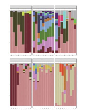
BRIEF COMMUNICATION

Topical Oxygen Therapy Shifts Microbiome
Dynamics in Chronic Diabetic Foot Ulcers
Paul Hunter, BSc1; Elisa Greco, MD2; Karen Cross, MD, PhD1,3; and Julie Perry, PhD1,4

Abstract
Introduction. Bacterial biofilm in wounds prevents healing by acting as a physical barrier to wound closure and hyperactivating local
inflammatory processes, thus making its removal a high priority. The authors previously have shown that adding topical oxygen to standard
wound care increased healing of Texas Grade II and III diabetic foot ulcers (DFUs), which they hypothesized was a result of alterations of
the wound microbiome/biofilm. Objective. This study aims to determine the mechanism of action of topical oxygen in DFUs by examining
the diversity of bacterial genera present in DFUs treated with topical oxygen. Materials and Methods. Six patients with chronic DFUs had
their wounds swabbed weekly over an 8-week period of continuous topical oxygen treatment, and microbiome diversity was assessed by
metagenomic 16S rDNA sequencing using a next-generation sequencing platform. Results. The wound microbiome shifted toward a
diverse flora dominated by aerobes and facultative anaerobes with oxygen therapy in 5 healed wounds. In contrast, anaerobic flora
persisted in a single nonhealing ulcer in the present study cohort. Conclusions. Although the sample size was small, this study suggests
topical oxygen therapy may have the ability to encourage the growth of aerobic members of the wound microbiome and be an effective
alternative to antibiotics in this area.

Key words
microbiome, biofilm, diabetic foot ulcer, topical oxygen, foot ulcer

Index
Wounds 2020;32(3):81–85.

Human skin is normally colonized by a            with other wounds6 and harbors a greater          MATERIALS AND METHODS
consortia of bacteria, fungi, and viruses        number of opportunistic pathogens when            This study was approved by the Research
(together referred to as the skin micro-         compared with contralateral intact skin.7         Ethics Board at St. Michael’s Hospital in
biome), which occupies this niche to             The abundance of anaerobes may be due             Toronto, Canada. Following informed
prevent colonization of the skin by more         to host factors (eg, poor tissue perfusion)       consent, 6 patients with diabetes and
pathogenic bacteria.1 When a break in the        but also may be inherent to the biofilm           DFUs present for more than 4 weeks in
skin occurs, members of the skin micro-          mode of growth.                                   duration were recruited in accordance
biome may colonize the wound as part                Recognizing deep, 3-dimensional                with the inclusion and exclusion criteria
of a developing wound biofilm. Although          wounds are often anaerobic, the authors           listed in Table 1.
the clinical diagnosis of wound biofilm          recently introduced a topical oxygen deliv-          Patients were assessed weekly and re-
remains controversial, it is now recog-          ery device into their wound care practice         ceived best practice standard care tailored
nized that biofilms are present in 60% to        with excellent clinical results.8 The present     to their wound (ie, iodine-based dressings,
100% of chronic wounds,2-4 where they            study evaluated whether topical oxygen            sharp debridement, and offloading as need-
form a physical barrier to wound closure         therapy (TOT) causes shifts in the wound          ed) as well as continuous topical oxygen
and maintain a state of chronic inflamma-        microbiome from anaerobic species toward          via the NATROX Oxygen Delivery System
tion that can damage surrounding tissue.4        an aerobic community, thereby favoring            (ODS; Inotec AMD, Cambridge, UK). This
Biofilm removal is therefore a therapeutic       healing. Given that the wound microbiome          small, battery-operated oxygen generation
priority, but treatment options are limited      represents the building blocks of biofilm         device delivers a continuous stream of
due to the fact that biofilms are intrinsi-      formation, understanding its composition          humidified oxygen to the wound bed via a
cally tolerant to antibiotics.5 The diabetic     and response to treatment is the first step       small tube and sterile adhesive pad worn
foot ulcer (DFU) microbiome is enriched          toward improving treatment of biofilms in         under the dressing. Patients wore the ODS
in anaerobic bacteria when compared              chronic wounds.                                   device 24 hours per day for the full duration

                              Licensed and used with permission from HMP from March 1, 2020 through March 31, 2021.

81                                                                                                                    March 2020 | vol. 32, no. 3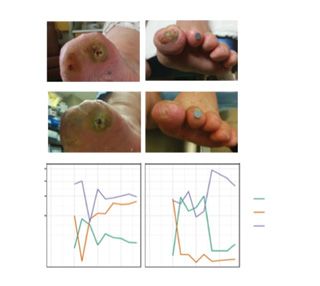
Hunter et al

                                               Table 1. Study inclusion and exclusion criteria
  INCLUSION CRITERIA                                                     EXCLUSION CRITERIA
  DFU >4 wk but 10mg
                                                                         prednisolone) or other second-line immunosuppressant
  Patients aged ≥18 y                                                    Need TCC
  Patients who understand the study, agree to adhere to the              Patients with a known sensitivity to any of the components of the
  treatment, and are able to give consent                                ODS device
  Patients who can be followed by the same investigating team            Patients with known or suspected malignancy in the ulcer or surrounding
  for the whole period of their participation in the study               tissue
                                                                         Patients who do not have the physical or mental capacity or a significant
                                                                         other with the ability to change the ODS battery pack on a daily basis
                                                                         Patients with >10% of the ulcer surface area covered in hard eschar
                                                                         Patients with ulcer surface area of >10cmx10cm
                                                                         Patients participating in another clinical study for ulcer management
                                                                         Patients with a known history of poor compliance with medical treatment
                                                                         Patients unable to understand the aims of the study and not provide
                                                                         informed consent
                                                                         Patients who are pregnant
  DFU: diabetic foot ulcer; wk: week; IV: intravenous; y: year; TCC: total contact cast; ODS: oxygen delivery system

   A                                                                                                               Patient 2        Patient 3
                     0.7                                                                                       A               B
                                                                                                      Week 0
Wound Volume (cm3)

                     0.6                                     Patient 1
                     0.5                                     Patient 4
                     0.4                                     Patient 5
                     0.3
                                                             Patient 6                                         C               D
                     0.2
                                                                                                      Week 8

                     0.1
                      0
                            0   1   2   3    4     5   6      7      8
                                            Week                                                     1.00 E                    F
                                                                                Relative Abundance

                                                                                                     0.75                                       Classification
   B                                                                                                 0.50                                           Aerobic
                                                                                                     0.25                                           Anaerobic
                     4.0                                                                                                                            Facultative
Wound Volume (cm3)

                     3.5
                     3.0                                     Patient 2
                     2.5                                     Patient 3
                     2.0                                                                                           Time (wk)       Time (wk)
                      1.5
                                                                               Figure 2. (A) Patient 2: nonhealed DFU compared with (B) a similarly sized
                     1.0
                                                                               healed wound (patient 3); (C, D) 16S rDNA sequencing was used to probe the
                     0.5
                                                                               microbiome over time while wounds were treated with continuous (24 h/d)
                       0
                                                                               topical oxygen, and the bacteria identified by sequencing were classified into
                            0   1   2   3     4    5    6      7         8
                                                                               aerobic, anaerobic, or facultatively anaerobic categories; (E) anaerobes were
                                            Week
                                                                               the most abundant organisms at the outset in both wounds, and persisted
Figure 1. Clinical results of topical oxygen therapy on wound healing. Wound   in the nonhealed wound; and (F) in contrast, topical oxygen induced a rapid
volume in cm3 was plotted for (A) 4 smaller wounds and (B) 2 larger wounds.    transition to a flora dominated by aerobes in healed wounds.

woundsresearch.com                                                                                                                                            82
Oxygen in Wound Biofilms
Taxa Bar Chart: Genus of the 8-week study so that their DFUs
Patient 1 Patient 2 Patient 3 were continuously exposed to higher than
1.00 ambient concentrations of oxygen. Ulcers
Staphylococcus were photographed and swabbed using
Streptococcus Levine’s technique at each weekly visit.
0.75
Shigella spp/Escherichia coli Genomic DNA was extracted from
Corynebacterium_1 wound swabs as described by Stearns et al,9
0.50 Anaerococcus and the variable region 3 of the 16S rRNA
Haemophilus gene was amplified.10 Positive amplicons
Helcococcus were normalized using the SequalPrep
Relative Abundance (Genus)
0.25
Pseudomonas Normalization Plate Kit (Thermo Fisher
f_Enterobacteriaceae Scientific, Waltham, WA) and sequenced
0.00 Mycoplasma on a MiSeq System (Illumina, San Diego,
0 1 2 3 4 5 6 7 8 0 1 2 3 4 5 6 7 8 0 1 2 3 4 5 6 7 8
Finegoldia CA). Reads were filtered and trimmed
Patient 4 Patient 5 Patient 6 using Cutadapt11 and processed using
Enterococcus
1.00
Neisseria DADA2.12 Taxonomy was assigned using the
urkholderia-Caballeronia-
B
SILVA database version 1.3.2 (Max Planck
0.75 Paraburkholderia Institute for Marine Microbiology and
Peptoniphilus Jacobs University, Bremen, Germany).
Porphyromonas
0.50 RESULTS AND DISCUSSION
Brevibacterium
Citrobacter Outcomes of DFUs
0.25 f_Burkholderiaceae Of the 6 DFUs, 5 healed over 8 weeks of
Other TOT (Figure 1). The DFU that did not
heal in the cohort was 1 of 2 ulcers that
0.00 was significantly larger than the oth-
-1 0 1 2 3 4 5 6 7 8 -1 0 1 2 3 4 5 6 7 8 -1 0 1 2 3 4 5 6 7 8
ers at the outset (Figure 2). Clinically,
Figure 3. Sequencing of the diabetic foot ulcer microbiome over 8 weeks of topical oxygen therapy. the nonhealed ulcer (patient 2; ~2 cm3)
Results are shown for the most abundant 99% of bacterial genera in each sample and expressed as a was on the plantar surface of the first
relative abundance measure.
metatarsal head and had been wrapped
in a compression dressing with acetic
InvSimpson Shannon acid washes for suspected Pseudomonas
10.0
aeruginosa for the 8-week study period
7.5
(Figure 1B). The largest healed ulcer
2.0
(patient 3; ~2.4 cm3) in the study cohort
5.0 was on the first digit (hallux tip) (Figure
1.0 1B), which was offloaded using a DARCO
2.5 shoe (DARCO International, Inc, Hun-
Patient 1 tington, WV) and intermittently cleansed
Diversity
0
-2.5 0 2.5 5.0 7.5 Patient 2 with povidone-iodine when deemed
Simpson Patient 3
clinically necessary. Despite being larger
Patient 4
0.75 Patient 6
than patient 2’s DFU at the outset of
treatment, patient 3 healed completely by
0.50 week 7 of TOT. The clinical course of all 6
wounds in the current study is presented
0.25 in Figure 1.
0
-2.5 0 2.5 5.0 7.5 Microbiome results
Week Sequencing identified typical DFU
pathogens (ie, Staphylococcus, Strepto-
Figure 4. Diversity indices of wound microbiome samples. The diversity of each sample was plotted
coccus, Pseudomonas) as well as some
over time, where higher numbers indicated less diversity. In all diversity measures used, the nonhealing
ulcer of patient 2 was stable and had little change over time. Healing wounds showed large fluctuations less commonly encountered genera (ie,
in diversity over time. Anaerococcus, Helcococcus, Mycoplasma,
83 March 2020 | vol. 32, no. 3Hunter et al
Haemophilus) in all ulcers in the present
study (Figure 3). There was no obvious Table 2. Classification of the most abundant 1% bacteria
association between the presence of a identified by 16S rDNA sequencing
specific pathogen and the duration/sever-
ity of an ulcer. In keeping with the results AEROBES FACULTATIVE ANAEROBES ANAEROBES
of Loesche et al,13 it was found that the Pseudomonas Streptococcus Porphyromonas
microbiome of the 5 healing DFUs in the
Corynebacterium Staphylococcus Peptoniphilus
present study cohort diversified and tran-
Burkholderia-Caballeronia- Mycoplasma Finegoldia
sitioned over time (Figures 3, 4). In con-
Paraburkholderia
trast, the microbiome of the 1 nonhealing
ulcer in the present cohort (patient 2) Brevibacterium Helcococcus Anaerococcus
was the least diverse and did not change Haemophilus
over time despite standard care plus TOT Shigella spp/Escherichia coli
(Figures 3, 4). The most abundant 1% of
organisms identified according to oxygen
utilization were classified (Table 2), and
their abundance over the course of treat-
ment was plotted (Figures 2, 5). Averaged
results from all 6 ulcers demonstrated 0.15
a transition from a flora dominated by
anaerobes at the outset of monitoring to a 0.10
flora rich in aerobic species after 8 weeks
Total Relative Abundance
of continuous (24 hours/day) topical ox-
ygen. In contrast, the wound microbiome Classification
of the nonhealed DFU remained anaero- 0.05 Aerobic
bic, with the exception of a timepoint in Anaerobic
Facultative
which the patient missed their scheduled
acetic acid wash (Figure 2). Although it
is recognized this is a single data point,
it is speculated acetic acid washes may
have sterilized the commensal aerobic
flora that were able to proliferate in other
patients’ healed ulcers.
-3 0 3 6
Week
LIMITATIONS
This study, to the best of the authors’ Figure 5. Mean abundance of each class of microbe across all patients. Wounds were sampled for
knowledge, is the first to document sequencing beginning 3 weeks (-3) before topical oxygen therapy commenced (week 0) to ensure the
changes to a chronic wound microbiome changes in abundance of aerobes were in fact due to TOT and not random fluctuations in the micro-
with TOT. However, the sample size was biome. We found that indeed, there was a clear increase in abundance of aerobic species within 1–2
weeks of topical oxygen therapy with a corresponding decrease in anaerobes.
limited to 6 patients due to the longi-
tudinal nature of the study. Because of
the small sample size, the authors were
unable to determine keystone species flora that more closely resembles that of acknowledgments
common to healing versus nonheal- intact skin. These changes may con- Note: The authors wish to thank Laura Rossi and J.C.
ing wounds. The authors are currently tribute to healing through suppression Szamosi for their expert technical assistance.
conducting a similar, larger trial to map of dysregulated inflammatory cascades Affiliations: 1Division of Plastic Surgery, St. Michael’s
changes in the microbiome on a finer induced by chronic wound colonization. Hospital, Toronto, Canada; 2Division of Vascular
scale and calculate the statistical signifi- Understanding the mechanism-of-action Surgery, St. Michael’s Hospital; 3Associate Scientist,
Keenan Research Centre for Biomedical Science,
cance of the findings. of topical oxygen may help in determin- Toronto, Canada; and 4Faculty of Dentistry, University
ing which patients will benefit most from of Toronto, Toronto, Canada
CONCLUSIONS its use and guide treatment decisions
Correspondence: Julie Perry, PhD, Senior Research
Topical oxygen therapy can enrich aer- by distinguishing responders from Associate, St. Michael’s Hospital, 30 Bond Street,
obes to drive the microbiome toward a non-responders earlier. Toronto, Ontario M5B 1W8; julie.perry@utoronto.ca
woundsresearch.com 84Oxygen in Wound Biofilms
Disclosure: This work was partially funded by Inotec online April 1, 2011]. Appl Environ Microbiol.
AMD (Cambridge, UK). The authors disclose no other 2011;77(11):3846–3852.
conflicts of interest.
11. Martin M. Cutadapt removes adapter sequences
from high-throughput sequencing reads. EMB-
references net.j. 2011;17(1):10–12.
1. Johnson TR, Gómez BI, McIntyre MK, et al. 12. Callahan BJ, McMurdie PJ, Rosen MJ, Han AW,
The cutaneous microbiome and wounds: new Johnson AJA, Holmes SP. DADA2: high-resolu-
molecular targets to promote wound healing. Int tion sample inference from Illumina amplicon
J Mol Sci. 2018;19(9):2699. data [published online May 23, 2016]. Nat
2. Malone M, Bjarnsholt T, McBain AJ, et al. The Methods. 2016;13(7):581–583.
prevalence of biofilms in chronic wounds: a sys- 13. Loesche M, Gardner SE, Kalan L, et al. Temporal
tematic review and meta-analysis of published stability in chronic wound microbiota is associ-
data. J Wound Care. 2017;26(1):20–25. ated with poor healing [published online August
3. Hurlow J, Blanz E, Gaddy JA. Clinical investiga- 24, 2016]. J Invest Dermatol. 2017;137(1):237–244.
tion of biofilm in non-healing wounds by high
resolution microscopy techniques. J Wound
Care. 2016;25 Suppl 9:S11–22.
4. Schultz G, Bjarnsholt T, James GA, et al.
Consensus guidelines for the identification and
treatment of biofilms in chronic nonhealing
wounds [published online December 12, 2017].
Wound Repair Regen. 2017;25(5):744–757.
5. Sønderholm M, Bjarnsholt T, Alhede M, et al.
The consequences of being in an infectious
biofilm: microenvironmental conditions
governing antibiotic tolerance. Int J Mol Sci.
2017;18(12):2688.
6. Citron DM, Goldstein EJ, Merriam CV,
Lipsky BA, Abramson MA. Bacteriology of
moderate-to-severe diabetic foot infections
and in vitro activity of antimicrobial agents
[published online July 3, 2007]. J Clin Microbiol.
2007;45(9):2819–2828.
7. Gontcharova V, Youn E, Yan S, Wolcott RD,
Dowd SE. A comparison of bacterial composi-
tion in diabetic ulcers and contralateral intact
skin [published online March 17, 2010]. Open
Microbiol J. 2010;4:8–19.
8. Yu J, Lu S, McLaren AM, Perry JA, Cross KM.
Topical oxygen therapy results in complete
wound healing in diabetic foot ulcers [published
online November 2, 2016]. Wound Repair Regen.
2016;24(6):1066–1072.
9. Stearns JC, Davidson CJ, McKeon S, et al. Cul-
ture and molecular-based profiles show shifts in
bacterial communities of the upper respiratory
tract that occur with age [published online
January 9, 2015]. ISME J. 2015;9(5):1246–1259.
10. Bartram AK, Lynch MD, Stearns JC, More-
no-Hagelsieb G, Neufeld JD. Generation of
multimillion-sequence 16S rRNA gene libraries
from complex microbial communities by
assembling paired-end illumina reads [published
85 March 2020 | vol. 32, no. 39.4%
US adult population
with diabetes1
13%
US prevalence of
diabetic foot ulceration2
33%
Diabetics over 50 will have
peripheral arterial disease³ 100%
Portable device delivering
continuous humidified oxygen
directly to the wound bed
CLEARED
To find out more
Call: 1 888 354 9772 or email: info@natroxwoundcare.com
AD_US_02/20 (V2)
www.natroxwoundcare.com
1. Centers For Diseases Control and Prevention (2017) National Diabetes Statistics Report. 2. Zhang P, Lu J, Jing Y, et al (2016) Global epidemiology of diabetic foot ulceration:
a systematic review and meta-analysis. Annal of Medicine 49(2) 106-116. 3. NICE Guideline NG19 (2015) Diabetic foot problems: prevention and management.
NATROX® is a registered trade mark of Inotec AMD Limited in EU, China, USA & UK
Ground Floor Unit 7340, Building 7300 | Cambridge Research Park | Waterbeach | Cambridge | Cambridgeshire | CB25 9PD | United KingdomYou can also read