Clinical Outcomes of U-blade Gamma3 Nails Used to Treat Patients with Trochanteric Fractures: Retrospective Multicenter Study
←
→
Page content transcription
If your browser does not render page correctly, please read the page content below
Print ISSN 2287-3260
               ORIGINAL ARTICLE                                                                                       Online ISSN 2287-3279
               Hip Pelvis 31(2): 95-101, 2019
               http://dx.doi.org/10.5371/hp.2019.31.2.95

  Clinical Outcomes of U-blade Gamma3 Nails
Used to Treat Patients with Trochanteric Fractures:
         Retrospective Multicenter Study
            Jehyun Yoo, MD, PhD, Sangmin Kim, MD, PhD*, Hojung Jung, MD, Jihyo Hwang, MD, PhD�
                  Department of Orthopaedic Surgery, Hallym University Sacred Heart Hospital,
                              Hallym University School of Medicine, Anyang, Korea
                Department of Orthopaedic Surgery, Korea University Guro Hospital, Seoul, Korea*
              Department of Orthopaedic Surgery, Hallym University Gangnam Sacred Heart Hospital,
                              Hallym University School of Medicine, Seoul, Korea�

   Purpose: This study was performed to assess the radiologic and clinical results of U-blade Gamma3 nail use for
   the treatment of trochanteric fractures.
   Materials and Methods: Between September 2015 and May 2018, all patients aged 65 years and older who
   underwent surgery with U-blade Gamma3 nails were analyzed. A total of 129 patients were selected based on
   having at least six months of follow-up. Image evaluations included bone quality (T-score), fracture classification
   on plain radiograph (AO/OTA), computed tomography configuration, union period, position of lag screw,
   anatomical reduction, tip apex distance (TAD), sliding extent of lag screw, change of neck shaft angle, and
   complications leading to reoperations were analyzed. Functional outcome were assessed using the Koval grade
   (ambulatory ability) at the final follow-up.
   Results: The mean time to union was 19.7 (range, 6-36) weeks. The screw position was centric (93 cases;
   72.1%) and anatomical reduction was achieved in 74 cases (57.4%). The mean TAD was 20.3 (range, 12.3-38.1)
   mm. The mean sliding length of the lag screws was 3.8 (range, 0.1-12.6) mm. The mean change of neck shaft
   angle was 3.4。(range, 0-12.8。). Reoperations were required in two cases (1.6%) due to the cutting out of the lag
   screw (n=1) and metal failure with U-blade bending (n=1). Finally, Koval grades for 49.8% of patients reached
   preoperative status.
   Conclusion: Overall, use of the U-blade Gamma3 nail led to favorable clinical results, suggesting that this system
   may be a good option for the treatment of trochanteric fractures.

   Key Words: U-blade, Gamma3 nail, Trochanteric fracture, Femur

Submitted: February 17, 2019 1st revision: April 23, 2019         This is an Open Access article distributed under the terms of the Creative
Final acceptance: May 1, 2019                                     Commons Attribution Non-Commercial License (http://creativecommons.
Address reprint request to                                        org/licenses/by-nc/4.0) which permits unrestricted non-commercial use,
Jihyo Hwang, MD, PhD                                              distribution, and reproduction in any medium, provided the original work is
(https://orcid.org/0000-0002-9141-9856)                           properly cited.
Department of Orthopaedic Surgery, Hallym University Gangnam
Sacred Heart Hospital, Hallym University College of Medicine, 1
Singil-ro, Yeongdeungpo-gu, Seoul 07441, Korea
TEL: +82-2-829-5165 FAX: +82-2-834-1728
E-mail: hwangjihyo7309@gmail.com

Copyright ⓒ 2019 by Korean Hip Society                                                                                                     95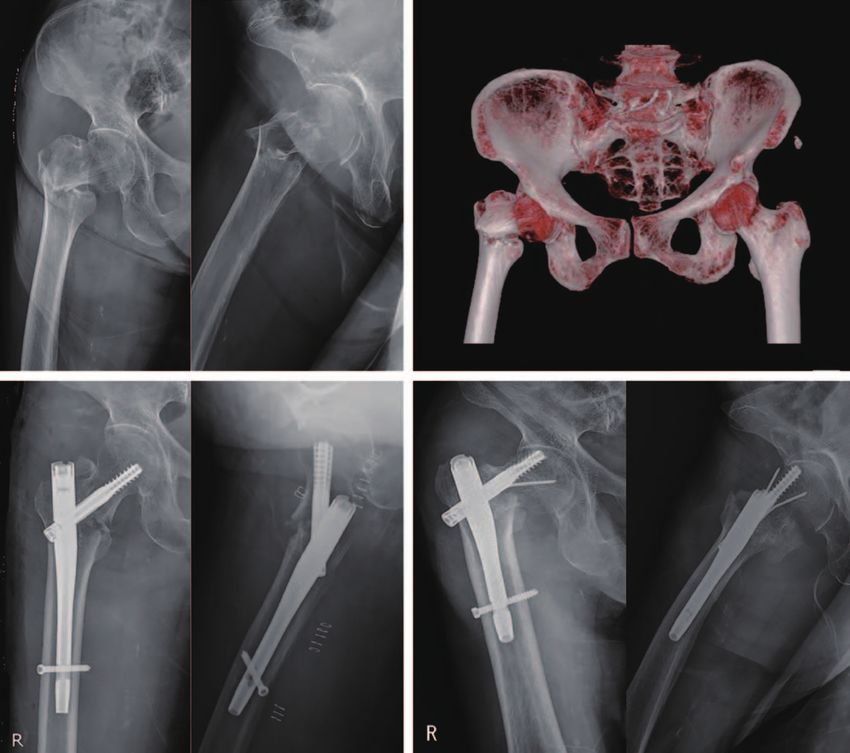
Hip Pelvis 31(2): 95-101, 2019

INTRODUCTION                                                     functional result of the trochanteric fracture patients who
                                                                 underwent internal fixation using the U-Blade Gamma3
  Trochanteric fractures are generally associated with           lag screw in the orthopedic departments of the three
osteoporosis and old age, therefore early treatment, accurate    different hospitals between September 2015 and May
reduction, and internal fixation are crucial to maximize         2018 were investigated. In total, data from 129 patients
clinical results. In particular, functional recovery through     (61, 48, and 20 patients at each hospital) were retrospectively
early weight-bearing is an important prognostic indicator        collected. The inclusion criteria were: 1) patient greater
of a favorable prognosis. Compression hip screws have            than 65 years of age, 2) patients had at least six months
less favorable results compared with intramedullary nail         of follow-up periods, 3) non-pathologic fractures except
fixation and are known to have fewer biomechanical               osteoporosis, 4) low energy fractures (e.g., fall from a
advantages than intramedullary nail, particularly as noted       standing height), and 5) preinjury independent walker
with instability and the risk of severe osteoporotic             (Koval grade,
Jehyun Yoo et al. Clinicall Results of U-blade Gamma3 Nail

comminution, and 3) basal neck type fractures. Radiological        continuity of the medial cortex and the anterior cortex.
outcomes assessed were: 1) union, 2) lag screw position and        When anatomical reduction was not maintained, positive
change, and 3) basal neck type fracture (Fig. 2). For functional   medial cortex5) and valgus alignment in anteroposterior
outcome, the recovery rate of functional ability (using Koval      fluoroscopic and posteriorly angulation than anterior
grade) was analyzed. The lag screw position in the femoral         angulation in lateral C-arm were accepted. When the
head was measured using the tip apex distance (TAD) and            fracture gap or step displacement remained, or when the
described as centric and eccentric. Centric position was           medial cortex gap was expanded during the insertion of the
defined as a lag screw that was simultaneously positioned          lag screw, the lag screw driver thumbwheel was gently
in the center in the anteroposterior and lateral views. TAD        tightened to compress the gap, and consequently, apposition
value, position, and reduction quality were evaluated from         was achieved. After the surgery, when the subjects could
the anteroposterior and lateral images taken immediately           already bear the pain in a sitting position, they were asked
after the surgery. The sliding distance and neck shaft angle,      to try to stand using a tilt table mostly one to three days
which can represent shortening and varus changes of the            after the surgery. Walking was allowed when the pain
fracture site, were calculated as a difference between the         became tolerable. Various weight-bearing training exercises
initial and final plain radiographs. All parameters which          were accomplished according to the subject’s pain level
were obtained in the plain radiographs were adjusted by            and fracture type. Follow-up occurred at 1, 3, 6, and 12
the calibration of magnification of real size and angle of the     months postoperatively and yearly thereafter.
inserted implants. For fracture union, a clinical union was
determined to be achieved when the pain scale improved             RESULTS
during the follow-up period and the absence of pain or
tenderness during weight-bearing. Radiological union was             The mean follow-up period was 12.8 months (range,
determined to be achieved when the fracture line was lost          6-34 months). Of the 129 patients included, 90 (69.8%)
and three or more external or internal calluses were formed        were females and their mean age was 77.4 years (range,
in the anteroposterior and lateral radiological images taken       65-93 years). The mean time lapse between admission and
during the follow-up period2). For functional outcome, ability     surgery was 2.9 days (range, 0-32 days); however, 40
status was evaluated using the Koval grade3,4) (Table 1). The      patients (31.0%) underwent operations within one day.
preinjury walking ability before surgery was compared to           Fractures were classified according to the AO/OTA fracture
the postinjury walking ability at the final visit. The recovery    classification scale revealing the following ratio: A1
rates were calculated.                                             (n=40), A2 (n=76), and A3 (n=13). A stable fracture (A11-

1. Surgical Method

  Operations were carried out in a general manner. When
the manual reduction was unsatisfactory, a long Kelly,
bone hook or Hohmann retractor was used to compress the
lateral cortex or the anterior cortex or pulling the medial
cortex for the reduction (Fig. 3). No invasive reductions
were required. Manual reduction was used to maintain

Table 1. Categories of Ambulatory Ability
 1. Independent community ambulator
 2. Community ambulator with cane
 3. Community ambulator with walker/crutches
 4. Independent household ambulator
 5. Household ambulator with cane
 6. Household ambulator with walker/crutches
 7. Nonfunctional ambulator*
                                                                   Fig. 3. Fluoroscopic image of pulling the displaced medial
* Used only after fracture ambulation.                             cortex using a bone hook.

www.hipandpelvis.or.kr                                                                                                     97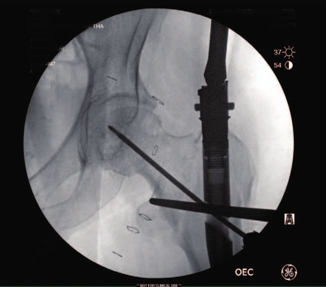
Hip Pelvis 31(2): 95-101, 2019
21) was observed in 59 cases (45.7%), and an unstable delay in union; however, healing was accomplished without
fracture (A22-33) in 70 cases (54.3%). Upon CT evaluation, any additional medication or procedures. The mean fracture
GT comminution was noted in 39 cases (30.2%) and basal union period was 19.7 weeks (range, 6-36 weeks). The mean
neck type fractures were noted in 29 cases (22.5%). For TAD was 20.3 mm (range, 12.3-38.1 mm) and 64 (49.6%)
osteoporosis assessments, quantitative CT and dual-energy had TAD of 10 to 20 mm. The screws were coded as being
X-ray absorptiometry (DEXA) were used. The mean body in the centric position in 93 cases (72.1%); the remaining
mass index was 22.21 kg/m2 (range, 13.67-33.73 kg/m2). 36 were in eccentric positions. Anatomical reductions were
The mean T-score was –2.68 (range, –0.9 to –5.5). The achieved in 74 (57.4%) cases; the remaining achieved non-
pre-operative American Society of Anesthesiologists anatomical reductions. The mean sliding distance was 3.8
(ASA) status was 2.3 (range, 1-4). The mean operative mm (range, 0.1-12.6 mm), and only six (4.7%) patients were
time from the skin incision to the nail insertion and the over 10 mm; there were no additional operations required
skin suture was 57.4 minutes (range, 25-160 minutes); due to over sliding. The mean change of neck shaft angle
the mean estimated blood loss was 255.6 mL (range, 50- was 3.4。(range, 0-12.8。). All subjects have varus changes.
1,000 mL), and the mean blood transfusion amount was A significant change was defined as a change of 10。or
0.09 L (range, 0-0.94 L). In total, 95 patients were moved more, but no complications (significant shortening, nail
to general wards after their surgeries and 34 (26.4%) breakage, non-union) were observed, so even a 10。or greater
patients were transferred to the surgical intensive care unit. varus deformity was not included as a complication. Re-
All of the ASA 3 and 4 patients and some of ASA 2 surgery was required in two subjects (1.6%). One presented
patients were transferred to ICU. No subjects died during with cut out of the femoral head at three months after
the hospital course (or admission periods). Bone union was operation (Fig. 4) and the other was a metal failure due
achieved in 127 subjects (98.4%). One patient experienced a to secondary injury at one month after surgery (Fig. 5);
A B
C D
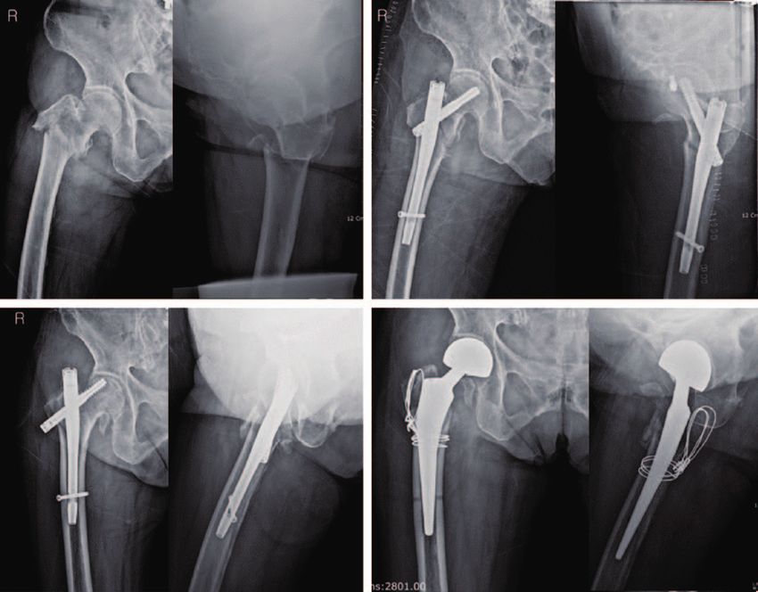
Fig. 4. (A
A) An 83-year-old woman suffered from a fall from height at home; the fracture type was reverse oblique (A32). (B
B)
The fragment was reduced using a U-blade Gamma3 nail. (C C) The lag screw was cut out of the head three months
D) The reoperation was converted to hemiarthroplasty.
postoperatively. (D
98 www.hipandpelvis.or.krJehyun Yoo et al. Clinicall Results of U-blade Gamma3 Nail
A B
C D
Fig. 5. (A
A) A 93-year-old woman presented with a stable fracture (A11). (B B) The three dimensional computed tomography
C) The reduction was controlled using a U-blade Gamma3 nail (170 mm, 125。). (D
scan revealed basilar neck fracture type. (C D)
The lag screw was cut out and failure of U-blade occurred at one month postoperatively.
both were converted to hemiarthroplasties. According to clinical experience, its initial model is associated with
the pre-operative fracture classification, there was one complications such as femoral shaft fracture, cut out of
case of a stable fracture (A11), basal neck fracture type the lag screw from the femoral head, and femoral pain1,8,9).
with T score of –3.5; and the other one case was unstable Kim et al.10) reported that the conventional Gamma3 nail
(A32) with T score of –1.9. The functional ability for walking was not significantly better at treating intertrochanteric
was compared to the preoperative status to the status of fractures than other available options (e.g., DLT [Dyna
final visits. The mean Koval grade at final visits was 2.7 Locking Trochanteric Nail] or PFNA [Proximal Femoral
(range, 1-7). The overall recovery rate of ambulatory status Nail Antirotation]). The recent U-blade Gamma3 nail is
was 49.8% (Table 2). one of the most innovative nails among the nail systems,
including the Asia-Pacific-type nails which were introduced
DISCUSSION in 2010 in South Korea. The U-blade Gamma3 nail was
introduced in 2015, and was designed to prevent rotation;
The initial Gamma nail model, a screw-type intramedullary however, no clinical trials describing the risk-benefit profile
nail, has a minimal bending stress applied to the internal have been published11). While the current study is not a
fixator, allowing for rigid stability without anatomical comparison study, the clinical outcomes appear favorable.
reduction of the posteromedial cortex, and subsequent early Various implants have been designed to improve rotational
weight-bearing6,7). Although the Gamma nail has been used stability in unstable femur fractures12,13). For the effort of
for almost 30 years and has thus garnered accumulated reducing the implant related complications such as lag
www.hipandpelvis.or.kr 99Hip Pelvis 31(2): 95-101, 2019
Table 2. Clinical Outcomes Associated with Use of U-blade surgery. In the case of the distal locking screw, the short
Gamma3 Nails metal nail caused less discrepancy with the jig, and wrong
Variable U blade Gamma3 (n=129) or difficult insertion of the distal locking screw did not occur.
Accordingly, complications such as stress concentration
Age (yr) 77.4 (65-93)
Sex (female: male) 90:39 fractures around the tips of the nails or the interlocking
AO classification screws can be avoided. Compared with the typical 240-,
31 A1 40 (31.0) 200-, and 180-mm nails, the surgical procedure is more
31 A2 76 (58.9) convenient and has fewer complications. In a study by
31 A3 13 (10.1) Rosenblum et al.7), a decrease in the stability of the fracture
Computed tomography
GT comminution 39 (30.2)
site reduces the calcar femoral load and thus increases
Basal neck type fracture 29 (22.5) the concentration of the distal portion of Gamma nail.
Body mass index (kg/m2) 22.21 (13.67-33.73) Furthermore, the excessive reaming required for larger
BMD (T-score) –2.68 (–0.9 to –5.5) nails weakens the femoral shaft, which is vulnerable to
ASA 2.3 (1-4) fracture during the operation or postoperative period.
Operation time (min) 57.4 (25-160)000
According to Halder6), when reaming is excessively conducted
EBL (mL) 255.6 (50-1,000)00.
Transfusion (L) 0.09 (0-0.9)
for Gamma nail insertion, stress shielding occurs to cause
ICU transfer 34 (26.4) a fatigue fracture in distal portion of nail, and it takes long
Union (wk) 19.7 (6-36) period for surgeons to be skillful with this method. The
TAD (mm) 20.3 (12.3-38.1)0 Gamma nail is prone to be implant-related femur fractures18).
Position of lag screw In our study, there were no reports of peri-implant fracture
Centric 93 (72.1)
or thigh pain. The short length (170 mm) of the Gamma3
Eccentric 36 (27.9)
Reduction quality nail is advantageous relative to this potential complication.
Anatomical 74 (57.4) In a biomechanical study, Wang et al.19) demonstrated the
Non-anatomical 55 (42.6) superiority of one lag screw design over the two lag screws
Sliding distance (mm) 3.8 (0.1-12.6)0 design regarding cut out and antirotation. Recently, Chinzei
Neck shaft angle (�) 3.4。(0-12.8。)00. et al.13) reported that the gliding nail, with its specific non
Fixation failure 02 (01.6)
cylindric blade design, provides the most favorable
Cause 1: cut-out, 1: metal failure
Recovery of Koval grade (%) 49.8 antirotation results compared to cylindric helical blade or
lag screws. According to the clinical experience of the
Values are presented as median (range) or number (%).
authors, it has been a recognized disadvantage of the U-
GT: greater trochanter, BMD: bone mineral density, ASA:
American Society of Anesthesiologists, EBL: estimated blade Gamma3. This system may be more time consuming
blood loss, ICU: intensive care unit, TAD: tip apex distance. due to the management of additional U-blade. The metal
failure related to the U-blade was reported as a case report
screw cutout, cut thorough and pull out, many different by Choi et al20). In this case, the reduction quality was poor,
companies have developed novel lag screw designs. The the TAD was outside of the normal range, and the lag screw
U-blade Gamma3 nail is the most evolved option and has was too low in the anteroposterior view of plain radiograph
the most clinical experience. Although though there is no and anterior in the lateral plain radiograph, so these factors
objective comparative data in this study to support its may contribute the lag screw failure. Unlike the typical
superiority to other options, only two cases (1.6%) of metal nails, the modified nails were made of titanium and
fixation failure suggest that the new U-blade Gamma3 were 170 mm long (down from 180 mm), and the distal
design is associated with favorable results and that it may diameter decreased from 11 to 10 mm. As a result, a less
successfully replace the previous Gamma3 nail design. invasive surgical procedure could be achieved, the levels
In particular, the femoral shaft fracture is the most of the post-operative thigh pain and the stress riser fracture,
serious peri- and post-operative complication associated which were caused by the abutment between the metal
with wrong procedures and poorly designed nails14). The nails and the femoral internal bone, decreased21). However,
incidence of post-operative shaft fractures has been when used for fractures extended to the subtrochanteric
reported to range from 0% to 12%9,15-17); however, in this area, it may be unstable after the surgery due to the shortened
study, no shaft fractures developed during or after the length of 170 mm, due to which a refracture may develop.
100 www.hipandpelvis.or.krJehyun Yoo et al. Clinicall Results of U-blade Gamma3 Nail
In these cases, long Gamma nails must be used, another key element in stability reconstruction for the unstable
potential disadvantage of this system. pertrochanteric hip fractures. Arch Orthop Trauma Surg.
2015;135:811-8.
We acknowledge major limitations in this study. First,
06. Halder SC. The Gamma nail for peritrochanteric fractures.
the number of patients was limited and it was difficult to J Bone Joint Surg Br. 1992;74:340-4.
follow-up because the patients were geriatric. If a patient 07. Rosenblum SF, Zuckerman JD, Kummer FJ, Tam BS. A
died or could not be followed properly, treatment outcomes biomechanical evaluation of the Gamma nail. J Bone Joint
could not be fully evaluated. Therefore, the results of Surg Br. 1992;74:352-7.
08. Williams WW, Parker BC. Complications associated with
this analysis might not be sufficiently suitable for the the use of the gamma nail. Injury. 1992;23:291-2.
interpretation of clinical results. Second, surgeons were 09. Hesse B, Gachter A. Complications following the treatment
in different hospitals, so any bias may remain and interfere of trochanteric fractures with the gamma nail. Arch Orthop
with an accurate interpretation of results. Lastly, the data Trauma Surg. 2004;124:692-8.
10. Kim SS, Lee KY, Kim CH, et al. Comparison of the Dyna
from the plain radiographs (e.g., neck shaft angle, sliding locking trochanteric nail, proximal femoral nail antirotation
distance, TAD, reduction quality, or screw position) are and gamma 3 nail in treatment of intertrochanteric fracture
continuous variables which may be associated with inter- of the femur. Hip Pelvis. 2013;25:211-9.
observer variability. 11. Lang NW, Arthold C, Joestl J, et al. Does an additional
antirotation U-Blade (RC) lag screw improve treatment of
AO/OTA 31 A1-3 fractures with gamma 3 nail? Injury. 2016;
CONCLUSION 47:2733-8.
12. Mereddy P, Kamath S, Ramakrishnan M, Malik H,
The U-blade Gamma3 nail can be used to treat proximal Donnachie N. The AO/ASIF proximal femoral nail antirotation
(PFNA): a new design for the treatment of unstable proximal
femoral trochanteric fracture with favorable results. When femoral fractures. Injury. 2009;40:428-32.
using this nail, accurate reduction, an appropriate lag screw 13. Chinzei N, Hiranaka T, Niikura T, et al. Comparison of the
length, and accurate positioning of the lag screw in the sliding and femoral head rotation among three different
femoral head can be achieved. A 1.6% cut out rate is very femoral head fixation devices for trochanteric fractures.
Clin Orthop Surg. 2015;7:291-7.
encouraging despite the metal failure by secondary trauma.
14. Chevalley F, Gamba D. Gamma nailing of pertrochanteric
Analysis of additional cases with longer follow-up will and subtrochanteric fractures: clinical results of a series
help provide a more comprehensive evaluation of this of 63 consecutive cases. J Orthop Trauma. 1997;11:412-5.
newly designed U-blade Gamma3 nail. 15. Goldhagen PR, O'Connor DR, Schwarze D, Schwartz E. A
prospective comparative study of the compression hip screw
and the gamma nail. J Orthop Trauma. 1994;8:367-72.
CONFLICT OF INTEREST 16. Friedl W, Colombo-Benkmann M, Dockter S, Machens
HG, Mieck U. [Gamma nail osteosynthesis of per- and
The authors declare that there is no potential conflict subtrochanteric femoral fractures. 4 years experiences and
of interest relevant to this article. their consequences for further implant development]. Chirurg.
1994;65:953-63. German.
17. Forthomme JP, Costenoble V, Soete P, Docquier J. [Treatment
REFERENCES of trochanteric fractures of the femur using the gamma nail
(apropos of a series of 92 cases)]. Acta Orthop Belg. 1993;
01. Stapert JW, Geesing CL, Jacobs PB, de Wit RJ, Vierhout 59:22-9. French.
PA. First experience and complications with the long Gamma 18. Robinson CM, Adams CI, Craig M, Doward W, Clarke MC,
nail. J Trauma. 1993;34:394-400. Auld J. Implant-related fractures of the femur following
02. Corrales LA, Morshed S, Bhandari M, Miclau T 3rd. Variability hip fracture surgery. J Bone Joint Surg Am. 2002;84-A:
in the assessment of fracture-healing in orthopaedic trauma 1116-22.
studies. J Bone Joint Surg Am. 2008;90:1862-8. 19. Wang CJ, Brown CJ, Yettram AL, Procter P. Intramedullary
03. Koval KJ, Skovron ML, Aharonoff GB, Meadows SE, femoral nails: one or two lag screws? A preliminary study.
Zuckerman JD. Ambulatory ability after hip fracture. A Med Eng Phys. 2000;22:613-24.
prospective study in geriatric patients. Clin Orthop Relat Res. 20. Choi K, Kim Y, Zhou S, Hwang J. Failure of a rotation control
1995;(310):150-9. Gamma 3 lag screw used to treat a trochanteric fracture.
04. Koval KJ, Aharonoff GB, Rosenberg AD, Bernstein RL, Hip Pelvis. 2018;30:129-33.
Zuckerman JD. Functional outcome after hip fracture. Effect 21. Loch DA, Kyle RF, Bechtold JE, Kane M, Anderson K,
of general versus regional anesthesia. Clin Orthop Relat Res. Sherman RE. Forces required to initiate sliding in second-
1998;(348):37-41. generation intramedullary nails. J Bone Joint Surg Am. 1998;
05. Chang SM, Zhang YQ, Ma Z, Li Q, Dargel J, Eysel P. 80:1626-31.
Fracture reduction with positive medial cortical support: a
www.hipandpelvis.or.kr 101You can also read