Suppression of Adrenal Function by Low-dose Prednisone: Assessment with 24-hour Urinary Steroid Hormone Profiles - A Review of Five Cases
←
→
Page content transcription
If your browser does not render page correctly, please read the page content below
Adrenal Suppression                                                                                           Case Study

         Suppression of Adrenal Function by
               Low-dose Prednisone:
      Assessment with 24-hour Urinary Steroid
      Hormone Profiles - A Review of Five Cases
                                                   Patrick N. Friel, BS; Thomas Alexander MD;
                                                                        Jonathan V. Wright, MD

Abstract                                                  significant adverse consequences in terms of sex
The impact of the synthetic glucocorticoid                hormone production, bone health, endocrine and
prednjsone on adrenal steroid hormone                     immune system function, and neuropsychiatric
production was examined using 24-hour urinary             status. Studies of DHEA replacement in patients
steroid hormone profiling. Five women, who                taking prednisone for lupus demonstrate
were chronically taking low-dose prednisone,              amelioration of some of these adverse effects.
were tested, and the relevant literature was              {Altern Med Rev 2006;11 (1 ):40-46)
reviewed. As expected, adrenal glucocorticoid
production, measured by urinary terminal                  Introduction
cortisol and cortisone metabolites, was markedly                   The synthetic glucocorticoid prednisone is
suppressed compared to reference range values             used for its anti-inflammatory properties in the treat-
(p=0.03). Urinary cortisol and cortisone, reflecting      ment of many different conditions, including rheu-
circulating gtucocorticoids, were decreased to            matologic, hormonal, allergic, respiratory, and other
a lesser extent than their terminal metabolites.          disorders. Prednisone administration results in nu-
Urinary dehydroepiandrosterone{DHEA)excretion             merous side effects., a number of which are the result
was dramatically suppressed (p=0.03), while the           of its suppression of adrenal cortex function. After
downstream androgen metabolites androsterone              prednisone administration, adrenal production of the
and etiocholanolone were suppressed to a lesser           endogenous glucocorticoids, cortisol and cortisone,
extent. Aldosterone and tetrahydrocorticosterone          declines. This can result in marked hypocortisolism
production demonstrated modest suppression                after discontinuation of high dose prednisone thera-
after prednisone administration, but allo-                py, especially in seriously ill patients.'
tetrahydrocorticosterone, which is highly
                                                                   Because prednisone suppresses adrenal
sensitive to adrenocorticotropic hormone (ACTH)
                                                          function by reducing adrenocorticotropic hormone
secretion, was suppressed to a greater extent.
                                                          (ACTH) secretion by the anterior pituitary gland.
Prednisone administration results in a decrease
in ACTH secretion by the anterior pituitary,
suppressing synthesis of glucocorticoids, DHEA,
and DHEA metabolites. Decreased glucocorticoid            Patrick N. Friel, BS - Forensic Toxicologist, Washington State Toxicology
synthesis is adaptive, because prednisone is active       Laboratory.
                                                          Correspondence address: 2203 Airport Way South, Seattle. WA 98134
at the glucocorticoid receptor, but suppression of        E-mail: Pat.Friel@wsp,wa.gov
DHEA synthesis is not mitigated by prednisone.            Thomas Alexander, MD - Private practice, Tahoma Clinic, Renton, WA; Clinical
DHEA is an important sex hormone precursor,               Consultant. Meridian Valley Laboratory, Renton, WA.
neurosterold, and endocrine and immune                    Jonathan V. Wright, MD - Medical Director, Tahoma Clinic, Renton, WA; Medical
modulator; therefore, DHEA depletion may have             Director, Meridian Valley Laboratory, Renton, WA.

Page 40                                           Alternative Medicine Review • Volume 11, Number 1 • 2006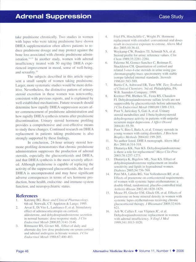
Case Study Adrenal Suppression
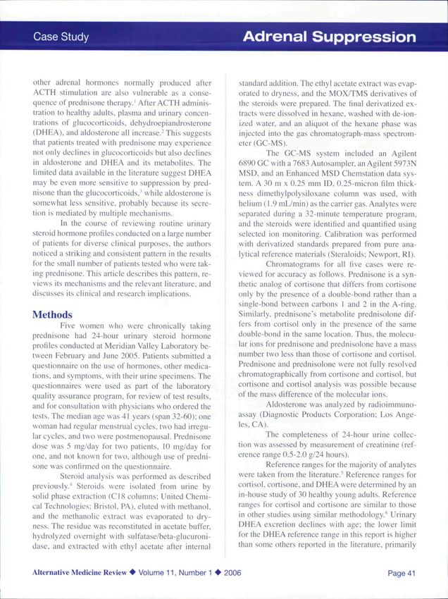
other adrenal hormones normally produeed after standard addition. The ethyl acetate extract was evap-
ACTH sthnulation are also vulnerable as a conse- orated to dryness. and the MOX/TMS derivatives of
quence of prednisone therapy.' After ACTH adminis- the steroids were prepared. The final derivatized ex-
tration to healthy adults, plasma and urinary concen- tracts were dissolved in hexane. washed with de-ion-
trations of glucocorticoids. dehydroepiandrosterone ized water, and an aliquot of the hexane phase was
(DHEA). and aldosterone all increase.- This suggests injected into the gas chromatograph-mass spectrom-
that patients treated with prednisone may experience eter (GC-MS).
not only declines in glucocorticoids but also declines The GC-MS system included an Agilent
in aldosterone and DHEA and its metabolites. The 6890 GC with a 7683 Autosampler. an Agilent 5973N
limited data available in the literature suggest DHEA MSD. and an Enhanced MSD Chemstation data sys-
may be even more sensitive to suppression by pred- tem. A 30 m X 0.25 mm ID. 0.25-micron film thick-
nisone than the glucocorticoids.' while aldosterone is ness dimethylpolysiloxane column was used, with
somewhat less sensitive, probably because its secre- helium (1.9 mL/min) as the carrier gas. Analytes were
tion is mediated by multiple mechanisms. separated during a 32-minute temperature program,
in the course of reviewing routine urinary and the steroids were identified and quantified using
steroid hormone profiles conducted on a large number selected ion monitoring. Calibration was performed
of patients for diverse clinical purposes, the authors with derivatized standards prepared from pure ana-
noticed a striking and consistent pattern in the results lytical reference materials (Steraloids; Newport. RI).
for the small number of patients tested who were tak- Chromatograms for all five cases were re-
ing prednisone. This article describes this pattern, re- viewed for accuracy as follows. Prednisone is a syn-
views its mechanisms and the relevant literature, and thetic analog of cortisone that differs from cortisone
discusses its clinical and research implications. only by the presence of a double-bond rather than a
single-bond between carbons I and 2 in the A-ring.
Methods Similarly, prednisone's metabolite prednisolone dif-
Five women who were chronically taking fers from cortisol only in the presence of the same
prednisone had 24-hour urinary steroid hormone double-bond in the same location. Thus, the molecu-
profiles conducted at Meridian Valley Laboratory be- lar ions for prednisone and prednisolone have a mass
tween February and June 2005. Patients submitted a number two less than those of cortisone and cortisol.
questionnaire on the use of hormones, other medica- Prednisone and prednisolone were not fully resolved
tions, and symptoms, with their urine specimens. The chromatographically from cortisone and cortisol. but
questionnaires were used as part of the laboratory cortisone and cortisol analysis was possible because
quality assurance program, for review of test results, of the mass difference of the molecular ions.
and for consultation with physicians who ordered the Aldosterone was analyzed by radioimmuno-
tests. The median age was 41 years (span 32-60); one assay (Diagnostic Products Corporation; Los Ange-
woman had regular menstrual cycles, two had irregu- les, CA).
lar cycles, and two were postmenopausal. Prednisone The completeness of 24-hour urine collec-
dose was 5 mg/day for two patients. 10 mg/day for tion was assessed by measurement of creatinine (ref-
one. and not known for two. although use of predni- erence range 0.5-2.0 g/24 hours).
sone was confirmed on the questionnaire. Reference ranges for the majority of analytes
Steroid analysis was performed as described were taken from the literature."* Reference ranges for
previously.^ Steroids were isolated from urine by cortisol. cortisone, and DHEA were determined by an
solid phase extraction (C18 columns: United Chemi- in-house study of 30 healthy young adults. Reference
cal Technologies: Bristol. PA), eluted with methanol. ranges for cortisol and cortisone are similar to those
and the methanolic extract was evaporated to dry- in other studies using similar methodology.^ Urinary
ness. The residue was reconstituted in acetate buffer, DHEA excretion declines with age; the lower limit
hydrolyzed overnight with sulfatase/beta-glucuroni- for the DHEA reference range in this report is higher
dase. and extracted with ethyl acetate after internal than some others reported in the literature, primarily
Alternative Medicine Review • Volume 11, Number 1 • 2006 Page 41
Adrenal Suppression                                                                                             Case Study

      Figure 1. Metabolic Pathways for Adrenal Steroids

           Mineralocorticoid                        Corticosteroid                        Sex Hormone
               Pathway                                Pathway                               Pathway

             Pregnenolone                         17-OH Pregnenolone                            DHEA

                              3-(i dehydrogenase

             Progesterone                          17-OH Progesterone                     Androstenedione

                                21-hydroxylase

       11 -Deoxy-Corticosterone                     11[i-Deoxycortisol                      Testosterone

                                 11-hydroxylase

             Corticosterone                                 Cortisol                    Estradiol

          Aldosterone                              Cortisone                                           Androsterone
                                                                                                    Etiocholanolone

                  Tetrahydrocorticosterone                         Tetrahydrocortisol
                                                                              +
               allo-Tetrahydrocorticosterone                    allo-Tetrahydrocortisol

                                               Tetrahydrocorttsone

            Compounds measured for this study are In bold type, and synthetic enzymes are in italics

because this laboratory's reference range reflects val-                         Urinary adrenal steroid hormones arc uni-
ues found in young adults. The reference range for                     formly lower in women taking prednisone than in un-
aldosterone was taken from the radioimmunoassay                        medicated patients of similar age. Steroid hormone
kit package insert and is specific for individuals with                data for the patients taking prednisone were organized
a normal sodium intake. Statistical analysis was con-                  into three groups: glucocorticoids. sex hormones, and
ducted using the sign test.                                            mineralocorticoids (Figure 1). As expected, glucocorti-
                                                                       coid production was suppressed, as shown in Table 1.
Results                                                                         Results for prcgnanetriol. which is elevated
        All five patients had normal creatinine excre-                 in adrenal hyperplasia. were within the reference
tion (median 1.1 g/24 hours, span 0.8-1.8 g/24 hours;                  range (100-1500 pi^l2A hours) in three patients and
reference range 0.5-2.0 g/24 hours).                                   below the lower limit in two.

Page 42                                                     Alternative Medicine Review • Volume 11, Number 1 • 2006
Case Study                                                  Adrenal Suppression

         Table 1. Glucocorticoid Levels in Subjects on Prednisone

             Glucocorticoid                Reference Range             Women taking
                                              (Mg/24 hr)          Prednisone (median, span)
             Cortisol                           30-170                        25 (8-38)
             Cortisone                          31-209                       18 9-56
             Tetrahydrocortisone               1700-4200                  191 (24-1628)*
             Tetrahydrocortisol                900-2600                    116(17-579)*
            'p=0.03 (patient values compared to lower limit of reference range)

         DHEA was excreted at remarkably low lev-                   Steroids derived from corticosterone demon-
els: two terminal DHEA metabolites, androsterone           strated a milder pattern of suppression after predni-
and etiocholanolone. were consistently below reter-        sone administration (Table 3). Results for aldo.sterone
enee range values as well, as summarized in Table          and tetrahydrocorticosterone were within the refer-
2. Androsterone and etiocholanolone results for one        ence range for a majority of patients, but results for
patient were excluded from Table 2 because she was         allo tetrahydrocorticosterone were below the lower
taking testosterone. In this ease, urinary testcster-      limit of the reference range for all but one patient.
one excretion was 66 /^g/24 hr (reference range 5-35                The results in the tables are summarized in
//g/24 hr). DHEA I /Yg/24 hr. androsterone 456 /(g/24      Figure 2. in which a vertical bar represents the span
hr. and etiocholanolone 530 /(g/24 hr. Androsterone        of values for each steroid: a horizontal bar intersect-
and etiocholanolone are metabolites of testosterone        ing the span represents the median.
as well as DHEA. Because of the exclusion of one
patient's values for androsterone and etiocholano-
lone. statistical comparisons of patient values and the
lower limit of the reference range were significant at
the p=0.06 level.

         Table 2. DHEA and Metabolites in Subjects on Prednisone

            Androgen                     Reference Range             Women taking
                                              (Mg/24 hr)        Prednisone (median, span)
            DHEA                               100-2000                     7 1-8*
            Androsterone                       500-3200                   160(30-333)
            Etiocholanolone                    500-5000                   177(70-384)
           'p=0.03 (patient values compared to lower limit of reference range)

Alternative Medicine Review • Volume 11, Number 1 • 2006                                                Page 43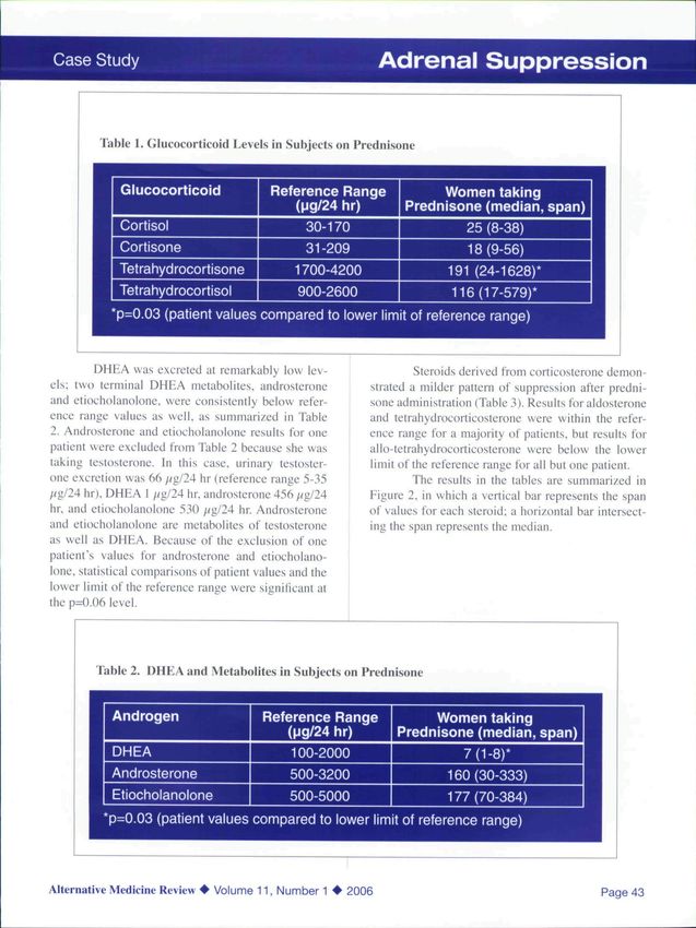
Adrenal Suppression                                                                                 Case Study

     Table 3. Corticosterone Metabolites in Subjects on Prednisone

        Corticosterone Metabolite                Reference Range              Women taking
                                                    (|jg/24 hr)          Prednisone (median, span)
        Aldosterone                                      6-25                     6(4-18)
        Tetrahydrocorticosterone                        30-240                   66(6-111)
        allo-Tetrahydrocorticosterone                   130-600                  38(6-218)
       *p=0.03 (patient values compared to lower limit of reference range)
                                                                                                                1
Discussion                                                              A second potential confounding factor is that
          The suppression of ACTH secretion after              depressed DHEA synthesis in the women studied
prednisone administration has a profound Impact on             could have been caused by their medical condition,
adrenal steroid production. The cortisol metabolites           rather than prednisone administration. The medi-
tetrahydrocortisone. letrahydrocortisol. and allo-tetra-       cal complaints included a diagnosis of fibromyalgia
hydrocortisol account for approximately 5i) percent            in two patients and "severe aches and pains" in two
of daily cortisol biosynthesis.^ The marked decline of         patients. Four patients also complained of headaches
these metabolites in these women taking prednisone             - two severe, one moderate, and one mild. However,
reflects the decrease in cortisol production that results      other studies of prednisone administration have also
from prednisone administration. Urinary cortisol and           demonstrated the same decline in DHEA production.
cortisone, which reflect circulating glucocorticoids.          For example, a study of daily versus alternate-day,
were also diminished after prednisone. but not to the          low-dose prednisone in hirsute women found that.
same extent as the terminal metabolites (Figure 2).            after both regimens, basal serum DHEA and DHBA-
          DHEA excretion was very low in this group            sulfate concentrations were suppressed to a greater
of women, which is consistent with the ACTH media-             degree than serum cortisol concentrations.^ Studies
tion of DHEA production. One potential confounding             in adolescents and adults, but not young children,
factor is that DHEA production declines with age.              demonstrate marked suppression of DHEA synthesis
The subjects ranged in age from 32-60 years, and the           after prednisone administration.'^ The prednisone-as-
laboratory reference range lower limit in this report          sociated reduction in ACTH release by the anterior
(100 //g/24 hr) reflects values for young adults. A            pituitary offers a compelling mechanism for the de-
comprehensive study by Weykamp et al determined                cline in DHEA production.
urinary DHEA excretion rates across the lifespan in                      The mineralocorticoid steroids aldosterone
males and females." They reported a reference range             and tetrahydrocorticosterone demonstrated modest
lower limit of 58 ;/g/24 hr for women ages 17-50                suppression after prednisone administration. This is
years and the same lower limit for women ages 51-70             consistent with the reports that aldosterone produc-
years. Even if this somewhat lower reference range              tion is mediated by multiple mechanisms, with ACTH
limit is used, the urinary DHEA values for subjects             stimulation responsible for approximately one-half of
in this study are remarkably low (1-8 //g/24 hr). The           aldosterone output.' The results suggest that suppres-
downstream DHEA metabolites androsterone and                    sion of aldosterone synthesis after prednisone admin-
etiocholanolone were also consistently below the ref-           istration may only be of concern in patients consum-
erence range lower limit, although not to the same              ing a low-salt diet, when demands for aldosterone
degree as DHEA values (Figure 2).                               synthesis are high. Allo-tetrahydrocorticosterone
                                                                production was suppressed to a greater extent than

Page 44                                                 Alternative Medicine Review • Volume 11, Number 1 • 2006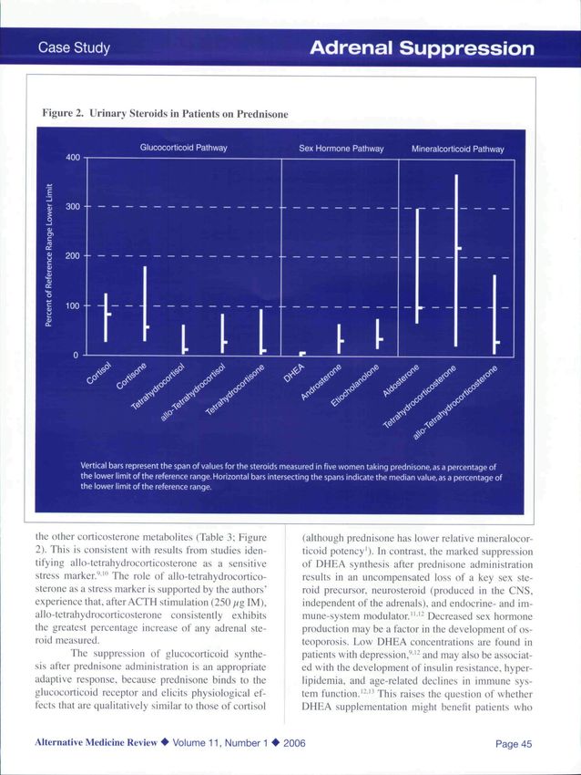
Case Study                                                   Adrenal Suppression

 Figure 2. Urinary Steroids in Patients on Prednisone

the other corticosterone metabolites (Table 3; Figure       (although prednisone has Iower relative mineralocor-
2). This is consistent with results from studies iden-      ticoid potency'). In contrast, the marked suppression
tifying allo-tetrahydrocortieosterone as a sensitive        of DHEA synthesis after prednisone administration
stress marker."'" The role of allo-tetrahydrocortico-       results in an uncompensated loss of a key sex ste-
sterone as a stress marker is supported by the authors'     roid precursor, neurosteroid (produced in the CNS,
experience that, after ACTH stimulation (250 //g IM),       independent of the adrenals), and endocrine- and im-
allo-tetrahydrocortieosterone consistently exhibits         mune-system modulator." '' Decreased sex hormone
the greatest percentage increase of any adrenal ste-        production may be a factor in the development of os-
roid measured.                                              teoporosis. Low DHEA concentrations are found in
         The suppression of glucocorticoid synthe-          patients with depression."^'- and may also be associat-
sis after prednisone administration is an appropriate       ed with the development of insulin resistance, hyper-
adaptive response, because prednisone binds to the          lipidemia. and age-related declines in immune sys-
glucocorticoid receptor and elicits physiological ef-       tem function.'-" This raises the question of whether
fects that are qualitatively similar to those of cortisol   DHEA supplementation might benefit patients who

Alternative Medicine Review • Volume 11, Number 1 • 2006                                                  Page 45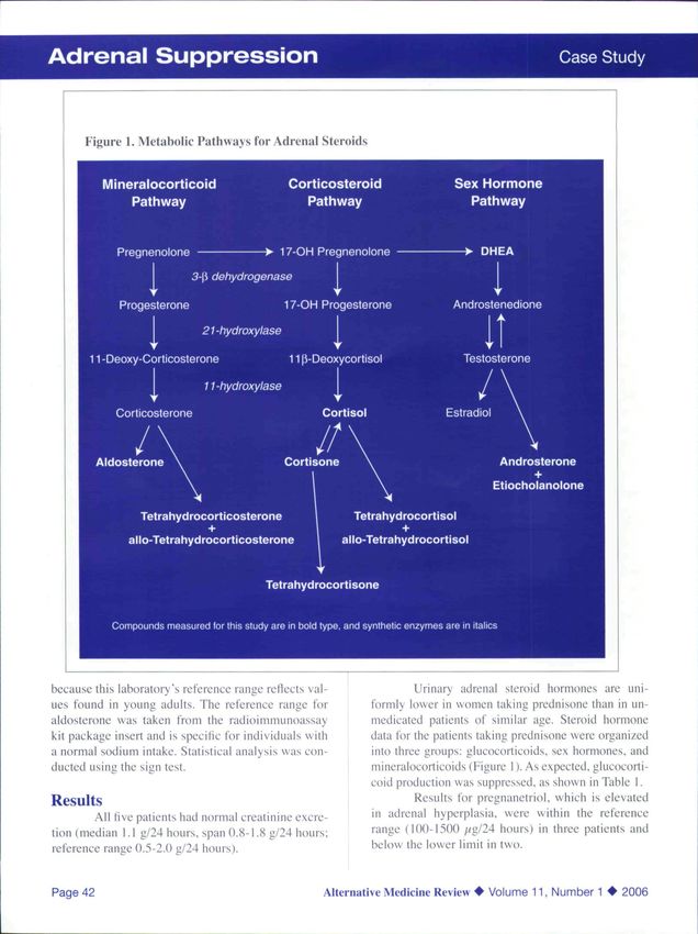
Adrenal Suppression                                                                                     Case Study

take prednisone chronically. Two studies in women              4.     Friel PN. Hinchcliffe C. Wright JV. Hormone
with lupus who were taking prednisone have shown                      replacement with estradiol: conventional oral doses
                                                                      result in excessive exposure to estrone. Altern Med
DHEA supplementation often allows patients to re-
                                                                      /eci 2005;10:36-4l.
duce prednisone dosage and may protect against the             5.     Weykamp CW. Penders TJ. Schmidt NA. et al.
bone loss associated with chronic prednisone admin-                   Steroid profile for urine: reference values. Clin
istration.'""^ In another study, women with adrenal                   Chem 1989:35:2281-2284.
insufficiency treated with 50 mg/day DHEA expe-                6.     Palermo M. Gomez-Sanehez C. Roitman E.
rienced improvement in mood, sense of well-being.,                    Shackleton CH. Quantilation of cortisol and
                                                                      related 3-oxo-4-enc steroids in urine using gas
and sexuality."'
                                                                      chromatography/mas^ speclrometry wiih stable
          The subjects described in this article repre-               isotope-labeled internal standards. Steroids
sent a small sample of women taking prednisone.                        1996:61:583-589.
Larger, more systematic studies would be more defm-            7.     Burtis CA. Ashwood FR. Tiet? NW. Tietz Textbook
itive. Nevertheless, the distinctive pattern of urinary               of Clinical ChemiMrw 3rd ed, Philadelphia. PA:
                                                                      W.B. Saunders Company; 1999.
steroid excretion in these women was noteworthy,
                                                               8.     Kreitzer PM. Blethen SL. Festa RS. Chasalow
consistent with previous reports, and is explained by                 Fl. Dehydroepiandrosterone sulfate levels are not
well-established mechanisms. Future research should                   suppressible by glucocorticoids before adrenarche.
determine how rapidly DHEA suppression occurs af-                     J Clin Endocrinol Metab 1989:69:1309^ L3 11.
ter commencement of prednisone administration and              9.     Poor V. Juricskay S. Gati A. et al. Urinary
how rapidly DHEA synthesis returns after prednisone                   steroid metabolites and 1 lbeta-hydroxysteroid
                                                                      dehydrogenase activity in patients with unipc)lar
discontinuation. Urinary steroid hormone profiling
                                                                      recurrent major depression. J Affect Disord
provides a comprehensive and cost-effective means                     2(K}4:81:55-59.
to study these changes. Continued research on DHEA              10.   Poor V, Biro i. Bufa A. et al. Urinary steroids in
replacement in patients taking prednisone is also                     young women with eating disorders. J Biochem
strongly supported by these findings.                                 Biophys Methods 2()()4:61:199-205.
                                                                11.   No author listed. DHEA monograph. Altern Med
         In conclusion. 24-hour urinary steroid hor-
                                                                      /?tn 2(K)I;6:3I4-3I8.
mone profiling demonstrates that chronic prednisone             12.    Dhatariya KK. Nair KS. Dehydrtxipiandrosterone:
administration suppresses the production of adrenal                    is there a role for replacement? Mayo Clin Proc
steroids, especially the glucocorticoids and DHEA.                    2003:78:1257-1273.
and that DHEA synthesis is the most severely affect-            13.    Dhatariya K. Bigelow ML, Nair KS. Effect of
ed. Although prednisone is capable of replacing the                   dehydroepiandrosterone replacement on insulin
                                                                      sensitivity and lipids in hypoadrenal wt)men.
activity of the suppressed glucocorticoids. the loss of               D/(//7(Vi'.v 2005:54:756-769.
DHEA is uncompensated and may have significant                  14.   Petri MA. Lahita RG. Van Vollenhoven RF et al.
adverse consequences in terms of sex hormone pro-                      Effects of prasterone on corticosteroid requirements
duction, bone health, endocrine- and immune-system                    of women with systemic lupus erythemalosus: a
function, and neuropsychiatric status.                                double-blind, randomized, placebo-controlled trial.
                                                                      Arthriti.s Rheum 2002-A6\\H2l)-\^29.
                                                                15.    Mease PJ. Ginzler FM, Gluck OS, et al. Effects of
References                                                             prasterone on bone mineral density in women with
1.    Katzung BG. Ba.sic and Clinical Pharniacoloi^y.                  systemic lupus erythematosus receiving chronic
      6th ed. Norvvalk. CT: Appleton & Laiige; 1995.                  glucocorticoid therapy. J Rheumato! 2005:32:616-
2.    Arvat E. Di Vito L. Lanfranco F. et ai. Stimulatory             621.
      effect of adrenocorticotropin on cortisol,                16.    Arlt W, Callies F van Vlijmen JC. et al.
      aldosterone, and dchydroepiaiidroslerone secretion               Dehydroepiandrosterone replacement in women
      in normal humans: dose-response study. J Clin                    with adrenal iiisunicieney. N Eni^l J Med
      Endocrinol Metab 20{}0;^5:i\4\-314(i.                            1999:341:1013-1020.
3.    Rittmaster RS. Givner ML. Effect of daily and
      alternate day low dose prednisone on scrum cortisol
      and adrenal androgens in hirsute women. J Clin
      Endocrinol Metah I988;67:40()-403.

Page 46                                                 Alternative Medicine Review • Volume 11, Number 1 • 2006
You can also read