Seven Deadly Sins of Volar Distal Radius Fracture Fixation
←
→
Page content transcription
If your browser does not render page correctly, please read the page content below
Seven Deadly Sins of Volar Distal Radius
     Fracture Fixation
     Allicia O. Imada, MD1; Jorge Orbay, MD2; Deana Mercer, MD1
     Department of Orthopaedics & Rehabilitation, The University of New Mexico Health Sciences Center,
     1

     Albuquerque, New Mexico
     2
         Miami Hand and Upper Extremity Institute, Miami, Florida
     Corresponding Author Allicia O. Imada, MD. Department of Orthopaedics & Rehabilitation,
     1 University of New Mexico, Albuquerque, NM 877131 (email: alimada@salud.unm.edu).

     Funding The authors received no financial support for the research, authorship, and publication of this article.

     Conflict of Interest The authors report no conflicts of interest.

     ABSTRACT                                                       implant failure, and complex regional pain syndrome.8-10
                                                                      We have identified seven preventable outcomes,
     Distal radius fractures are common injuries, accounting
                                                                    the seven “deadly sins,” in distal radius volar plating.
     for 17.0% of all emergency department visits. Operative
                                                                    We propose strategies for avoiding these potential
     treatment is an option when indicated. Volar plating
                                                                    outcomes.
     has become the most frequently used mode of fixation.
     Although fixed angle volar locking plates allow for            I. Inadequate Exposure
     reliable and stable fixation, complications have been          Inadequate exposure can lead to difficulty with fracture
     reported. Complications include flexor and extensor            visualization and may result in malreduction and
     tendon rupture, intra-articular screw penetration,             difficulty with accurate implant placement. In subacute
     malreduction, loss of reduction, carpal tunnel                 injuries, dorsal callus and hematoma formation can be
     syndrome, implant failure, and complex regional pain           considerable and make adequate reduction of fractures
     syndrome. We propose seven principles to avoid these           difficult.
     preventable outcomes and tips to succeed.                          The flexor carpi radialis (FCR) approach is often used
                                                                    to gain adequate exposure. The FCR tendon sheath
     Keywords: Radius, Wrist, Hand
                                                                    should be released past the trapezial ridge distally
     INTRODUCTION                                                   (Figure 1A). The radial septum is delineated, and the
                                                                    first dorsal extensor compartment is released to free the
     Distal radius fractures are common injuries, accounting
                                                                    radial styloid fragment. The brachioradialis is step-cut for
     for 17.0% of all emergency department visits.1,2 In
                                                                    later repair and also releases the radial styloid fragment.
     people over the age of 50, the mechanism of injury is
                                                                    The pronator quadratus is elevated from the watershed
     most often a fall onto an outstretched hand. Closed
                                                                    line at the tuberosity just proximal to the lunate facet
     reduction and immobilization are options for stable,
                                                                    and elevated radial to ulnar to the level of the distal
     extra-articular fractures.3
                                                                    radial ulnar joint (DRUJ). The ulnar cortical border of
        In unstable, intra-articular distal radius fractures,
                                                                    the distal radius is visualized and used for adequate
     surgical options include closed reduction and
                                                                    reduction of the distal fragment. The watershed line
     percutaneous fixation, external fixation, intramedullary
                                                                    should be clearly visible at the proximal reflection of
     nailing, open reduction internal fixation (ORIF) with
                                                                    the carpal bursa (Figure 1B). After releasing the radial
     volar or dorsal plating, and ORIF with fragment
                                                                    septum, including the first extensor compartment
     specific fixation.4 The goal of surgical fixation is to
                                                                    and brachioradialis, the next step is pronation of the
     re-establish radial inclination, volar tilt, length, and
                                                                    proximal fragment. Pronation of the proximal fragment
     articular congruity to improve range of motion,
                                                                    facilitates dorsal periosteal and callous release, which
     comfort, function, and potentially decrease the risk of
                                                                    frees the distal fragment to facilitate reduction (Figure
     post-traumatic arthritis.5,6 Volar plating has become the
                                                                    1C). Adequate exposure facilitates visualization of the
     most frequently used mode of fixation in the treatment
                                                                    surrounding nerves, arteries, and tendons and decreases
     of unstable distal radius fractures.7 Fixed angled
                                                                    the force of retraction across these structures. This
     anatomic volar locking plates provide stable fixation
                                                                    exposure provides excellent visualization and facilitates
     in osteoporotic and comminuted fractures with the
                                                                    fracture reduction.11
     goal of avoiding extensor tendon and other soft-tissue
     injuries common in dorsal plating. Complications have          II. Anatomical Structures within the Surgical Field
     been reported, including flexor and extensor tendon            Anatomical structures to be aware of include the
     rupture, intra-articular screw penetration, malreduction,      median nerve, palmar cutaneous branch of the median
     loss of reduction, carpal tunnel syndrome (CTS),               nerve, and radial artery. The surgeon should be aware

24          REVIEW ARTICLES • WJO VOL. 10 • 2021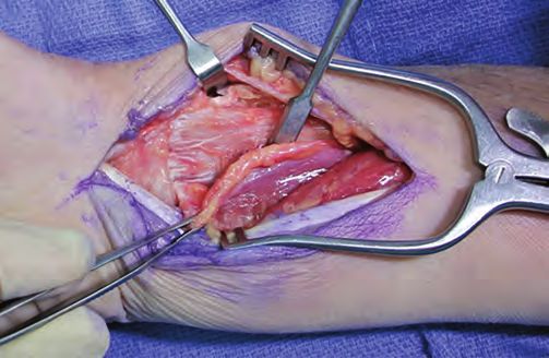
A
A
B
B
C
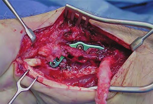
Figure 2. Photographs of dissections in the extended
flexor carpi radialis (FCR) approach. A) The arrow points
to the median nerve that lies medial to the FCR tendon
encased in fatty tissue. B) Tenotomies identifying a
C branch of the radial artery distal near the radial septum.
C) Forceps identifying the palmar cutaneous branch of
Figure 1. A) A photograph of dissection in the extended the median nerve ulnar to the FCR tendon.
flexor carpi radialis (FCR) approach. B) Release of FCR
sheath to the scaphoid tuberosity distally. C) Complete
exposure with final removal of pronator quadratus. do not improve with closed fracture reduction. If
Example of pronation of the proximal fragment in the there is a concern for compartment syndrome caused
extended FCR approach for volar fixation of distal by accompanying severe forearm or hand injury,
radius fractures. fasciotomies and carpal tunnel release should be
performed. In patients who have underlying CTS,
accompanying surgical release of the carpal tunnel
of the standard approach and proximity of these
may be considered. The median nerve is protected
structures to retractors and other instruments.
by applying retraction across the FCR tendon in the
Median nerve injury and CTS are known to occur
extended FCR approach (Figure 2A).
acutely after distal radius fractures. An article
The palmar cutaneous branch of the median nerve
describing postoperative median nerve injuries
typically branches off the radial side of the median
after distal radius fixation has been reported in the
nerve 5 cm to 6 cm proximal to the wrist crease, runs
literature.12 Carpal tunnel release should be performed
with the median nerve for 2 cm to 3 cm, and then runs
if patients present acutely with CTS symptoms that
REVIEW ARTICLES • WJO VOL. 10 • 2021 25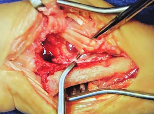
along the ulnar border of FCR (Figure 2B). The anatomy        often owing to the healing of the distal radius fracture in
     of the palmar cutaneous nerve is variable. Care should        a malreduced position. Contralateral wrist radiographs
     be taken to sharply incise the volar ulnar aspect of the      can be helpful and used as a guide to re-establish
     deep FCR sheath because the palmar cutaneous branch           anatomical ulnar variance, owing to the variability
     is typically deep to the sheath and lies ulnar.13             across individuals. The extended tangential view can
         The radial artery branch that traverses the distal        help assess intra-articular reduction of the sigmoid
     radius volarly is at risk during release of the radial        notch, DRUJ alignment, and screw breakthrough into
     septum distally in the extended FCR approach                  the DRUJ.19,20 Distal radius malunion can restrict forearm
     (Figure 2C). One case report in the literature reports        rotation and alter DRUJ kinematics, causing pain and
     a radial artery pseudoaneurysm after volar distal radius      instability.21,22
     ORIF following postoperative collapse and dorsal
                                                                   IV. Plate Placement
     displacement of the fracture.14
                                                                   Plate placement may lead to flexor tendon irritation.
     III. Malreduction                                             Rupture can lead to intra-articular screw penetration
     Malreduction can lead to injury of the flexor tendons,        because the plates are anatomical. Improper plate
     particularly the flexor pollicus longus (FPL) and             placement can also contribute to the loss of fixation
     flexor digitorum profundus (FDP) of the index finger.         because the distal fragments are not well stabilized. One
     In general, acceptable closed reduction parameters            of the benefits of volar plating is the decreased risk of
     include radial height of 8 mm to 12 mm, radial inclination    tendon irritation and rupture, unlike dorsal plating. The
     of 21° on anteroposterior view and a neutral tilt of          correct technique should be employed to maximize this
     10° volar tilt on lateral, and intra-articular step-off of    benefit.
     less than 2 mm.15 Many of the newer volar fixed angle
     plates are designed to fit on the volar surface of distal
     radii. The plate can be used to facilitate anatomical          A
     reduction because the fracture can be reduced to the
     plate contour. Plates may not fit well when anatomic
     reduction is not achieved, leading to problems such as
     intra-articular screw penetration and loss of fixation
     if the screws are not placed subchondral. Adequate
     reduction using fixed angle devices is key to fixation
     of distal radius fractures. The extended FCR approach
     provides excellent visualization of the volar distal radius
     and facilitates the reduction of complex distal radius
     fractures.
         Loss of reduction is most often the result of
     inadequate fixation of the multiple distal fracture
     fragments. Malreduction of the fracture can also lead
     to inadequate fixation because the volar plates are
     designed to capture the fragments in near anatomical           B
     reduction. Loss of fixation presents most commonly
     with dorsal collapse, loss of reduction of the volar
     lunate facet, or radial shortening.8 A cadaveric study
     showed that radii with distal screws placed over 4 mm
     proximal to the subchondral bone had significantly more
     radial shortening than when fixed with screws closer
     to the subchondral bone.16 Fragment-specific fixation
     can be particularly useful in volar marginal fragment
     fractures, in which capturing the volar-ulnar corner is
     critical for radiocarpal stability.17 Using the anatomical
     plate as a guide followed by reduction and fixation into
     the proximal shaft, the distal fragment first technique
     can be used where distal locking screws are placed to         Figure 3. A) In the extended flexor carpi radialis
     capture the distal fragment. This technique helps correct     approach: Identification of the watershed line (I), flexor
     volar tilt.18 Surgeons should aim for anatomic reductions     digitorum profundus (FDP) to the index finger, and
     with the subchondral placement of screws and should           flexor pollicis longus (FPL) in relation to a volar fixed-
     consider various reduction techniques such as using the       angle distal radius plate. B) FPL rupture in a patient who
     plate as a reduction tool.                                    was taken back to the operating room for exploration
         After treatment of distal radius fractures, DRUJ          after inability to extend their thumb after distal radius
     problems can be a considerable source of morbidity,           volar open reduction internal fixation.

26        REVIEW ARTICLES • WJO VOL. 10 • 2021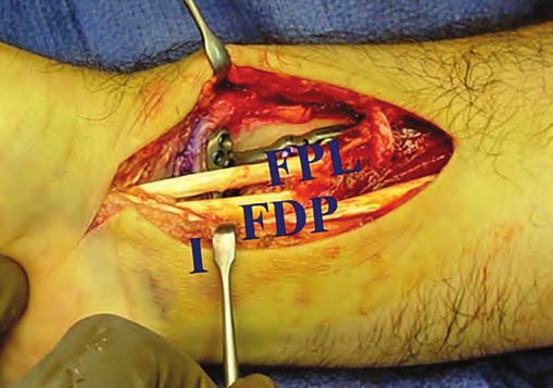
The watershed line is a ridge on the distal radius
                                                            distal screws unicortically instead of bicortically. In a
just distal to the pronator quadratus and proximal to
                                                            biomechanical study, Wall et al28 showed unicortical
the volar ligaments of the wrist. Plates placed distal to
                                                            locking screws of at least 75.0% length produced similar
this line have been shown to have increased rates of
                                                            construct stiffness to bicortical screws. Pegs or screws
flexor tendon injury, most commonly to the FPL and
                                                            will lock into the plate, minimizing the risk of projection
FDP of the index finger (Figure 3A and 3B).23,24 Screw-
                                                            past the dorsal cortex.
back out has also been reported to cause flexor tendon
                                                               While extensor tendon rupture is much less common
problems.16,25,26 Re-establishing adequate volar tilt and
                                                            with volar fixation of distal radius fractures compared
repairing the pronator quadratus may also aid in the
                                                            to dorsal fixation, extensor tendon rupture and synovitis
protection of the flexor tendons because the pronator
                                                            have been reported.29-32 Prominent dorsal screws and
quadratus can be used to cover the entire distal portion
                                                            drill tip penetration have been thought to contribute
of the plate. Anatomic fixed angle plates should be
                                                            to extensor tendon rupture. Lister’s tubercle can
placed proximal to the watershed line. Care should
                                                            make it challenging to identify dorsal screws that are
be taken to restore volar tilt and repair the pronator
                                                            prominent. Live fluoroscopy, the dorsal tangential,
quadratus to minimize the risk of flexor tendon injury.
                                                            or the skyline view can aid in the evaluation of screw
V. Screw Trajectory/Placement                               lengths dorsally.33 Fracture type has been shown to be
Screws can be placed with a trajectory that may             associated with extensor pollicis longus (EPL) rupture
lead to intra-articular breakthrough, loss of fixation      risk. A retrospective review of patients with distal
if not placed subchondral, or rupture of the extensor       radius fractures treated at a single institution with
tendons. Screws can inadvertently be placed into the        volar locking plates found a 5.7% EPL rupture rate.32
joint (Figure 4), as reported in a series by Arora et       EPL rupture was found to have a high association
al.25 The goal of fixed angle, volar locked plating is      to patients with fracture extension into or through
plate placement proximal to the watershed line and          Lister’s tubercle.32 Similarly, Lee et al34 found a higher
the subchondral placement of screws. The 30° lateral        association of EPL rupture in patients with “displaced
allows a tangential view of the radiocarpal joint and       dorsal beak fracture of Lister’s tubercle.” Callus
is the best method of assessing subchondral screw           formation, even without screw or drill penetration, can
placement because it provides excellent visualization of    narrow the EPL groove.
the articular surface.27 Screw lengths should be checked
                                                            VI. Postoperative Management
and shortened if needed based on tangential lateral
                                                            Inadequate pain management and delayed rehabilitation
fluoroscopy. The extended tangential view can also
                                                            can lead to poor outcomes after distal radius fracture
be used to evaluate screw lengths. Collapse of intra-
                                                            fixation.
articular fractures can also result in penetration of the
                                                               Mobilization and rehabilitation after distal radius
joint by screws.
                                                            ORIF have been studied, and most surgeons advocate
    Fixed angle anatomic distal radius plates rely on
                                                            for a period of postoperative immobilization.35 A
subchondral placement. Bicortical fixation of distal
                                                            recent randomized clinical trial of 133 adults found
pegs or screws is not required. One technique to avoid
                                                            that immobilization of 1 to 3 weeks after ORIF resulted
tendon or intra-articular complications involves drilling
                                                            in superior function, range of motion, and pain
                                                            management.36 Lozano-Calderón et al37 studied smaller
                                                            groups of patients and found no significant differences
                                                            in two groups of 30 patients who began wrist range of
                                                            motion 6 weeks after volar plate fixation versus 2 weeks.
                                                            While the exact length of immobilization is debated, we
                                                            recommend up to 2 weeks of immobilization, depending
                                                            on the fracture and surgeon preference. Hand therapy
                                                            should be started soon after immobilization has been
                                                            discontinued in patients with clinical stiffness. Finger
                                                            and thumb range of motion needs to be emphasized
                                                            whether or not the fractures are treated with surgery.
                                                            Elevation of the hand and wrist can be helpful to minimize
                                                            postoperative and post-injury swelling, which can
                                                            adversely affect outcomes because swelling limits motion.
                                                               While distal radius ORIF and other orthopaedic bony
                                                            procedures are known to be painful, the prescription
            Figure 4. A sagittal                            of narcotic pain medications has become a large topic
            computerized tomography                         of debate in recent times. In a prospective series
            scan of volar distal radius                     of patients undergoing upper extremity surgical
            plate with distal intra-articular               procedures, Kim et al38 found that their surgeons were
            extension of screw.                             prescribing on average 24 opioid pills, and patients

                                                                              REVIEW ARTICLES • WJO VOL. 10 • 2021        27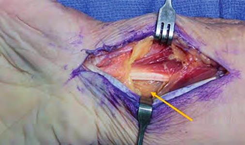
consumed on average 8.1 pills over an average of 3.1          pain postoperatively. Vitamin C may be prescribed for
     days. Teunis et al39 found that male sex and greater          50 days after the initial fracture. If patients present
     dorsal angulation of the articular surface on lateral         with concerning signs of tendonitis or tendon rupture,
     radiograph were associated with requesting a second           plates should be removed, and patients with ruptures
     opioid prescription after locking plate fixation of distal    should be treated with appropriate tendon procedures.
     radius fractures. We recommend prescription of no more        Avoiding the “seven deadly sins” proposed above can
     than 20 opioid pills to patients after distal radius ORIF     lead to fewer complications in the surgical treatment of
     over 3 days to 2 weeks. Patients should be counseled          distal radius fractures.
     to expect some postoperative pain and that additional
     narcotics are not recommended after the first 2-week          REFERENCES
     postoperative visit.                                          1.    Bonafede M, Espindle D, Bower AG. The direct and
        Complex regional pain syndrome (CRPS) may                        indirect costs of long bone fractures in a working age
     occur after distal radius ORIF and can be difficult to              US population. J Med Econ. 2013;16(1):169-178. doi:
     treat.40 Various studies have suggested that if started             10.3111/13696998.2012.737391.
     on the date of injury, vitamin C supplementation for          2.    MacIntyre NJ, Dewan N. Epidemiology of distal radius
     50 days may decrease the risk of CRPS after distal                  fractures and factors predicting risk and prognosis.
                                                                         J Hand Ther. 2016;29(2):136-145. doi: 10.1016/j.
     radius fracture, while follow-up studies have shown no
                                                                         jht.2016.03.003.
     benefit.41-43 While the evidence is controversial, vitamin
                                                                   3.    Vannabouathong C, Hussain N, Guerra-Farfan E, et
     C supplementation is quite benign, and we recommend
                                                                         al. Interventions for distal radius fractures: a network
     500 mg daily to our patients for 50 days post-injury.
                                                                         meta-analysis of randomized trials. J Am Acad
     VII. Removal of Implants                                            Orthop Surg. 2019;27(13):e596-e605. doi: 10.5435/
     Volar-locked plates rarely need to be removed, but if               JAAOS-D-18-00424.
     there is evidence of tendonitis or tendon rupture, one        4.    Sammer DM, Fuller DS, Kim HM, et al. A comparative
     should not hesitate. Patients may describe a sudden loss            study of fragment-specific versus volar plate
     of active range of motion of a finger, indicating tendon            fixation of distal radius fractures. Plast Reconstr
     rupture. In tendonitis, patients often present with dorsal          Surg. 2008;122(5):1441-1450. doi: 10.1097/
     swelling proximal and distal to the extensor retinaculum.           PRS.0b013e3181891677.
     Early removal of symptomatic plates is important in           5.    Knirk JL, Jupiter JB. Intra-articular fractures of the
     preventing tendon rupture.                                          distal end of the radius in young adults. J Bone Joint
                                                                         Surg Am. 1986;68(5):647-659.
        The rate of plate removal ranges from 3.0% to 10.0%
                                                                   6.    Gruber G, Zacherl M, Giessauf C, et al. Quality of
     in the literature, most commonly for flexor tendon
                                                                         life after volar plate fixation of articular fractures of
     rupture or irritation.44,45 Timeframe of rupture is on
                                                                         the distal part of the radius. J Bone Joint Surg Am.
     average 6 months to 26 months postoperatively.
                                                                         2010;92(5):1170-1180. doi: 10.2106/JBJS.I.00737.
     Patients should be counseled about warning signs of
                                                                   7.    Mattila VM, Huttunen TT, Sillanpää P, et al. Significant
     flexor tendon irritation, including difficult and painful
                                                                         change in the surgical treatment of distal radius
     flexion of the thumb, fingers, and volar wrist synovitis.46         fractures: a nationwide study between 1998 and 2008
     If patients do present with any of these warning signs              in Finland. J Trauma. 2011;71(4):939-942; discussion
     or symptoms, they should be offered removal of the                  942-943. doi: 10.1097/TA.0b013e3182231af9.
     implant.                                                      8.    Berglund LM, Messer TM. Complications of volar
                                                                         plate fixation for managing distal radius fractures.
     CONCLUSION
                                                                         J Am Acad Orthop Surg. 2009;17(6):369-377. doi:
     Distal radius fractures are common. Fixed angle volar               10.5435/00124635-200906000-00005..
     locking plates are the most common fixation method            9.    Johnson NA, Cutler L, Dias JJ, et al. Complications
     for distal radius fractures, with excellent outcomes if             after volar locking plate fixation of distal radius
     done technically well. Complications of volar plating               fractures. Injury. 2014;45(3):528-533. doi: 10.1016/j.
     include flexor and extensor tendon injury, malreduction,            injury.2013.10.003.
     intra-articular screw penetration, nerve and artery injury,   10.   Navarro CM, Pettersson HJ, Enocson A.
     and pain and stiffness. The extended FCR approach                   Complications after distal radius fracture surgery:
     can aid in visualization, reduction, and proper plate               results from a Swedish nationwide registry study.
     placement proximal to the watershed line, with the                  J Orthop Trauma. 2015;29(2):e36-42. doi: 10.1097/
     goal of anatomic reduction and distal screw placement               BOT.0000000000000199.
     subchondral and extra-articular. Tangential lateral,          11.   Orbay JL, Badia A, Indriago IR, et al. The extended
     extended tangential, and live fluoroscopy can aid in                flexor carpi radialis approach: a new perspective for
     the examination of screw lengths dorsally. A period of              the distal radius fracture. Tech Hand Up Extrem Surg.
                                                                         2001;5(4):204-211. doi: 10.1097/00130911-200112000-
     up to 2 weeks of immobilization should be followed
                                                                         00004.
     by encouraged range of motion and hand therapy.
     Opioids should be prescribed for no more than 2
     weeks. Patients should be counseled to expect some

28        REVIEW ARTICLES • WJO VOL. 10 • 2021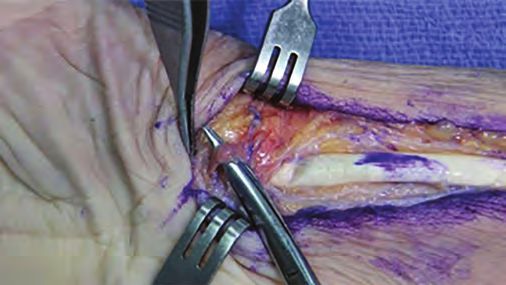
12. Stern PJ, Derr RG. Non-osseous complications               26. Bhattacharyya T, Wadgaonkar AD. Inadvertent
    following distal radius fractures. Iowa Orthop J.              retention of angled drill guides after volar locking
    1993;13:63-69.                                                 plate fixation of distal radial fractures: a report of
13. Samson D, Power DM. Iatrogenic injuries of the                 three cases. J Bone Joint Surg Am. 2008;90(2):401-
    palmar branch of the median nerve following volar              403. doi: 10.2106/JBJS.G.00939.
    plate fixation of the distal radius. J Hand Surg           27. Kumar D, Breakwell L, Deshmukh SC, et al. Tangential
    Asian Pac Vol. 2017;22(3):343-349. doi: 10.1142/               views of the articular surface of the distal radius-aid
    S021881041750040X.                                             to open reduction and internal fixation of fractures.
14. Dao KD, Venn-Watson E, Shin AY. Radial artery                  Injury. 2001;32(10):783-786. doi: 10.1016/s0020-
    pseudoaneurysm complication from use of AO/ASIF                1383(01)00155-3.
    volar distal radius plate: a case report. J Hand Surg      28. Wall LB, Brodt MD, Silva MJ, et al. The effects of
    Am. 2001;26(3):448-453. doi: 10.1053/jhsu.2001.24138.          screw length on stability of simulated osteoporotic
15. Lichtman DM, Bindra RR, Boyer MI, et al. Treatment             distal radius fractures fixed with volar locking plates.
    of distal radius fractures. J Am Acad Orthop                   J Hand Surg Am. 2012;37(3):446-453. doi: 10.1016/j.
    Surg. 2010;18(3):180-189. doi: 10.5435/00124635-               jhsa.2011.12.013.
    201003000-00007.                                           29. Benson EC, DeCarvalho A, Mikola EA, et al. Two
16. Drobetz H, Bryant AL, Pokorny T, et al. Volar fixed-           potential causes of EPL rupture after distal radius
    angle plating of distal radius extension fractures:            volar plate fixation. Clin Orthop Relat Res.
    influence of plate position on secondary loss of               2006;451:218-222. doi: 10.1097/01blo.0000223998.
    reduction--a biomechanic study in a cadaveric model.           02765.0d.
    J Hand Surg Am. 2006;31(4):615-622. doi: 10.1016/j.        30. Failla JM, Koniuch MP, Moed BR. Extensor pollicis
    jhsa.2006.01.011.                                              longus rupture at the tip of a prominent fixation screw:
17. Hozack BA, Tosti RJ. Fragment-specific fixation in             report of three cases. J Hand Surg Am. 1993;18(4):648-
    distal radius fractures. Curr Rev Musculoskelet Med.           651. doi: 10.1016/0363-5023(93)90310-Y.
    2019;12(2):190-197. doi: 10.1007/s12178-019-09538-6.       31. Al-Rashid M, Theivendran K, Craigen MA. Delayed
18. Im JH, Lee JY. Pearls and pitfalls of the volar                ruptures of the extensor tendon secondary to the use
    locking plating for distal radius fractures. J Hand            of volar locking compression plates for distal radial
    Surg Asian Pac Vol. 2016;21(2):125-132. doi: 10.1142/          fractures. J Bone Joint Surg Br. 2006;88(12):1610-1612.
    S242483551640004X.                                             doi: 10.1302/0301-620X.88B12.17696.
19. Klammer G, Dietrich M, Farshad M, et al.                   32. Cha SM, Shin HD, Lee SH. "Island-shape" fractures
    Intraoperative imaging of the distal radioulnar joint          of lister's tubercle have an increased risk of delayed
    using a modified skyline view. J Hand Surg Am.                 extensor pollicis longus rupture in distal radial
    2012;37(3):503-508. doi: 10.1016/j.jhsa.2011.12.009.           fractures: after surgical treatment by volar locking
20. El Naga AN, Jordan ME, Netscher DT, et al. Reliability         plate. Injury. 2018;49(10):1816-1821. doi: 10.1016/j.
    of the dorsal tangential view in assessment of distal          injury.2018.08.019.
    radioulnar joint reduction in the neutral, pronated,       33. Taylor BC, Malarkey AR, Eschbaugh RL, et al. Distal
    and supinated positions in a cadaver model. J Hand             radius skyline view: how to prevent dorsal cortical
    Surg Am. 2020;45(4):359.e1-359.e8. doi: 10.1016/j.             penetration. J Surg Orthop Adv. 2017;26(3):183-186.
    jhsa.2019.08.004.                                          34. Lee JK, Bang JY, Choi YS, et al. Extensor pollicis
21. Abe S, Oka K, Miyamura S, et al. Three-dimensional             longus tendon rupture caused by a displaced
    in vivo analysis of malunited distal radius fractures          dorsal "beak" fragment of Lister's tubercle in distal
    with restricted forearm rotation. J Orthop Res.                radius fractures. Handchir Mikrochir Plast Chir.
    2019;37(9):1881-1891. doi:10.1002/jor.24332.                   2019;51(3):199-204. English. doi: 10.1055/a-0826-4731.
22. Nishiwaki M, Welsh MF, Gammon B, et al. Effect of          35. Quadlbauer S, Pezzei C, Jurkowitsch J, et al.
    volarly angulated distal radius fractures on forearm           Rehabilitation after distal radius fractures: is there
    rotation and distal radioulnar joint kinematics. J             a need for immobilization and physiotherapy? Arch
    Hand Surg Am. 2015;40(11):2236-2242. doi: 10.1016/j.           Orthop Trauma Surg. 2020;140(5):651-663. doi:
    jhsa.2015.07.034.                                              10.1007/s00402-020-03367-w.
23. Orbay JL, Touhami A. Current concepts in volar fixed-      36. Watson N, Haines T, Tran P, et al. A Comparison of the
    angle fixation of unstable distal radius fractures. Clin       effect of one, three, or six weeks of immobilization
    Orthop Relat Res. 2006;445:58-67. doi: 10.1097/01.             on function and pain after open reduction and
    blo.0000205891.96575.0f.                                       internal fixation of distal radial fractures in adults: a
24. Bell JS, Wollstein R, Citron ND. Rupture of flexor             randomized controlled trial. J Bone Joint Surg Am.
    pollicis longus tendon: a complication of volar plating        2018;100(13):1118-1125. doi: 10.2106/JBJS.17.00912.
    of the distal radius. J Bone Joint Surg Br. 1998;80(2):    37. Lozano-Calderón SA, Souer S, Mudgal C, et al. Wrist
    225-226. doi: 10.1302/0301-620x.80b2.8351.                     mobilization following volar plate fixation of fractures
25. Arora R, Lutz M, Hennerbichler A, et al. Complications         of the distal part of the radius. J Bone Joint Surg Am.
    following internal fixation of unstable distal                 2008;90(6):1297-1304. doi: 10.2106/JBJS.G.01368.
    radius fracture with a palmar locking-plate. J
    Orthop Trauma. 2007;21(5):316-322. doi: 10.1097/
    BOT.0b013e318059b993.

                                                                                 REVIEW ARTICLES • WJO VOL. 10 • 2021          29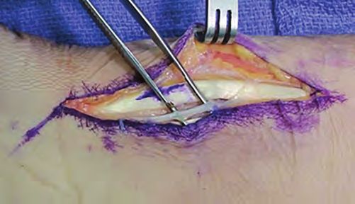
38. Kim N, Matzon JL, Abboudi J, et al. A prospective           43. Evaniew N, McCarthy C, Kleinlugtenbelt YV, et
         evaluation of opioid utilization after upper-extremity          al. Vitamin C to prevent complex regional pain
         surgical procedures: identifying consumption patterns           syndrome in patients with distal radius fractures:
         and determining prescribing guidelines. J Bone Joint            a meta-analysis of randomized controlled trials. J
         Surg Am. 2016;98(20):e89. doi: 10.2106/JBJS.15.00614.           Orthop Trauma. 2015;29(8):e235-241. doi: 10.1097/
     39. Teunis T, Stoop N, Park CJ, et al. What factors are             BOT.0000000000000305.
         associated with a second opioid prescription after          44. Lutsky KF, Beredjiklian PK, Hioe S, et al. Incidence
         treatment of distal radius fractures with a volar locking       of hardware removal following volar plate
         plate? Hand (N Y). 2015;10(4):639-648. doi: 10.1007/            fixation of distal radius fracture. J Hand Surg Am.
         s11552-015-9767-6.                                              2015;40(12):2410-2415. doi: 10.1016/j.jhsa.2015.09.017.
     40. DeGeorge BR Jr, Brogan DM, Becker HA, et al.                45. Snoddy MC, An TJ, Hooe BS, et al. Incidence and
         Incidence of complications following volar locking              reasons for hardware removal following operative
         plate fixation of distal radius fractures: an analysis of       fixation of distal radius fractures. J Hand Surg Am.
         647 Cases. Plast Reconstr Surg. 2020;145(4):969-976.            2015;40(3):505-507. doi: 10.1016/j.jhsa.2014.11.022.
         doi: 10.1097/PRS.0000000000006636.                          46. Asadollahi S, Keith PP. Flexor tendon injuries following
     41. Aïm F, Klouche S, Frison A, et al. Efficacy of vitamin C        plate fixation of distal radius fractures: a systematic
         in preventing complex regional pain syndrome after              review of the literature. J Orthop Traumatol.
         wrist fracture: a systematic review and meta-analysis.          2013;14(4):227-234. doi: 10.1007/s10195-013- 0245-z.
         Orthop Traumatol Surg Res. 2017;103(3):465-470. doi:
         10.1016/j.otsr.2016.12.021.
     42. Ekrol I, Duckworth AD, Ralston SH, et al. The influence
         of vitamin C on the outcome of distal radial fractures:
         a double-blind, randomized controlled trial. J Bone
         Joint Surg Am. 2014;96(17):1451-1459. doi: 10.2106/
         JBJS.M.00268.

30        REVIEW ARTICLES • WJO VOL. 10 • 2021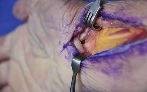
You can also read